A. O. Vet. Fak. Derg. 33 (3) : 416-433, 198(,
HlSTOPATHOLOGICAL CHANGES IN THE nSSl'ES OF THE FRESH WATER S;\ıAIL GYRAULUS LAEVIS (ALDER) PRODUCED BY
PARAi\1PHISTOML'M LARVAL STAGES
Erian G. Kame)l Ayşe Burgu~
Paramphistomum gelişme dönemlerinin tatlısu sümüklüsü Gyraulus lae\is (Alder) ooku-rarında meydana getirdiği histopatoiojik değir-iklik!er.
Ö7-et: Tatltsu SÜ111iilclüsüGyraulus laevİs (Alder) ve Paramphis-tomum serlcer/eri ile ilgili seri araştırma/ann ikincisi olan bu çaltşma, Paramphistomu!1l gelişme dönemlerinin sümiikliideki lokalizasyonianm ve özellik/e slimüklü doku/amıda neden olduklan histopatolojik değişik-lik/eri saptamağa yljneliktir.
Binokü/er disseksiyon mikroskobu ku/lamlarak yapi/an kontrollerde saydam o/an kabuk yapi/amıdan ötürii sümüklü içersindeki sporokist, redi, serker gibi gelişme dönemlerini görerek enfekte G. laevİs' lerin ayrl/11I mümkün olabilmektedir.
Histopatolojik yok/ama/ar için aynlan örnekler sert kabuk k1S1111-lan uzak/aştm/dıktan sonra Bouin sıvısmda tespit, alkol serilerinden geçirilerek dehidre edilmiş ve ksilolde saydam/aştmldiktan sonra para/in bloklara alznnllştır. Bu blok/ardan 8-12 (J.111kalmhğmda kesitler yapi/mış ve hematoksi/en-eozin ile boyamnlştır.
Kesitlerin incelenmesinde,. Paramphistomum/ann sporokist, redi ve serker gibi gelişme dönem/erine başltea sindirim bezi ve ovotestis'lerde rast/anl1l1ştır. Bu lokalizasyonlara bağli olarak sindirim bezi tüplerinin sayısında belirgin bir azalma olduğu, kaybolan doku klSlmlannlıı yerinde parazİt gelişme dönemlerinin bulunduğu gözlenmiştir. Ovotestis'i çevre-leyen kan sinüs/erinde de Paramphistoli1um gelişme dönemlerinin yer-leştiği, foliküller içerisinde sporokist ve redilere rast/andığı, dolayısıyla cinsiyet hücre/erinin de zarar gördüğü saptanmıştır. Bunlara ilaveten,
i Associate Professor, Ain Shams Un:versity, Women College For Arts, Science and Education, Cairo, Egypt.
HlSOPATHOlOGICAL CGANGES IN THE TISSLJES... 117
ayak ve höhrek te daha az olarak ParamphistolJlum gelişme dönemlerine rastlanmıştır. Paramphistomum gelişme dönemleriyle enfekte sümük-tülerde, sindirim bezi ve ovotestislerin ençok zarar gören iki organ 01-olduğu saptanmıştır.
Summary: The present paper is the second in the series dealt with
the fresh water snail Gyraulus laevis (Alder) and Paramphistomum larval stages. This ıvork was direeted toıvards aıı investigatioıı of the oeeurrenee of the Paramphistomum larval stages withiıı the .mail G. laevis and more particulady their histopathological effeets on the .mail tissue.>.
Gyraulus laevis parasitized with ParmnphistomUln larval stages eould be positively identified by deteeting the diflerellt developing stages of the parasite such as sporocysts, rediae aııd eercariae through the transparent shell using binoeular dissecting microseope.
For histopathological examinations, representative snails were rixed using Bouin's fluid, dehydrated, eleared and paraffin embedded. Seetions were eut at 8-12 mierons iıı thiekness and stained with haema-toxyliıı and eosin.
The results indicated that the majority of the Paramphistomu//1 larval stages, sporoeysts, rediae and eercariae, parasitized G. laevis were located maiııly in the region of the digestive gland and ovotestis. These two organs I1weseverely damaged hy the presence of Paramphis-tomU//1 larval stages. There ıvas a considerahle reduction ilZ the number
of the digestive gland tubules. Tlıeir tissues l1we replaced hy the parasile. On the other hand Paramphistomunı larval stages settled in the blood sinuses surrounding the ovotestis. Sporoeysts and rediae were oceurred iliside the tubules and invaded tire adni damaging the sex eells. In addi-tion foot, and kidney were affeeted by Paramplıistomum larval stages.
Introduction
The vital activities of an animal whose body is İnvadcd by other organism become very greatly disturbed. One can expect to find signi-ficant changes induced by the foreign organism, such alternation from the normal can be expected as mechanical, histopathologicaJ, physİ-ological, biochemical, eve n sometimes morphoJogical changes (2,14,
4Jıı E.G. KAMEL - A. BURGU
Mechailical changes are recognized as the local strueture distur-bance; morphologieal changes such as abnormal proliferation of the tissues and eyst formation; physiologieal and bioehemieal effects are expressed in the Iimitation or modifieation of the normal proeesses of the host, the histopathologieal changes described as the damages which the foreign organisms produee in their hosts tissues (5,17,18,25).
The mollusca is a partieularly large and remarkable diverse phylum in the animal kingdom. Many speeies of gastropod moIluses suffer digenetie trematode larval infeetions. The histopathologieal effeets of the parasite on the snail host have been studied by many authors (i ,9,10,1 I, i5, i6,22).
One is impressed by the degree of damage which the larval trema-todes produce in the snail hosts, and at the same time one is equaIly impressed by the toleranee of the snail to thesc infeetions.
The sporoeysts and rediae of trematodes affeet the genitalia, albumen gland, the prostate, the uterus, the sperm duet, and indireetly affect the reprod uctive eapaeity of the parasitized snail (l5,16).
The damages that larval flukes cause are produce when the larval stages migrate through the various tissues or by the consumption of the larvae of absorbed nutritiye materia\. The amount of these dama-ges depend on whether the infection is light, moderate or heavy and by aecumulation of large amount of toxie waste produets exereted by the parasite. Digenetie larval trematodes are differ in the extent of the harm theyeause to the host. Some cause little damage on the host, others are very destmetive and the pathologieal changes in the host tissues are very pronouneed. lt can be stated in general that the trema-to des whieh posses aredial stages in the life cycle are more injurious than others to their snail hosts (2,6,25,26).
Cheng and Snyder (7) reported the destmetive effeet on the diges-tive gland of Heliosoma trivolvis infeeted with Glyptlıelmins pennsylva-niensis. Bouros (3) indicated that larval trematodes parasitized Lym-naea stagnalis harm their host to the extent of inhibiting its growth and shortening its life span. Sehmid et a\. (23) on infeeted Planorbis planorbis with Paranıplıistomum cervi larval stages showed an inere-ased in mortality and deerease in fertility. Histopathologieal studies were made on the details of sporoeysts, rediae and cereariae of Pa-ramphistomuJ17 cervi in the snail host Planorbis planorbis and its
reac-L_
HlSOPATHOLOGıCAL CGANGES ıN THE TlSSUES... ,llı)
tions on host tİssues. Damages of the host eaused by the parasite were found mainly in the oyotestİs whereby its funetion was severely af-feeted (8).
Reeently Kamel et ai. (13) on the oeeurrenee of xiphidioeereariae and theİr histopathologieal effeets on the tissues of the fresh water snail Lanis/es carina/us, showed that in heavily parasitized snails the gonad were eompletely destroyed and replaeed by the parasite, the digestive gland was highlv redueed in addition the etenidia and kidncy were also affccted by xiphidiocercariae.
The present study is the second of a series that deals with the fresh water snail Gyraulus laevis (Alder) and the Paramphistomum larva! stages.
The present invcstigation was earried out to observe the loeation and the histopathologieal effeets of Paramphistomum larval stages on the tissues of the snail host Gyraulus laevis.
Materİals and Methods
Gyrau/us lael'is snails were eoııeeted from Eskişehir Çifteler State Farm, west of Ankara, Turkey. The snails were transported to the iaboratory and they were kept in plastie aquaria eontaining dechlo-rinated tap water. Snails were fed on frcsh lettuee leaves.
Parasitized spceimens with Paramphistomum larval stages eould be positively identified and scparated from unparasitized ones by de-teeting the different developing stages of the parasite such as sporoeysts, rediae and eereariae through the transparent sheıı of the snail G. laevis using binocular disseeting mieroscope, or by keeping individual snail in a container half filled with deehlorinated water for one hour exposed to light to allow mature cereariac to emerge (13,21).
For histop:.ı.thologieal examinations, representative specimens were fixed using 80uin's fluid, whieh proved to be satisfactory for this work. The material was then dehydrated, eleared and paraffin embed-ded. Seetions werc eut at 10-12 mİcrons in thiekness and stained with haematoxylin and eosİn.
Results
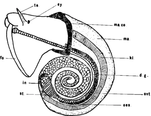
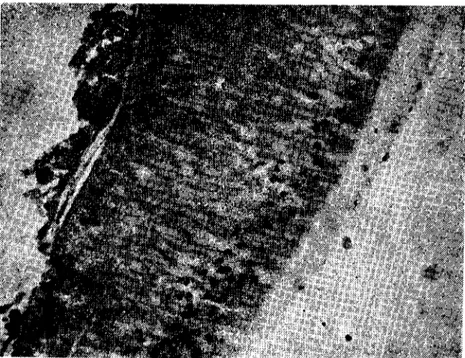
In the fresh water snail Gyraulus laevis the digestive gland is in the form of a spirally coiled eo ne, which extends down to the posterior
420 E.G. KAMEL - A. BURGU
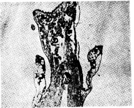
end of the visceral mass, and occupics the greater part of the visceral hump which fills the shell spire above the body whole. it is light gre-yish or rather blackish in colour. The stomach lies embedded in the digestive gland, while the ovatestis extend over the right side and can be differentiated from the digestive gland by its colour and structure. (Fig. I).
ma.
ki.
oes.
Fig. ı.Left side of Gyraıılus laevis (Alder) removed from the shell (Kabuksuz Gyraulııs tael'is (Alderrin sol yanı)
d.g.
ovt.
d.g.-digesıiye gland (sindirim bezi) fO.-:oot (ayak)
ki.-kidney (böbrek)
ma.eo.-manıle eollar (manto yakası) Oyt.-oyolestis (oyotesıis) te.-tentac1e (ıenıakü!) ey.-eye (göz) in.-inıesline (baı'sak) ma.-mantle (manto) oes.-ocsophagus (özefagus) st.-slomach (mide)
HlSOPATHOLOGICAL CHANGES IN THE TlSSUES... 421
Normally the digestive gland consists of collecting tubules. Each tubule consists of two types of ce lls, the digestive cells (absorptive) and the excretory cells. The digestive cells are numerous and contains basely placed nuclei with the cytoplasm firly evenly distributed throug-hout the cells. These cells have been defined by various authors as pre-fixes liver-ferment, glandular and absorptive. On the other hand, the excretory cells are less abundant and occur between the digestive cells. The nuclei of the secretory cells are relatively large, the cells contain densely granular cytoplasm. The function of the excretory cell is to extract metabolic excretory products from the visceral haemocael and to pass them into the digestive gland tubule lumen, from where they are passed into the alimentary canal (Figs.2 and 3).
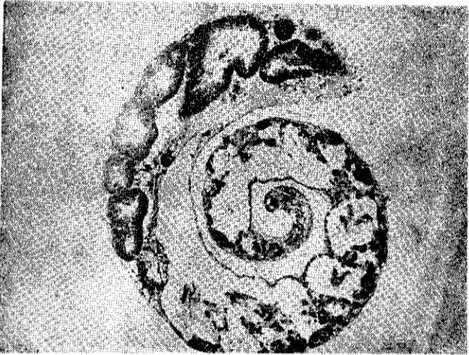
Fig. 2. Digestive gland and ovotestis of unparasitizcd G.laevis (x 40). (Parazitsiz G. laevis'İn sindirim bezi ve ovotestis'i)
In parasitized G. laevis with Paramphistomum larval stages the
growth and development of Paramphistomum larval stages in the
inter-lobular spaces of the digestive gland leads to displacement of the lobes and loss of their branched structure with subsequent degeneration
E~. KAMEL-A. BURGU
Fig. 3. A digestive gland tubule of unparasitized G.lacvis (x 200). (Parazitsiz G.lacvis'in sii1dirim bezi tüpü)
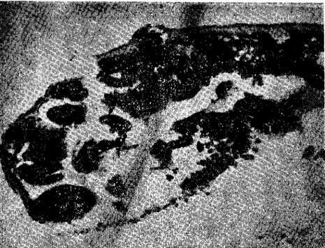
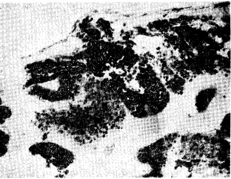
of the epithelium lining the tubules. There was a considerable reduction İn the number of the digestive gland tubules. The nuclei of the digestive ceııs migrate away from the cell-base and there was a decrease in the contained granular food store. The distal walls of these cells of ten break down and some of these ceııs content were released into the lu men of the tubules (Figs.4,5 A and B).
The pulmonates snails Gyrau!us taevis are hermaphrodite. The ovotestis, which is the reproductive organ in the hermaphroditic snails produces both cggs and sperms in compartment ealled acini, İs the other organ in the visceral mass situated at the end of the spiral anima! (Fig. I). The oyotestis or hermaphroditic gland eonsists of se-vera! follicles or acİnİ at the end of the spiral of the snail. Each acinus İs enveloped in a sheath of squamous epithelium and a thin connective tissuc lies between the acini and the covering epitIıelium of the mantle.
In each acİnus of the ovotestis, a number of immature oocysts, a few mature ova, and bundles of sperms can be detected (Figs. ı,2 and 6).
HISOPATHOLOGICAL CGANGES IN THE TlSSUES... 423
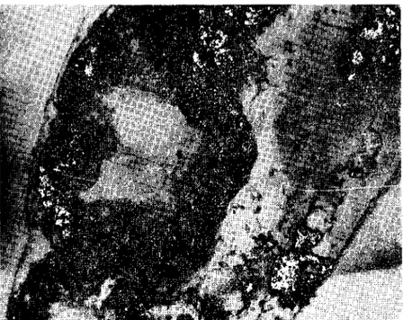
Fig. 4. Moderate parasitized digeslive g!and of G.lael'is (x 100). (G./aevis'in orta derecede parazitli sindirim bezi)
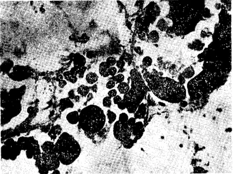
lt was observed during the present investigation that the majority of Paramphistomum larval stages, sporocysts, rediae and cercariae, parasitized G. laevis wcrc located in the region of the digestive g!and and ovotestis. The pathological changes of the ovotestis in parasitized specimens can be summarized in the following steps: Paramphisto-mum larva stages sett!ed in the blood sinuses surrounding the tubules of the ovotestİs. In some cases the rediae occurred inside the persisting tubules and İnvaded the acİni damaging the sex cells. The parasite inhibited the normal gametogenesİs through the disruptİon of normal vascularization, crowding and may be toxicity (Fig.7). The shape of the eggs within the acini were not uniform and the lumen of the tubules becomes filled wİth degenerated protoplasm and nuclei. The tissue of the ovotestis in parasitized G. laevis with Paramphistomum larva! stages is most severely damage when rediae are present. The ovotestis of most G. laevis speeimcns examined during the present study were greatly destroyed and rep!aeed by the larval stages. it is of interest to point out that the aeeessory genital organs in heavily parasitized snai! beeame redueed and in extreme cases became invisib!c.
-Fig. 5. A) Rep!acement of the digestive gland tubulP.s by the parasite (x 200). (Sindirim b::zi tüplerinin yerini alan parazitler).
Fig. 5. B) Disappzarance of most of the digestive gland tubules (x 200). (Sindirim bezi tüplerinin çoğunun kayboluşu)
Fig. 6. Ova ar.d sp~rıns of unparasitized ovatestİs (x 200). (Parazİtsiz OVGtcstis'te yumurta ve spermler)
Fig. 7. Sporocysts and rediae invading the acİni of the ovatestis (x 200). (Ovatestis foliklillerinde sporokist ve rediler)
426 ~G. KAMEL-A. BURGU
The headfoot of the snail Gyrautus taevis is surrounded by an epithelial layer of columnar ceııs. The goblet cells are found beneath the columnar ceııs, but open bctween them. The core of the foot ;s made of a large number of thin muscle fibers running singIy in various direction throughout a dense vascular connective tissue (Figs. i and 8).
Fig. 8. Hc:ıdfool of uııpara:;itized G./aevis (x 100) (Parazilciz G.lanis'in başayağı)
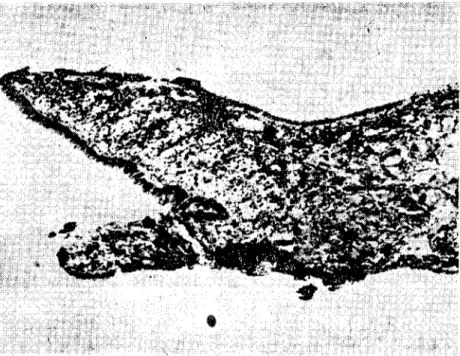
In parasitized G. taevis with Paramphistomum larval stages, it is dear (Fig.9) that the parasite located in the region of the headfoot The sporocyst in the headfoot causes swelling and deformations which are particularly easy to detect. In compact tissues the sporocysts may bring about localized degenerative changes due to the pressure upon the muscular tissues of the foot (Fig.9).
The healthy kidney of the fresh water snail Gyrautus taevis is ad-hering to the inner surface of the mantle and extending distally to the pericardium. it is e10ngated and is as Jong as the respiratory cavity. The kidney terminates in a short, thick, tubular ureter, which curves on itself and opens into the respiratory cavity near the mantle collar.
ı;ıSOPAT!-IOLOGICAL CHANGES IN THE TISSUES... 427
Fig. 9. ;>osition of the parasite in the h::adfoot (x 40).
(Başayağiılda pai'azitl::rin lokalizasyonu)
The epithelial part of the kidncy consists of columnar to cuboidal cells resting on a basement membrane and is supported by alayer of connce-tive tissue and ınuscle fibers. VanıaJes are round in the cpithelial kidney cells (Figs.
ı
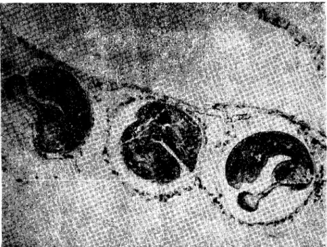
and 10).Within the kidney of parasiti:~cd G. laevis with Paramphistomum larval stages, little disturbance was raund during the present investi-gation. In same cases of parasitized snails, the sporocysts and rediae were carried in the haemolymph to the kidney and lodged near its tissue causes mechanical disturbance by pressing up on the tissue of this organ (Fig. i ı).
During the present investigation it is very important to note that sporocysts, rediae and ccrc<ıriae of Paramphistomum parasitized G. laevis \Yere also detected in the respiratory cavity and around the digcstive tract particularly oesophagus and sometimes in the blood sinuses iı,the region of the geniı,-'.! wıcts. tt seC;~1Slikcly that the rcdiae are transferred by the haemolymph to every organ in the visceral mass of the snail G. [aevis (Figs.
ı
2 andı
3). .Fig. 10. Unparasitized kidney of G.laevis (x 200). (G laevis'in parazitsiz böbregi)
Fig. 11. The location of the parasite in kidncy (x 2(0). (Böbrckte parazit lokalizasyonu)
Fig. 12. The locaıion of rediae in the manıle cavily (x 100). (Manto boşluğunda redilerin lokalizasyonu)
Fig. 13. Rediae of Paramphistomum (x 200). (Paramphistomum redileri)
E.G. KAMEL-A. BURGU
As a result of infection in G. laevis it was observcd that there is tt
heavy prodııetion and agglomcration of excretory granules, neerosis of eells of the digestive gland tııbules and proliferation on intertubules connective tissues.
Discussİon ~md Conclıısİon
The effects of digenetic larval trcrnatodes on theİr moLluscan hosts moderately we!! doeumentd (25). Some larval tr~~l1atodes are known to attac!< the hosts tissues directly. Othcrs deveIop in ce,'tain organs and stil! athers merely exert an indireet influenee on the host tissues. Inereasing!y more inve3tigatofs are studying the cffects of larval tre-matodes on their mollusean hosts. This topic has bcen reviewed by some aııthors (2,5,7.25). The range of effcets varies from none to conside-rable, the lattcr bcing the role.
The effects of larval trematades on their mollusean hosts can be eategorizcd in four groups. The cffeet on host's Jigestivt: gland, the effeet on the host's gonad and rcproduetive stnıeture, the effeet on the general physiology and biochemistry state of the host, and cellular responses on the part of the host.
During the course of the present investigation the infection ot G. laevis with Paramphistomunı larval stages, sporoeysts, rediae and cercariae, has profoıındly damaging effeets on the tissues of the viseeral mass of G. laevis. The tissues of the digestive gland and ovotestis in G. laevis were most severely damage by the presenee of sporoeysts, rediae or eereariae of Paramphistomıım. The histopathology of infee-ted gastropod digestive gland has been the subject of great number of authors and reviewed by Wright (25) anel Beeker (2). The destruetive damages described in the present studyare in full agreement with the histopathological changes described by previous authors (I O,i i, i2,22). it was observed during the present study that there are some dif-ferenees in the effeets produeed by sporocysts and rediae, beeause, while in the former all of the uptakc of nutrier-ts takes place through the tegument, in rediae this method is supplemented by active feeding with the pharynx and eonsequently more mechanical damage is done. The present investigation indieated that the rediae of Paramphistomıım attaek and eonsume the host's oyotestis very quickly after the infec-tion was established. Infection wİth Paramphıstomum, whieh posses-ses redial stages, caused most direct damage espeeİally as these tended
HISOPATHOLOGICAL CGANGES IN THE TISSUES... 4Jl
to be evenly distributed throughout the dİgestive gland and ovotestis of G. laevis. it seems likely that the rediae feed on bat h ovatestis and digestive gland tissues. Rees (20) mentİoned that when female Littori-na littorea are infected with cercariae of Himasthla seconda, rediae seem to be full of eggs and yolk sucked fr0111the ovarian tubııles by theİr powerfu! muscıılar pharynx.
Wright (26) mentioned that the miracidia which enter the more compact muscıılar tissues of the foot are less successfııl in establishing an infection than those which get into the open spaccs of the headfoot regions and the mantle. It \Vas also obsen'ed by Burgıı (4) that Param-phistomum cervi miracidia çenetreted the İntermediate snail host Pla-narhis planorhis only through the mantle cavity and not from the headfoot or tcntaclcs.
it is of interest to note thnt the present investigation indicated that the Paramphİstomum miracidia penetrate the muscular tissues of the headfoot of the snaJ G. laevis and successfully established the infec-tion. The results presented İn this study Olay be due to the fact the snails G. laevis are smaIl in size and the tissues of the headfoot regions are soft and less compact.
The present study indicated that the parasite tend to become con-centrated in the lower part of the visceral mass of G. laevis where they form a '"blocking layer" this layer isolates the upper part of the visceral mass and deprives most of the digestivc gland and oyotestis of their
normal haeınolymph supplics with consequent dcgencration due to
starvation and accuınulation of excretory prodııcts.
it seems likely that during the development of the Paramphisto-mum larval stages within the snail G. !aevis, the parasite pass from one stage to another and migrate into all parts of the snail tissues causing great damage to most of the host tissues.
Finally, intermolluscan larval treınatodes do deplete theİr host carbohydrate, lipid and amino acid (2,25) whİch causes great harm to the hos1. In fact, ınfected molluscs generally do not survİve as long as uninfected ones.
Acknow ledgements
The authors wish to express their deep gratitude to Professor Nevzat Güralp, Faculty of Veterinary Medicine, Ankara University, Turkey, for the interest taken in this work and for his valuable advice.
EG. KAMEL-A. BURGU
References
ı. Agersı>org, H.P.K. (1924), Studies on ılıe e//ccı oj'parasiıism UpOIlıissues f. Wiılı
.\'/K-eial refafnce lo cerraiıı Kasırop:Jd ıııollııses.Q. JI. microsc. Sci., 63, 361-401.
2. Becker, W. (ı930). Meıabolie inrerrelaıianslıip oj' parasilie rreıııal()(/es and //loılııse.\', especially Selıisıosoıııa /ilaıısoııi in Biomplıalaria glabrara. Z. Parasitenkd., 63, iO
ı-iıi.
3. Bourns, T.K.R. (19()3). Larval treıııarodes para,5I1l1igL)'II'lıaea stagııalis apperessa (Say,' in Oııra!'h) ıı'ith e:nplnısis 011mil/tip/e in/eelioıı. Can. J. Zool., 41, 937-9~ı. 4. Burgu, A. (1982). Slııdies 011ılıe biology of Paramplıisloll1l1111ceni Selırank, 1790 in
sJ:eep iıı the disırict of ESkişelıir Çijieler Stale Farııı. A.Ü. Ve!. Fak. Derg .• 2R. 50-71. 5, Cheng, T.C (i967). Mariııe n:olluses as hasts for symbioses: wiıh o rerieı,' of known
parasiıes of commercialfı' imp:Jrl{{l11species. Adv. mar. Biol., 5, 1-24. 6. Cheng, T.C. (1973). "Geııeral Parasiıology". Academic Press New York.
7, Cheng, T.C. and Snyder, R.W. (1962). Srudies all hosı-p:ırasire relarioIlShl;)s bctll'ee:ı I:ırval ırematodes aııd their hosrs.1- A rel'iew./1- Hosr glycogeıı urilizatioıı by the
iıı-tamollııscaıı lanae of Gfvprhelmins pennsy/ı'aniensis Cheng and relared plıenoıııena. Trans. Amcr. Microsc, See., 81, 327-331.
8, Erich, K.(i983). Histologisclıe Vlltersııc!lıl11gelıaıı Sporozysreıı, Redieıı ııııd Zerkarien voil Paramphistoıııuııı cerı'i, Zeder 1790 iıı der Zll'isc!ıeıı- Wirtsc!ıııecke Plaııorbis pla-norbis. Vet. med. Diss., München.
9. Faust, E.C. (l920~. Parhological chaııges in the Gastropod liı'er prodııced by flııke iıı-fecrioıı. Jahns. Hopkins. Hosp. Bull., 31, 79-84.
LO. Jame~, B.L (1965). Tlıe e!fecı ofparasitism by ları'al digeııea 011tlıe digeslİFe g/wıd of tlıe imerıidal prosobranclı Lirtariııa sm;atilis (Oliı'i) subsp. tenebrosa (Montagıı). Parasitology, 55, 93-114.
II. Kamel, E.G. (i979). Tlıe physiological e/fects of platylıelmimlı parasites on Urtorina littorea (L.), P:ı. D. Thesis, Manchester University, England.
12. Kamel, E.G., Abd, E!-Rchhı, L., Mohamed, A.M. and Hanna, M.Y. (986). Tlıe oecıır-reııce of xiplıidiocercariae and rheir Iıisıop:ırlıalogieal elfeets 011rhe tissues of the freslı waler snail L.aııistes cariıı"rııs Olider (Gasıropoda-Pr,)sobraııchia). Proc. Zool. Soc. Egypt., in press.
13, Kamel, E.G. and Burgu, A. (l9R6). First record of ılıe /resıı warer sııail Gyraulus learis (Alder) ııatıırally iııfeered IFirh l'araıııplıistoıııııııı cercariae Foııı Turkey. A.Ü. Vet.
Fak. Derg., in Pre~s.
14. Lebour, M.V. (1911). A re"iew of the Briıish ıııariııe eercari"e. Parasit., 4, 416-456. 15. Malck, E.A. (ı955). Aııatoıııy of Bioıııplıalaria b:Jissy as re/a!ed to its iııfectioıı with
Sc!ıistosoma maıısoııi. Amer. Midl. Natur.. 54, 394-404.
lG. Malck, E.A. (1958). Factors coııdilioııiııg tlıe Iıabitat of billıarziasis interıııediate hosts of rhe family plaııorbidae. Bujı' W.H.O., 18, 785-818.
HISOPATHOLOGICAL CHANGES IN THE T1SSUES ...
17. Malek, E.A. (1962). "Laboraıory gııide aııd nOles for medical malacology". Bur£css Publishing Company. U.S.A.
18. Male", E.A. and Cheng, T.C. (t974). ",Hedieal and Ecoııomieal Malacology". Acadc-mic Press, New York, London.
ı9. Pelsen~er. E. (1906). Tremalodes "arasiıes de mollıısqııes marino5.Bull. Sei. Fr. Bclg.,
40, 161-186.
20. Rccs, W.J. (1936). T!ıe effeeı of parasi/üm by 101'1'01 Irenıalodes 011ı!ıe lissııes of
Uı-ıorina li/forea (Unıı!:). Proc. Zool. Sac. Lond., Part i,357-368.
2 I. Rees, F.G. (I 948). A sıııdy of ı!ıe effeeı of liglıı. ıemperolure awl saliniıyon ıhe emer-!Jelice of Cereorio pıır"ıırae Lf!bour from Nııcella lopel/ııs (L.). Parasitology., 38, 228-242.
22. Rob~on, E.M. and Williams, I'C (1971), Relaliolıshipo5o/same species ofdiKeııeo lI'il!ı ılıe mariııe prosobmııch U/fol'iııa Ii/forea (L.) 11- Tlıe e/fecI o/Ianal digeııea011 Ihe reprodııclire biolog)' (ıt L, li/forea ..i. Helminth., 45, 145-159
23. Sehmid, K., Rüekrich, H.U. und Boeh, J. (1981). Die Eıı/ıricklung ron Paramplıisle-mıım cervi 1'011 Mira::idiıım bis ::111' Meıa::erkarie. BerI. Münch, Ticrarzt!. Wsehr.,
94, 463-467.
24. Ward, H.B. (1907), The iııfıııence ofparasiıism 011 ıhe hosl. Scicnce (N.S.)., 25, 201-218.
25. Wright, CA. (i966). The paıhogeııesis of !ıelmim!ı iıı /Ilo/lusca, Hclminth. Abstr., 35(3), 207-224.
26. Wright, CA. (1971). "F/ııkes and Sııails". Scie:ıce of biological series No, 4. George Alien and Unwiıı, London.