Allergen specific IgE determination by in vitro allergy test in head and
facial feline dermatitis: A pilot study
Kerem URAL, Hasan ERDOĞAN, Mehmet GÜLTEKİN
Adnan Menderes University, Faculty of Veterinary, Department of Internal Medicine, Aydın, Turkey.
Summary: Pruritus is one of the most common clinical findings in cats which have hypersensitivity on head and face regions. The purpose of this small pilot study was to verify underlying causes of pruritus in cats with head and facial dermatitis by using in vitro Veterinary Polycheck allergy tests which specifically detect Immunoglobulin E (IgE) concentrations against 20 different allergens. Ten cats were introduced to the clinic with varying degrees of head and facial dermatitis along with primary/secondary skin lesions and pruritus. At first, haematological, parasitological, microbiological and clinical examinations were performed. Six healthy cats without dermatitis (neither infection nor hypersensitivity) were also evaluated as a control group. Afterwards, 0.2 ml of blood was taken from all cats for specific IgE analysis against 20 different allergens. Regarding allergen specific IgE levels (kU/l) in cats with head/facial dermatitis and in healthy cats, in vitro tests gave a positive reaction most frequently against flea, Acarus siro, D. farinae, Ragweed (Ambrosia), Lambs quarter and Tyrophagus. There was a statistical significance between two groups for all allergens as follows; Lepidoglyphus (p=0.031), Alternaria/Cladosporium (p=0.011), Stinging nettle (p=0.011) Lambs quarter (p=0.031), Sorrel (p=0.003) and flea (Ctenocephalides) (p=0.031). In the present study, all positive reactions on the Polycheck test were thought to indicate underlying allergens of facial/head dermatitis in cats. It has to be mentioned that, in vitro allergy tests cannot be used as a sole method for distinguishing hypersensitivity from healthy cats due to the clinically irrelevant findings regarding morphology in healthy cats. Positive reactions in healthy cats might indicate a subclinical hypersensitivity state or the requirement for investigating other relevant factors.
Keywords: allergy, dermatitis, feline, immunoglobulin E.
Baş ve yüz dermatitli kedilerde in vitro alerji testi ile alerjen spesifik IgE tayini: bir pilot çalışma
Özet: Aşırı duyarlılığı olan kedilerde baş ve yüzde kaşıntı en sık görülen klinik bulgulardan biridir. Bu pilot çalışmanın amacı, İmmunoglobulin E (IgE) konsantrasyonlarını 20 farklı allerjene karşı spesifik olarak saptayan in vitro Polycheck Veteriner alerji testi kullanılarak baş ve yüz dermatitis bulunan kedilerde kaşıntının altta yatan nedenlerini açıklığa kavuşturmaktır. Baş ve yüz dermatiti yanında çeşitli derecelerde primer/sekonder deri lezyonları ve kaşıntısı bulunan 10 kedi kliniğe getirildi. İlk aşamada hematolojik, parazitolojik, mikrobiyolojik ve klinik muayeneler gerçekleştirildi. Aynı zamanda karşılaştırmalı olarak dermatit bulguları olmayan (enfeksiyöz ya da hipersensivite) 6 sağlıklı kedi kontrol grubu olarak değerlendirildi. Daha sonra tüm kedilerden 20 farklı allerjene karşı spesifik IgE analizi için 0.2 ml kan örneği alındı. Baş/yüz dermatiti olan kedilerdeki allerjen spesifik IgE düzeyleri (kU/l) sağlıklı kediler ile karşılaştırıldığında in vitro testlerde en sık pire, Acarus siro, D. farinae, Ragweed (Ambrosia), sirken otu ve Tyrophagus’a karşı reaksiyon belirlendi. İki gruptaki sonuçlar karşılaştırıldığında Lepidoglyphus (p=0.031), Alternaria / Cladosporium (p=0.011), ısırgan otu (p=0.011) sirken otu (p=0.031), kuzukulağı (p=0.003) ve pire’ye (Ctenocephalides) (p=0.031) karşı elde edilen değerlerde istatistiksel olarak anlamlı bir fark vardı. Bu çalışmada, Polycheck testinde belirlenen pozitif reaksiyonların kedilerdeki baş/yüz dermatitinin altında yatan alerjenleri gösterdiği düşünülmektedir. Bununla birlikte sağlıklı kedilerde klinik bulguların düzensiz morfolojisi nedeniyle in vitro allerji testlerinin hipersensitiviteyi ayırmada tek yöntem olarak kullanılamayacağı belirtilmelidir. Sağlıklı kedilerde belirlenen artışlar subklinik hipersensitivite durumunu veya diğer ilgili faktörlerin araştırılması gerekliliğini gösterebilir.Anahtar sözcükler: allerji, dermatit, immunoglobulin E, kedi.
Introduction
One may often encounter allergic diseases in a veterinary field. Together with elevated standards in veterinary practice, intradermal allergy testing and allergen immunotherapy were started to be applied in the small animal practice many years ago. These tests were then followed by serum testing for allergen‐specific IgE in
dogs and cats (20). Atopic dermatitis has also been received attention in cats. However, clinical findings of atopic dermatitis in cats differ from the same disease seen in humans (14). Although intradermal and serum IgE testing take place in feline medicine, researches investigating allergen types in this species are scarce (25).
Hypersensitivity and relevant dermatological disorders are foremost common causes of pruritus in cats. In general, affected cats exhibit head and neck excoriations, self-induced symmetrical alopecia, miliary dermatitis, or eosinophilic dermatitis. Environmental (i.e. Birch, alder, hazel, willow etc.) food, flea, and mite (Dermatophagoides farinae, Dermatophagoides pteronyssinus, Tyrophagus, Lepidoglyphus destructor, Acarus siro etc.) allergens are recognized as offending
factors (1).
To our knowledge, there is a limited amount of research on conducting allergy tests for feline head/neck dermatitis. Considering the relevant literature, participation of allergen-specific IgE in these conditions is likely to occur. However, this topic has not yet extensively been tested (4,21). Allergen-specific IgE might be found in both allergic and healthy/specific pathogen free cats (18,34).
The purpose of the present study was to identify most prevalent allergens affecting cats with facial/head dermatitis in Turkey by in vitro allergy test. Obtained results might contribute a new sight on relation between dermatitis and environmental conditions, which could direct and influence further treatment.
Material and methods
Demographic data: Serum samples from pruritic
cats (n = 10) at the age of 2 to 7 years, of both sexes (6 male, 4 female) were obtained. The retrospectively diagnosed cats with head and facial dermatitis were included in the study. None of the cats had prior diagnosis or treatment. All cats with dermatitis had health certificates from private veterinary clinics. Accordingly, parasitic control and frequent vaccination had already been applied to all cats. Privately owned 3 male and 3 female healthy cats witin the same age range, were enrolled as a control group. The cats in a healthy group also had health certificates. Informed consents were obtained from the owners. Healthy cats were only included in case that they were no presenting history of allergy signs (pruritus), or gastrointestinal/respiratory signs. Differential diagnosis was performed considering relevant tests for mycotic (Wood’s lamp examination, mycological isolation and identification) or parasitic (skin scraping, acetate tape impression) diseases and autoimmune conditions (anti-nuclear antibody test, skin punch biopsy). In cats with hypersensivity dermatitis (HD) caused by food allergens, a 6- to 8-week restriction of diet followed by a two-week challenge with the previous diet (namely a dietary restriction-provocation test) was carried out. Cats free of flea infestation giving respond entirely to the latter procedure were diagnosed as having food-induced hypersensitivity dermatitis as
reported previously (1). In cats with HD caused by flea saliva allergens, the diagnosis of flea bite hypersensitivity dermatitis was based on compatible clinical findings. They also well responded to flea treatment with fipronil. Cats with HD, based on unremarkable serum biochemistry, hematological results, clinical examination and those suspected to be sensitive against environmental and/or food allergens were included in the undetermined hypersensitivity dermatitis group as reported previously (1). If necessary, diagnosis was based on histopathological examination of skin punch biopsy.
Polycheck Feline Allergy Test principle: The present
test (Polycheck, Allergy test, Gmbh, Germany, Distributer RDA Group, Istanbul) is a relatively novel in vitro test which detects allergen-specific IgE in cat serum. This test is based on an immunoassay principle, i.e. via covered coated allergens and biotinylated monoclonal antibodies against cat-IgE. At initial step, namely incubation of serum, the allergen-specific IgE attaches to the corresponding allergen. Afterwards, washing step where the biotinylated antibody founds the bounded IgE, was conductes. The biotinylated antibody furtherreacts with another incubation step composed of streptavidin-alkaline- phosphatase conjugates. Following washing and including of 5-bromo-4-chloro-3-indolyl-1-phosphate/ nitro blue tetrazolium (BCIP/NBT) the enzymes cause a coloured precipitate. Available number of precipitate is linked to the specific IgE levels found in the present serum, which result in a significant/nonsignificant colouring of 20 relevant and different individual allergens.
Calculation of results: A well equipped computer
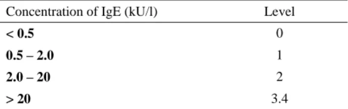
accompanied by a scanner and a user friendly Biocheck Imaging Software were used to evaluate the tests precisely. The cassettes placed on top of the scanner were read out. The latter specifical software programme analyzed and the cassette data were calculated. The levels of allergen-specific IgE for each allergen were given as relative kilo units per litre (kU/l) and classified according to the manufacturer and previous studies (Table 1).
Statistical analyses: Descriptive statistics of mean
and standard deviations of animals were performed in pruritic and healthy groups. The Mann-Whitney U test was used to determine the differences between the groups. A value of p<0.05 was considered statistically significant.
Table 1. Five different levels of allergic reactions in cats as detected by Polycheck allergy test.
Tablo 1. Kedilerde Polycheck alerji testi ile saptanan beş farklı alerjik reaksiyon seviyesi.
Concentration of IgE (kU/l) Level
< 0.5 0
0.5 – 2.0 1
2.0 – 20 2
Results
Clinical findings: Demographic data, etiological
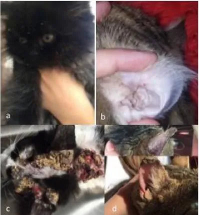
investigation and relevant selected test results were shown in table 2. All presented cats having primary and/or secondary skin lesions on head and facial regions as shown in Figure 1-3.
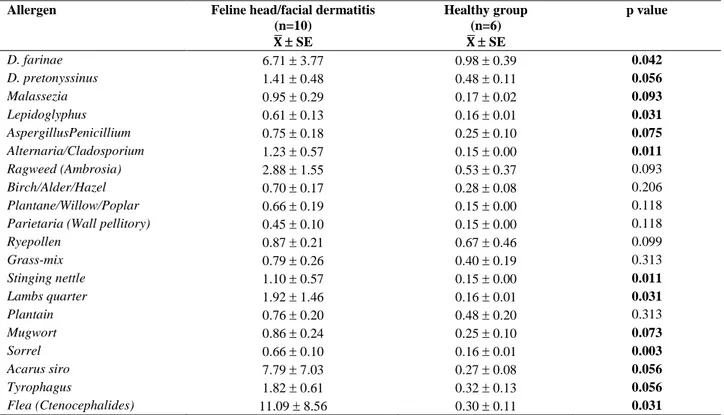
Allergen specific IgE levels and interpretation:
Allergen specific IgE levels (kU/l) along with meanstandard errors in cats with head/facial dermatitis and in healthy group of cats were shown in table 3. All of the 10 cats with head/facial dermatitis presented weak (n=4) to strong (n=5) or very strong (n=1) (mean standard error) IgE response against D. farinae 6.71 3.77
kU/l versus 0.98 0.39. These findings showed statistical significant differences with those in the healthy group of cats (p=0.042). Other relevant allergens having statistical significance between 2 groups were as follows; Lepidoglyphus (p=0.031), Alternaria/Cladosporium (p=0.011), Stinging nettle (p=0.011) Lambs quarter (p=0.031), Sorrel (p=0.003) and flea (Ctenocephalides) (p=0.031).
Among tested 20 different allergens the most commonly observed ones were (with descending order) flea (Ctenocephalides), Acarus siro, D. farinae, Ragweed (Ambrosia), Lambs quarter, Tyrophagus as shown in fig 4.
Table 2. Group and etiological data of involved cats with head and facial dermatitis. Tablo 2. Baş ve yüz dermatitli kedilerin grupları ve etiyolojik bulguları.
Feline head and facial dermatitis
Number of cats
Supportive diagnosis Polycheck in vitro Ig E test-top 3 allergen
Food-induced
hypersenstivity dermatitis
5 History, food-trial (1) D. farinae, Lepidoglyphus, Grass mix
Flea bite hypersenstivity dermatitis
1 Clinical findings (pruritus etc.), wet paper test, IgE test
Flea, A. siro, Tyrophagus
Cheyletiellosis 1 Skin scraping, acetate tape impression Flea, Stinging nettle, Lambs quarter Pemphigus foliaceus 1 Anti-nuclear antibody test, skin punch
biopsy
D. farinae, A. siro, Tyrophagus
Mycotic infection (Alternaria sp.)
1 Woods lamp examination, mycological isolation and identification
Ragweed (Ambrosia),
Alternaria/Cladosporum, Rye pollen
Undetermined
hypersensitivity dermatitis
1 History, clinical examination, suspicion of having allergy against environmental and/or food allergens (1)
D. farinae, D. pteronyssinus, Rye
pollen
Table 3. Allergen specific IgE levels (kU/l) within mean ± standard errors in cats with head/facial dermatitis and to those of healthy group of cats.
Tablo 3. Baş ve yüz dermatitli ve sağlıklı kediler arasındaki alerjen spesifik IgE seviyelerinin (kU/l) ortalama ± standart hata değerleri.
Allergen Feline head/facial dermatitis
(n=10) 𝐗̅ SE Healthy group (n=6) 𝐗̅ SE p value D. farinae 6.71 3.77 0.98 0.39 0.042 D. pretonyssinus 1.41 0.48 0.48 0.11 0.056 Malassezia 0.95 0.29 0.17 0.02 0.093 Lepidoglyphus 0.61 0.13 0.16 0.01 0.031 AspergillusPenicillium 0.75 0.18 0.25 0.10 0.075 Alternaria/Cladosporium 1.23 0.57 0.15 0.00 0.011 Ragweed (Ambrosia) 2.88 1.55 0.53 0.37 0.093 Birch/Alder/Hazel 0.70 0.17 0.28 0.08 0.206 Plantane/Willow/Poplar 0.66 0.19 0.15 0.00 0.118
Parietaria (Wall pellitory) 0.45 0.10 0.15 0.00 0.118
Ryepollen 0.87 0.21 0.67 0.46 0.099 Grass-mix 0.79 0.26 0.40 0.19 0.313 Stinging nettle 1.10 0.57 0.15 0.00 0.011 Lambs quarter 1.92 1.46 0.16 0.01 0.031 Plantain 0.76 0.20 0.48 0.20 0.313 Mugwort 0.86 0.24 0.25 0.10 0.073 Sorrel 0.66 0.10 0.16 0.01 0.003 Acarus siro 7.79 7.03 0.27 0.08 0.056 Tyrophagus 1.82 0.61 0.32 0.13 0.056 Flea (Ctenocephalides) 11.09 8.56 0.30 0.11 0.031
Figure 1. Four different cats with head/facial dermatitis with a common history of food allergy. a) bilateral periorbital scaling, erythema and alopecia along with muzzle crusting b) general alopecia on the head with extensive grooming and itching behaviour c) erythema and alopecia accompanying pruritus following dietary change d) severe crusting on unilateral periorbital region and muzzle with the history of extensive pruritius.
Şekil 1. Yaygın olarak gıda alerjisi geçmişi bulunan baş/yüz dermatitli dört farklı kedi. a) bilateral periorbital skuam, eritem ve alopesiyle birlikte burun bölgesinde kabuklanma b) yalama ve kaşıntıya neden olan baş bölgesinde yaygın alopesi c) gıda değişimini takiben görülen kaşıntıyla birlikte eritem ve alopesi d) periorbital bölgede tek taraflı şiddetli kabuklanma ve burun bölgesinde yaygın kaşıntı geçmişi.
Figure 2. Different cats with common history of pruritus associated with head/facial dermatitis. a) Cheyletiellosis with extensive scaling and pruritus b) early signs of allergy/hypersensitivity presenting erythema, crusting on inner auricular region c) extensive crusting, erythema due to Pemphigus foliaceus d) ear margin dermatosis due to food allergy and Alternaria sp. infection.
Şekil 2. Baş/yüz dermatitis ile ilişkili kaşıntı geçmişi bulunan farklı kediler. a) Cheyletiellosis ile birlikte yaygın kabuklanma ve kaşıntı b) alerji/hipersensitivite erken bulguları olan iç kulak bölgesinde kabuklanma ve ödem c) Pemphigus foliaceus’a ilişkin yaygın kabuklanma ve eritem d) gıda alerjisi ve Alternaria sp. Enfeksiyonuna ilişkin. kulak ucu dermatozu.
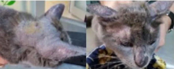
Figure 3. This cat with severe pruritus showed IgE against 18 out of 20 allergens (classified as weak, strong or very strong). There was very strong IgE response against Tyrophagus and Flea allergens.
Şekil 3. Şiddetli kaşıntı mevcut kedide 20 alerjenden 18’ine karşı IgE tespit edildi (zayıf, kuvvetli ya da çok kuvvetli olarak sınıflandırılacak şekilde). Tyrophagus akarı ve pireye karşı kuvvetli IgE yanıtı tespit edildi.
Figure 4. Spatial distribution of allergens and related specific IgE levels (kU/l) among cats with head/facial dermatitis or healthy cats without dermatitis.
Şekil 4. Sağlıklı ya da baş/yüz dermatiti bulunan kediler arasında spesifik IgE seviyeleri (kU/l) alerjenlerin bölgesel dağılımı.
Discussion and Conclusion
Feline facial dermatoses might be presented as a frequent disorder with several underlying diseases (2).
Facial dermatitis might be related to parasites [mite infestation such as Feline scabies (Notoedres), fleas, cheyletiellosis, is rare, Demodex gatoi’] mycoses
(dermatophytosis, secondary yeast overgrowth), feline herpesvirus-1 (26), allergy (food allergy, environmental allergy, or insect allergy), selected autoimmune skin disease (Pemphigus Foliaceus), feline acne, idiopathic facial dermatitis (“Dirty Face” in Persian cats) (8), or indolent ulcer (2). A total of 10 cats were enrolled in the present study, which were classified as those having food-induced hypersenstivity dermatitis (n=5), flea bite hypersenstivity dermatitis, cheyletiellosis, Pemphigus
complex mycotic infection and undetermined hypersensitivity dermatitis.
Atopic dermatitis might be dedicated as a skin disorder in association with hypersensitivity against environmental allergens (31). A genetic predisposition (23) to atopy was confirmed in cats mostly at the age of 6 months to 3 years (9). The prominent clinical sign is pruritus, followed by miliary dermatitis and eosinophilic granuloma (9, 17, 24,31). The typical dermatological signs exist on the head and neck regions (31). In the present study all diseased cats presented clinical signs (prurtius, crusting etc.) on the head and facial region, whereas it should not be easy to draw conclusion that all cats were atopic, due to lack of general diagnostic criteria permitting a diagnosis based on the clinical signs (33). On the other hand, all cats with head/facial dermatitis presented in this study were under 3 years of age, which might not be due to atopic conditions (9).
Hypersensitivity and related dermatological disorders in cats are denoted as nominates for in vitro measurement of allergen-specific IgE. On the other hand, it is not known whether the latter tests are important, type I hypersensitivity reactions and pathogenesis of IgE still remain unclear. Furthermore, allergen-specific IgE concentrations might be influenced both by hypersensitivity against food, flea and environmental allergens which cause dermatological diseases and endoparasitic agents (1). Someone might speculate that food allergy participated within the clinical signs of cats involved in this study. Furthermore 5 out of 10 cats were diagnosed with food allergy. A 6- to 8-week restriction diet followed by a two-week challenge with the previous diet (namely a dietary restriction-provocation test) was carried out, in which cats gave respond entirely to the latter procedure, were denoted as having food-induced hypersensitivity dermatitis as reported previously (1).
Little data exist regarding possible pathogenesis inducing pruritic dermatitis in cats after comsumption of food. Hence, food hypersensitivity in feline species is not always IgE mediated (35), in which the reaction might not have immunological back ground. It should therefore be suggested that IgE test are not be useful for diagnosis of adverse food reactions (1). Furthemore, it has been recognized that there was a poor correlation between serum food-specific IgE and adverse food reaction,
considering that high percent of healthy control cats might present food IgE (10). Based on a forementioned knowledge, we might not support the idea that food-induced hypersensitivity dermatitis in 5 cats in this study might have direct relationship with analyzed antigens. However, IgE production must be taken into consideration, at least for prevention and for prognosis.
In the present study frequent deworming had been applied toall cases which exclude the parasitic causes for elevated IgE concentrations. On the other hand, occurence of endoparasites both cause parasitic IgE formation and IgE to other relevant allergens (1). Given detected positive correlation between Toxocara infections in cats and dogs and sensitization to orally administered antigens (7), Belova et al. (1) suggested similar correlation between endoparasitic invasion and IgE occurence against environmental allergens. Endoparasitic infestation is related with decreasing clinical signs of hypersensitivity in humans and dogs (12, 22). However, no study investigated dewormed and non-dewormed cats comperatively (1).
In a previous study 36 cats with allergic skin disease were comperatively evaluated by using in vitro and in vivo tests. In that study, intradermal skin testing composed of sole flea extract (6 cats) or 40 inhalant allergy panel (30 cats) were used. Sera samples were analyzed by using IgE ELISA against flea extract and 36 inhalant allergens. Flea bite hypersensitivity (n=18), atopy (n=9) and food hypersensitivity (n=1) were diagnosed. Intradermal skin testing possessed positive predictive value of over 85 per cent and 100 per cent for flea allergy and atopy, respectively. The IgE ELISA test presented low predictive values for flea allergy and atopic disease. The researchers, thus, suggested that it was not a useful diagnostic test (5). Another study in cats, revealed strong agreement between a positive E-screen result and a positive finding on in vitro allergy testing (88%) (3). Out of 10 cats with head and facial dermatitis in that study, only 1 cat was diagnosed with flea hypersenstivity.
An ELISA was developed for investigating feline serum allergen-specific IgG against selected house dust, pollen and flea allergens. The latter assay was established comperatively for allergen-specific IgG concentrations in healthy and diseased cats [without dermatologic disorder, allergic or pruritus]. In the latter study, cats with allergy presented significantly more IgG directed against house dust mite, flea and ryegrass allergens in contrast to other cats. The researchers suggested that cats with allergic dermatitis have a backgorund TH2 lymphocyte response resulting in both allergen-specific IgG and IgE production (6). Another study was previously conducted to detect if cats with allergic dermatitis present detectable levels of serum IgE specific for antigens derived from the house dust mites Dermatophagoides farinae (DF) and
Dermatophagoides pteronyssinus (DP) by ELISA. Sera
samples among 59 cats with allergic skin disease and 54 clinically normal cats were monitorized. Feline allergic skin diseases were classified as: self-induced alopecia (group I), papulocrusting dermatitis (group II), eosinophilic granuloma complex (group III), papular/ulcerative dermatitis of head and neck/facial dermatitis (group IV), polysymptomatic clinical signs (group V) and control cats (group VI). In the latter study, there was no significant difference among groups for DF- and DP-specific IgE levels. Considering the ELISA results regarding house dust mite-specific feline IgE, the existence of allergen-specific IgE correlated poorly with clinical signs (34). In the present study regarding DF, there was a significant difference (p=0.042) among IgE levels (kU/l) between groups (6.71 3.77 vs. 0.98 0.39). On the other hand, other relevant allergens having statistical significance between 2 groups were as follows; Lepidoglyphus (p=0.031), Alternaria/Cladosporium (p=0.011), Stinging nettle (p=0.011) Lambs quarter (p=0.031), Sorrel (p=0.003) and flea (Ctenocephalides) (p=0.031) as detected by in vitro allergy testing.
From another point of view, although it is not easy to compare intradermal skin test with serum IgE determination, it might be better to briefly discuss relevant results. By using skin tests do diagnose allergy in cats with small-airway diseases, positive reactions to allergens of house dust mites (D. farinae, D. pteronyssinus) were common, followed by grass allergens and trees allergens (23). Some previous reports indicated that dust mites (D.
farinae, D. pteronyssinus, A. siro, T. putrescientiae) as
most common causes of atopic reactions in cats (27, 29, 33).In the present study, all clinical signs (exfoliative dermatitis, crusting, erythema and alopecia) showed a good correlation with IgE results. Five of the 10 cats involved in this study showed strong or very strong IgE specific for antigens derived from the house dust mite DF. Besides, 9 cats also showed weak or strong IgE specific for antigens derived from the house dust mite DP.
House dust mite antigens were detected in household cats, at a concentration of >2 mcg/g dust (19). Intradermal testing positively and more frequently detected in cats with allergic dermatitis than in nonallergic cats (30), whereas sera levels of dust mite-specific IgE among healthy cats were not different from allergic cats with dermatoses (34). Due to the lack of data regarding major/minor dust mite allergens in allergic dermatitis in cats and evidenced proof for the participation of storage mites in feline allergic diseases, it can not be easily drawn conclusion that house dust mite sor other relevant antigens as detected by IgE production took direct role in head and neck dermatitis. However, it may be suggested that there might be an underlying condition as those antigens, had an influence on the immune system.
In the present study, all positive reactions on the Polycheck test were thought to indicate underlying allergens of facial/head dermatitis in cats. It has to be mentioned that, in vitro allergy tests cannot be used as a sole method for distinguishing hypersensitivity from normal cats due to the clinically irrelevant findings considering morphology in normal cats. This kind of reactions might indicate a subclinical hypersensitivity state, or the requirement for other relevant factors other than mast cell-bound antibodies for the development of AD (barrier functional defect, mast cell function disorder, IgE receptor mutation, or up-regulated cytokines) (13,15). The cats in the present study, had been diagnosed with hypersenstivity based on history, clinical signs evaluated, serum IgE testing directed against 20 different allergens and with exclusion of other diagnostic criteria (parasitological, microbiological and other relevant tests). Therefore, the Polycheck allergy reactions of the cats in the present study might be related to the clinical signs of head/facial dermatitis.
Interpretation of allergen specific IgE determination will briefly be discussed. Based on human medicine data, available in vitro testing of allergens and related data might not be claimed as promoting the status that low IgE levels should be used randomly or in limited diagnostic aproach solely as allergy (16). Investigators suggested that, even at relatively high levels of allergen-specific IgE, “the most reasonable interpretation of a positive IgE antibody levels can be considered as a risk factor for allergy and not as a precise marker of the existence of allergy (11). On the other hand, elevated concentrations of allergen specific IgE are more likely in relation with clinical signs of allergy (28,32). In practice, investigators might suggest that lower levels of allergen specific IgE should be confirmator, even if the clinical signs are strongly suggestive (16).
In conclusion, it may not be unwise to draw preliminary suggestions that some of the factors causing allergen-specific IgE formation may be linked together, and multi-factors must be taken into account for interpretation and assessing correlations among aformentioned factors. Based on IgE results and related allergy tests desensitization (i.e. allergen specific immunotherapy and probiotherapy) should be initiated, which would be the purpose of our subsequent study.
References
1. Belova S, Wilhelm S, Linek M et al. (2012): Factors
affecting allergen-specific IgE serum levels in cats. Can J
Vet Res, 76, 45–51.
2. DeBoer DJ (2016): Feline Facial Dermatoses. South Euro Vet Conf, Granada, Spain, p. 45.
3. Diesel A, DeBoer DJ (2011): Serum allergen-specific
of a rapid screening immunoassay and complete-panel analysis. Vet Dermatol, 22, 39-45.
4. Foster AP (2002): Diagnosing and treating feline atopy. Vet Med Us, 97, 226-229.
5. Foster AP, O'Dair HA (1993): Allergy Testing for Skin
Disease in the Cat. In Vivo vs In Vitro Tests, 4, 111–115.
6. Foster AP, O'Dair HA, DeBoer DJ (1997):
Allergen-specific IgG antibodies in cats with allergic skin disease.
Res in Vet Sci, 63, 239-243.
7. Gilbert S, Halliwell REW (2005): The effects of
endoparasitism on the immune response to orally administred antigen in cats. Vet Immunol Immunopath,
106, 113–120.
8. Gross TL, Ihrke PJ, Walder EJ et al. (2005): Spongiotic
and vesicular diseases of the epidermis. Facial dermatitis of persian and himalayan cats. 112-115. In: LG Thelma, JI
Peter, JW Emily, K Verena (Eds), Skin diseases of the dog and cat. Clinical and histopathologic diagnosis. Blackwell, Oxford.
9. Guaguere E, Prelaud PA (1999): Practical guide to feline
dermatology. Merial, New York.
10. Guilford WG, Jones BR, Markwell PJ, et al. (2001): Food
hypersensitivity in cats with chronic idiopathic
gastrointestinal problems. J Vet Int Med, 15, 7–13.
11. Hamilton RG, Williams PB (2010): Human IgE antibody
serology: A primer for the practicing North American allergist/immunologist. J Allergy Clin Immunol, 126, 33–
38.
12. Helmer M, Epe C, Mueller RS (2008): The effect of
helminth administration on caninne AD: A pilot study. Vet
Dermatol, 19, 33.
13. Hill PB, DeBoer DJ (2001): The ACVD task force on
canine atopic dermatitis (IV): environmental allergens. Vet
Immunol Immunopath, 81, 169–186.
14. Hobi S, Linek M, Marignac G, et al. (2011): Clinical
characteristics and causes of pruritus in cats: a multicentre study on feline hypersensitivity-associated dermatoses. Vet
Dermatol, 22, 406–413.
15. Kim HJ, Kang MH, Park HM (2011): Common
allergens of atopic dermatitis in dogs: comparative findings based on intradermal tests. J Vet Sci, 12, 287–290.
16. Linden CC, Misiak RT, Wegienka G, et al. (2011):
Zoratti EM. Analysis of allergen specific IgE cutpoints to cat and dog in the suburban Detroit Childhood Allergy Study. Ann Allergy Asthma Immunol, 106, 153–158.
17. Locke PH, Harvey RG, Mason IS (1993): Manual of small
animal dermatology. British Small Animal Veterinary
Association, Gloucestershire.
18. Loft KE, Pedersen K (2007): The presence of pollen and
house dust mite allergen-specific IgE in serum of 15 SPF cats and prevalence of house dust mites allergens (Dermatophagoides pteronyssinus1, Dermatophagoides farinae1, and mite group 2) in their microenvironement. Vet
Dermatol, 18, 184.
19. Loft KE, Rosser EJJ (2010): Group 1 and 2 Der-
matophagoides house dust mite allergens in the microenvironment of cats. Vet Dermatol, 21, 152–158.
20. Marsella R, Girolomoni G (2009): Canine models of
atopic dermatitis: a useful tool with untapped potential. J
Invest Dermatol, 129, 2351-2357.
21. MCall CA, Steadmann KE, Bevier DE, et al. (1997):
Correlation of feline IgE, determined by FcEpsilonR1alfa-based ELISA technology, and IDST to Ctenocephalides felis salivary antigens in a feline model of flea bite allergic dermatitis. Suppl to Comp Cont Edu Pract Vet, 3, 29–32.
22. Moncayo AL, Cooper PJ (2006): Geohelminth infections:
Impact on allergic diseases. International J Bioch Cell Biol,
38, 1031–1035.
23. Moriello KA (2001): Case report: Feline atopy in three
littermates. Vet Dermatol, 12, 177-181.
24. Moriello KA, Masom I (1995): Handbook of small animal
dermatology. Elsevier Science Ltd, Oxford.
25. Mueller RS, Janda J, Jensen-Jarolim E, et al. (2016):
Allergens in veterinary medicine. Allergy, 71, 27-35.
26. Nagata M, Rosenkrantz W (2013): Cutaneous viral
dermatoses in dogs and cats. Comp Cont Edu Pract Vet, 35,
1-6.
27. Roosje PJ, Koeman JP, Thepen T, et al. (2004): Mast
cells and eosinophils in feline allergic dermatitis: a qualitative and quantitative analysis. J Comp Path, 131, 61–
69.
28. Sampson HA, Ho DG (1997): Relationship between
food-specific IgE concentrations and the risk of positive food challenges in children and adolescents. J Allergy Clin
Immunol, 100, 444–451.
29. Schenkel M, Bigler B, Jungi T (2000): The use of
fluorescein for intradermal skin testing in cats. Vet
Dermatol, 11, 14-40.
30. Schleifer SG, Willemse T (2003): Evaluation of skin test
reactivity to environmental allergens in healthy cats and cats with atopic dermatitis. Am J Vet Res, 64, 773–778.
31. Scott DW, Miller WH, Griffin CE (2001): Small Animal
Dermatology. W.B. Saunders Company, Philadelphia.
32. Simpson A, Soderstrom L, Ahlstedt S, et al. (2005): IgE
antibody quantification and the probability of wheeze in preschool children. J Allergy Clin Immunol, 116, 744–749.
33. Szczepanik M, Pomorska D, Wilkołek P (2016):
Diagnostic approach to atopy in cats. Bullet Vet Inst Pul,
52, 477-480.
34. Taglinger K, Helps CR, Day MJ, et al. (2005):
Measurement of serum immunoglobulin E (IgE) specific for house dust mite antigens in normal cats and cats with allergic skin disease. Vet Immunol Immunopath, 105,
85-93.
35. Verlinden A, Hesta M, Millet S, et al. (2006): Food allergy
in dogs and cats: A review. Crit Rev Food Sci Nutr, 46, 259–
273.
Geliş tarihi: 15.05.2017 / Kabul tarihi: 11.09.2017
Address for correspondence:
Prof. Dr. Kerem URAL Adnan Menderes Üniversitesi, Veteriner Fakültesi,
İç Hastalıkları ABD. 09017, Efeler, Aydın, Türkiye e-mail: [email protected]