Contents lists available atScienceDirect
Food and Chemical Toxicology
journal homepage:www.elsevier.com/locate/foodchemtoxFlaxseed extract induces apoptosis in human breast cancer MCF-7 cells
Tingyan Hu
a, Kegang Linghu
a, Siqi Huang
b,∗∗, Maurizio Battino
c, Milen I. Georgiev
d,
Gokhan Zengin
e, Defang Li
b, Yun Deng
f, Y.T. Wang
a, Hui Cao
g,f,∗aInstitute of Chinese Medical Sciences, State Key Laboratory of Quality Control in Chinese Medicine, University of Macau, Macau, China bInstitute of Bast Fiber Crops, Chinese Academy of Agricultural Sciences, Changsha, Hunan, China
cDepartment of Clinical Sciences, Faculty of Medicine, Università Politecnica Delle Marche, Ancona, Ancona, Italy
dLaboratory of Applied Biotechnologies, The Stephan Angeloff Institute of Microbiology, Bulgarian Academy of Sciences, Plovdiv, Bulgaria eDepartment of Biology, Science Faculty, Selcuk University, Konya, Turkey
fSchool of Pharmacy, Chengdu University of Traditional Chinese Medicine, Chengdu, 611137, China
gGuangdong-Macau Traditional Chinese Medicine Technology Industrial Park Development Co., Ltd, Hengqin New Area, Zhuhai, Guangdong, 519031, China
A R T I C L E I N F O Keywords: Flaxseed MCF-7 cells Apoptosis ROS
Mitochondrial membrane potential Dietary supplement
A B S T R A C T
Significant evidence indicated that flaxseed (Linum usitatissimum) possesses various positive health aspects such as reducing the risk of cancer and cardiovascular diseases. The fatty acids are considered to be responsible for these benefits of flaxseed. Herein, the in vitro effects of flaxseed extract on the growth and apoptosis of human breast cancer MCF-7 cells were investigated. The MCF-7 cells treated with flaxseed extract showed a dose-de-pendent decrease in cell viability. The flaxseed extract induced reactive oxygen species and the flow cytometric analysis demonstrated that flaxseed fatty acids triggered apoptosis of MCF-7 cells, which was also shown by the loss of mitochondrial membrane potential and caspase cascade reaction. Thus, the flaxseed extract regulated the growth of MCF-7 cells and induced apoptosis. Eventually, the flaxseed could be used as a dietary supplement to prevent breast cancer.
1. Introduction
Flaxseed (Linum usitatissimum) is an abundant prominent source of α-linolenic acid and lignans in the functional food (Doiron and Yu, 2017;El-Sayed and Ibrahim, 2017;Mohanan et al., 2018;Wang et al., 2017). Significant evidence indicated that flaxseed possesses various bioactivities such as anticancer activity, anti-obesity activity, anti-dia-betic activity, and so on. Epidemiological evidence indicated that flaxseed fatty acids reduced the risk of cancer and cardiovascular dis-eases (Oomah, 2001). In women, breast cancer is the most commonly diagnosed type of cancer and with the highest mortality rate (Jemal et al., 2011). Although the morbidity and mortality have been reduced significantly due to the effective treatment regimens in recent years, there are still many challenges that lie ahead (Sledge et al., 2014). Heterogeneity, ineffectiveness, resistance to all systemic therapies are the major problems in the treatment, therefore, flaxseed is considered as an alternative agent to combat breast cancer (Wiggins et al., 2015).
Although flaxseed is considered as a traditional drug and a kind of modern functional food, the effects of flaxseed on breast cancer cells and the underlying mechanisms are still not fully understood.
Apoptosis is a natural process of programmed cell death that occurs in multicellular organisms to remove unwanted or damaged cells (Call et al., 2008). Several factors can trigger apoptosis such as cytotoxic chemicals (Robertson and Orrenius, 2000), ROS (Simon et al., 2000), and bacterial pathogens (Weinrauch and Zychlinsky, 1999). Apoptosis can be ignited through one of two pathways, the extrinsic or death receptor pathway and the intrinsic or mitochondrial pathway (Elmore, 2007). The former one involves Fas, p55, and Apo3 (Ashkenazi and Dixit, 1998) and the latter one triggers apoptosis through several fac-tors such as cytochrome c and Apaf-1 (Reed, 1997; Thornberry and Lazebnik, 1998). The lipid peroxidation is a free radical-induced pro-cess that can trigger apoptosis, besides the activation of the intrinsic suicide pathway present within all cells (Gago-Dominguez et al., 2005). Because of the high concentration of polyunsaturated fatty acids
https://doi.org/10.1016/j.fct.2019.03.029
Received 12 December 2018; Received in revised form 9 March 2019; Accepted 16 March 2019
∗Corresponding author. Guangdong-Macau Traditional Chinese Medicine Technology Industrial Park Development Co., Ltd, Hengqin New Area, Zhuhai, Guangdong, 519031, China.
∗∗Corresponding author.
E-mail addresses:[email protected](T. Hu),[email protected](K. Linghu),[email protected](S. Huang),
[email protected](M. Battino),[email protected](M.I. Georgiev),[email protected](G. Zengin),[email protected](D. Li), [email protected](Y. Deng),[email protected](Y.T. Wang),[email protected](H. Cao).
Available online 21 March 2019
0278-6915/ © 2019 Elsevier Ltd. All rights reserved.
contained in the cell membranes, they are particularly susceptible to peroxidation, which is a critical mechanism leading to growth inhibi-tion and cell death (Deshpande et al., 2013). ROS is the product of normal cellular metabolism, which plays a dual role in the process of cell proliferation and apoptosis (Valko et al., 2007). The ROS generated by mitochondria as well as other intra- and extra-cellular factors can induce apoptosis, resulting in the mitochondrial membrane potential (△Ψm) loss (Cadenas and Davies, 2000). Loss of mitochondrial membrane potential is a crucial step, causing the release of a variety of proapoptotic signals including caspase activation and finally, leading to the cell death (Kroemer et al., 2007). Since the mitochondria integrate several proapoptotic signals, it is considered that mitochondrial mem-brane potential to be a key event in the induction of apoptosis.
Taken together, the aim of present study is to investigate the in vitro effect of flaxseed extract on the growth and apoptosis of human breast cancer MCF-7 cells and to explore the possible molecular mechanisms. The results of the present study provide molecular basis for under-standing the anticancer activities of flaxseed.
2. Materials and methods
2.1. Chemicals and flaxseed extract
Sample of whole flaxseed was collected from mainland of China and stored at 4 °C at the Institute of Chinese Medical Sciences, University of Macau (Macao). The HPLC gradient grade methanol was purchased from EMD Millipore Corporation (Merck, Darmstadt, Germany). The analytical reagent grade chloroform was purchased from DAMAO chemical reagent factory (DAMAO, Tianjin, China). The HPLC grade dimethyl sulfoxide was purchased from Alfa Aesar (MA, USA). The n-hexane was purchased from Aladdin (Shanghai, China) and boron tri-fluoride (BF3) in methanol (14%) was purchased from Aladdin (Shanghai, China). The GLC-461 reference standards were purchased from Nu-Chek Prep (Elysian, MN, USA), which consisted 32 different fatty acid methyl esters (FAMEs). Deionized water was purified by a Milli-Q purification system (Bedford, MA, USA). The air-dried and ground flaxseed was extracted with chloroform/methanol (2:1, v/v).
Briefly, 1.0 g flaxseed was finely ground and weighed into a 500 mL wide mouthed bottle, then mixed with 100 mL chloroform and 50 mL methanol. After being vortexed, the mixture was kept at room tem-perature for 24 h. After that, 100 mL H2O was added to promote dela-mination and then the mixture was centrifuged for 15 min at 3000 rpm. The lower chloroform layer was removed and concentrated under the flow of nitrogen gas. The residue was kept in the centrifuge tube at 4 °C. Working flaxseed extract solution was prepared in dimethyl sulfoxide before further dilution in the treatment medium.
2.2. Fatty acid profiles in flaxseed
A rapid, simple and highly sensitive method with some modifica-tions developed by Kang (Kang and Wang, 2005) was used, combining the extraction and methylation into a single step to isolate and purify fatty acids from flaxseed. The glass methylation tube was heated in MK200-2 dry bath incubator (Hangzhou, China). The vortex mixer (Thermo Fisher Scientific, MA, US) was carried out in the process of fatty acids methylation reaction. The Thermo Fisher Scientific trace 1300 gas chromatography instrument coupled to an ISQ LT single quadrupole mass spectrometry and Thermo Xcalibur software (Thermo, MA, US) were used for the fatty acids determination. The Omegawax™ 250 fused silica capillary column (30 m × 0.25 mm i.d., 0.25 μm film thickness, Supelco, Belletonte, PA) was used for isolation in the ex-periment.
In brief, the flaxseed was finely ground. 20.0 mg flaxseed powder was accurately weighted into a glass test tube. Chloroform/methanol (2:1, v/v) (3 mL) was added and mixed vigorously for 1 min then left overnight in the dark. Then 2 mL H2O were added to promote
stratification. After centrifugation, the lower chloroform phase con-taining fatty acids was collected and transferred to glass methylation tube before being concentrated under liquid nitrogen gas. Then, the remains were mixed with 1.5 mL of n-hexane, and 1.5 mL of 14% BF3/ methanol regent. After blanketed with nitrogen, the mixture was heated at 100 °C in dry bath incubator for 1 h and cooled down to room tem-perature. In this process, the mixture needed to be mixed for every 15 min. After the addition of 1 mL H2O to promote stratification, the mixture was centrifuged for 5 min at 3000 rpm. The fatty acid methyl esters were dissolved in the upper layer of hexane. The upper n-hexane layer was then removed and collected to be concentrated under liquid nitrogen gas. Finally, the residue was re-dissolved in 200 μL n-hexane for GC-MS analysis.
The following was the column temperature program involved: the initial temperature was set at 180 °C and last for 3 min; then the tem-perature ramped to 240 °C at 2 °C/min, held at 240 °C for 7 min. Split injection (10 μL) with a split ratio of 1:15 was used and the temperature of injector was set at 250 °C. The spectrometer was set as electron-im-pact (EI) mode with ionization energy of 70 eV, scan range was 35–550 atomic mass unit (amu) between 3 min and 40 min and scan rate was 0.34 s per scan. The quadrupole and ionization source tem-perature were 150 °C and 280 °C, respectively.
2.3. Cell culture
The MCF-7 cell (HTB-22, adenocarcinoma) was purchased from the American Type Culture Collection (Shanghai, China). Cells were cul-tured in Dulbecco's modified Eagle's medium (DMEM) (HyClone, Logan, USA) supplemented with 10% (v/v), fetal bovine serum (FBS) (Gibco, paisley, UK), 100 U/mL of penicillin, and 100 mg/mL of streptomycin (Gibco, Grand Island, USA), as recommended by the supplier. The cell lines were incubated at 37 °C in a humidified atmosphere of 95% air and 5% carbon dioxide. Cells were passaged by enzymatic digestion using 0.25% trypsin-EDTA (1X) solution (Gibco, Grand Island, USA) at 80–90% confluence.
Subsequent experiments of flaxseed extract were performed in so-lution series ranging from 0 μg/mL to 320 μg/mL. The flaxseed extract was dissolved in DMSO to make the stock solution at a concentration of 160 mg/mL. The stock solution was subsequently diluted with DMEM medium in a ratio of 1:500, yielding to a final DMSO-concentration of 2%. Starting from this solution, the dilution series in DMEM were prepared.
2.4. Cell viability assay
Cell survival rate was measured by Cell Counting Kit 8 assay (Beyotime, China). Briefly, MCF-7 cells (5 × 10³cells/well) were seeded into a 96-well plate (Thermo, Suzhou, China) and incubated at 37 °C for 24 h. Following overnight incubation, the flaxseed extract was added at different concentrations ranging from 0 to 320 μg/mL for 24, 48, and 72 h. After incubation, the CCK-8 reagent (10 μL) was added into each well of the 96-well culture plate and cells were incubated for another 1.5 h at 37 °C in an atmosphere of 5% CO2. Cell viability was assessed by the optical density (OD) value at a wavelength of 450 nm using a Microplate Reader (FlexStation 3, Molecular Devices, CA, USA).
2.5. Flow cytometric apoptosis assay
Cell apoptosis was determined with FITC Annexin V Apoptosis Detection Kit (Beyotime, China) according to the manufacturer's in-structions. The MCF-7 cells (2 × 10⁵cells/well) were seeded into twelve-well plates (Eppendorf, Hamburg, Germany), cultured for 24 h and treated with flaxseed extract (0–320 μg/mL) for 24 h. After treat-ment, the cells were washed with PBS and harvested, re-suspended in 195 μL Annexin V-FITC binding buffer. Next, the cells were stained with Annexin-FITC (5 μL) and PI (10 μL) and incubated at room temperature
(20–25 °C) for 10–15 min in the dark. Afterwards, cell apoptosis was analyzed with flow cytometer (C6, BD Accuri, USA). The double-stain procedure distinguished live cells (lower-left quadrant) from early-stage apoptotic cells (lower-right quadrant), late-stage apoptotic cells (upper-right quadrant) and necrotic cells (upper-left quadrant).
2.6. Determination of ROS generation
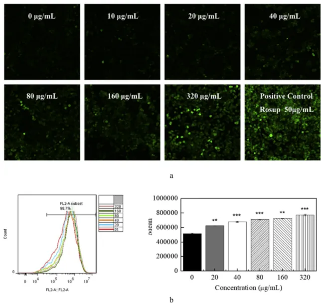
The effect of flaxseed extract on the accumulation of ROS in MCF-7 human breast cancer cells was measured by confocal laser microscope (TCS SP8, Leica, Germany) and flow cytometer (C6, BD Accuri, USA) using Reactive Oxygen Species Assay Kit (Beyotime, China) with a cell permeable fluorogenic dye DCFH-DA. MCF-7 cells (5 × 10⁵cells/well) were seeded into a six-well plate (Eppendorf, Hamburg, Germany) and allowed to adhere for 24 h. After that, the cells were treated with dif-ferent concentrations of flaxseed extract (0–320 μg/mL) for 24 h. Then the cells were loaded with 10 μM DCFH-DA in serum- and phenol red-free DMEM for 20 min at 37 °C in the dark. After being washed with PBS for 3 times, the cells were observed under a confocal microscopy. At the same time, the cellular fluorescence density was detected with flow cytometer at an excitation wavelength of 488 nm and an emission wavelength of 525 nm. The Rosup (50 μg/mL) was used as a positive control.
2.7. Evaluation of lipid peroxidation
Malondialdehyde (MDA) is the end product of lipid peroxidation. Here, the lipid peroxidation was estimated by testing the levels of MDA through lipid peroxidation MDA assay kit (Beyotime, China) on the base of reaction with thiobarbituric acid. The Thiobarbituric Acid Reactive Substances (TBARs) values were normalized by the total protein con-tent of each sample examined by BCA Protein Assay Kit (Beyotime, China). Briefly, after the cells (5 × 10⁵cells/well) were incubated with flaxseed extract (0–320 μg/mL) for 24 h, 0.1 mL supernatant from lysed cells was mixed with 0.2 mL MDA working solution, heated at 100 °C for 15 min, cooled, and centrifuged. The supernatant of each sample was transferred to a 96-well plate and the optical density was recorded at 532 nm.
2.8. Measurement of mitochondrial membrane potential
Mitochondrial membrane potential (MMP) of MCF-7 human breast cancer cells was evaluated with MMP assay kit with JC-1 (Beyotime, China). After the cells (5 × 10⁵cells/well) were stimulated by flaxseed extract of a series of concentrations (0–320 μg/mL) for 24 h, the cells were stained with fluorescent probe JC-1, which can enter mitochon-dria matrix selectively according to the level of MMP. Briefly, the cells were washed with PBS and incubated with JC-1 staining working so-lution at 37 °C for 20 min in the dark. After being fully rinsed with JC-1 washing buffer, the cells were immediately monitored by a confocal laser scanning microscope. The fluorescence ratio of JC-1 aggregates to monomers reflected the change of MMP. The carbonylcyanide-m-chlorophenylhydrazone (CCCP) (10 μM) was used as a positive control.
2.9. Western blotting
The MCF-7 cells were seeded (5 × 10⁵cells/well) into 6-well plate and treated by flaxseed extract (0–320 μg/mL) for 24 h. After being harvested, the cells were re-suspended in 30 μL cold radio-immunoprecipitation assay (RIPA) lysis buffer containing 1 mM phe-nylmethanesulfonyl fluoride and protease inhibitor cocktail, cen-trifuged at 15,000×g for 20 min at 4 °C. The protein concentration of each sample was examined by BCA Protein Assay Kit (Beyotime, China). The equal amounts of proteins (30–50 μg) were mixed with 5 × Sodium Dodecyl Sulfate (SDS)-loading buffer (4:1, v:v), boiled at 100 °C for 5 min, separated through 10% SDS-Polyacrylamide Gel Electrophoresis
(PAGE gel). Proteins were electrotransferred to a polyvinylidene fluoride (PVDF) membrane (Milipore Corp., Bedford, MA), and the membranes were blocked in 5% nonfat dry milk for 1 h. Then, the membranes were incubated with indicated primary antibodies (1:1000 dilution) at 4 °C overnight for the detection of p53, cleaved-caspase 3, cleaved-caspase 7, cleaved-PARP, GADPH (Cell Signaling Technology, MA, United States). The membranes were further incubated with HRP-conjugated antibodies (1:5000 dilution) at room temperature for 1.5 h. Blots were visualized using an enhanced chemiluminescence kit. Digital images of blots were produced by a Syngene Gel Imaging System (Bio-Rad) and quantified with Syngene software.
2.10. Quantitative real time polymerase chain reaction (qRT-PCR)
The MCF-7 cells (5 × 10⁵cells/well) were cultured in a six-well plate and treated with flaxseed extract (0–320 μg/mL) for 24 h. After the cells being harvested, the total RNA was isolated using TaKaRa MiniBEST Universal RNA Extraction Kit (TaKaRa Bio Inc., Kusatsu city, Japan) according to the manufacturer's instructions. The RNA con-centration was assessed using a NanoVue Spectrophotometer (Biochrom, Cambridge, UK). A total of 600 ng RNA of each sample was reverse-transcribed to cDNA using the PrimeScript RT Reagent Kit (TaKaRa Bio Inc., Kusatsu city, Japan) through a C1000 Touch Thermal Cycler (BIO-RAD, CA, USA) according to the manufacturer’ re-commended protocol. Primers (Sangon Biotech, Shanghai, China) for p53, MDM2, GADPH were designed for real-time RT-PCR. The primer sequences are provided inTable 1. The mRNA levels were evaluated by real time PCR using SYBR Green qPCR Master Mix (TaKaRa Bio Inc., Kusatsu city, Japan) with a Stratagene Mx3005P qPCR system (Agilent, CA, USA). The thermal cycling profile conditions were as follows: 30s of denaturation at 95 °C followed by 40 cycles at 95 °C for 5 s and 60 °C for 30–60 s. Melting curve analysis was carried out after amplification to verify the validity of the amplicon and one distinct peak was observed for each primer. The gene expression of each sample was normalized by the expression of GADPH as an internal standard, using the 2−ΔΔCt method.
2.11. Statistical analysis
The results are shown as mean ± SD for the all of representative experiments. Multiple comparisons between the experimental groups were distinguished by one-way ANOVA. The level of significance was set at p < 0.05.
3. Results
3.1. Fatty acid profiles in flaxseed
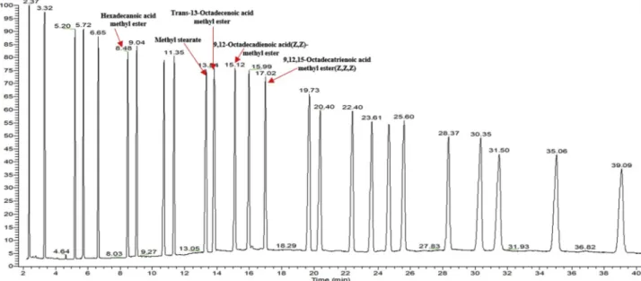
The reference standards were well separated on an Omegawax 250 column within 40 min (Fig. 1). Through the retention time and mass spectra, five peaks in the flaxseed extract were confirmed and char-acterized by comparison with the reference standards (Fig. 2). They are hexadecanoic acid methyl ester (9.52%), methyl stearate (9.25%), trans-13-octadecenoic acid methyl ester (22.54%), 9,12-octadecadie-noic acid (Z, Z)-methyl ester (19.02%), and 9,12,15-octadecatrie9,12-octadecadie-noic
Table 1
Primer sequences of the genes used in this study.
Gene name Primer sequence (5'→3′)
p53 F: TTCCTGAAAACAACGTTCTGTC R: AACCATTGTTCAATATCGTCCG MDM2 F: CTTCTAGGAGATTTGTTTGGCG R: ATGTACCTGAGTCCGATGATTC GADPH F: GCACCGCAAGGCTGAGAAC R: TGGTGAAGACGCCAGTGGA
acid methyl ester (Z, Z, Z) (39.67%). Among the five fatty acids, the most abundant was 9,12,15-octadecatrienoic acid, also known as lino-lenic acid, which accounted for about 39.67% of total fatty acids.
3.2. Flaxseed extract decreases MCF-7 cell growth
In this study, the MCF-7 cells were cultured with flaxseed extract at different concentrations (0–320 μg/mL) for 24, 48, and 72 h to identify the cell viability. It was observed that the flaxseed extract inhibited the growth of MCF-7 cells in a dose- and time-dependent manner (Fig. 3). In all three groups, there was a remarkable decrease in the cell viability (p < 0.01) at the dose of 160 μg/mL, whereas after 24 h, the cell
viability was found to decrease significantly (p < 0.05) at the rela-tively lower dose of 20 μg/mL. The cell viability showed a similar de-crease at 72 h, with 50% inhibition (IC50) of 367.28 μg/mL. These re-sults indicated the cytotoxic effects of flaxseed extract on MCF-7 cells.
3.3. Flaxseed extract induces apoptosis in MCF-7 cells
The fluorescein isothiocyanate-conjugated Annexin V and propi-dium iodide (PI) assay was used to determine the apoptosis of MCF-7 human breast cancer cells treated with flaxseed extract (0–320 μg/mL) for 24 h. The apoptosis (upper and lower-right quadrants) of MCF-7 human breast cancer cells was induced by flaxseed extract in a dose-Fig. 1. The GC-MS chromatograms of GLC-461 reference standards. The reference standards were well separated on the indicated column within 40 min.
Fig. 2. The GC-MS chromatograms of fatty acid methyl esters of flaxseed extract sample. The fatty acids represented as the corresponding methyl esters. They were identified as hexadecanoic acid, stearate, trans-13-octadecenoic acid, 9,12-octadecadienoic acid, and 9,12,15-octadecatrienoic acid.
dependent manner (Fig. 4a). The apoptosis cell percentage of control group was 0.791%, which was far less than the group of the highest concentration 15.09%. Also, significant differences were observed be-tween the control group (0.791%) and the 20 μg/mL group (5.99%) (Fig. 4b). Our results suggested that the flaxseed extract could be ef-fective in increasing the apoptosis of MCF-7 human breast cancer cells at the early and late stages, remarkably.
3.4. Generation of ROS in flaxseed extract-induced apoptosis
Intracellular ROS levels were measured by flow cytometry as well as by confocal laser scanning microscope, being stained with DCFH-DA fluorescent probe. At the presence of endogenous peroxidase, the DCFH was oxidized by ROS, changing to highly fluorescent compound DCF. This process was induced by the flaxseed extract in a dose-dependent manner, which indicates the generation of ROS in MCF-7 human breast cancer cells (Fig. 5a). Moreover, DCF was used as a fluorescent ROS indicator followed by the flow cytometry analysis (Fig. 5b). Significant differences (p < 0.01) were observed between the control group (0 μg/ mL) and the low concentration group (20 μg/mL) after the treatment of flaxseed extract for 24 h. As the concentration increased, the compar-ison between the experimental group (320 μg/mL) and the control group was much more obvious.
3.5. Flaxseed extract increases lipid peroxidation
The effect of flaxseed extract on MCF-7 cell line was analyzed with a lipid peroxidation kit. It was observed that the flaxseed extract in-creased the lipid peroxidation at all doses (Fig. 6). Significant difference (P < 0.001) was observed at the dose of 20 μg/mL, which increased the lipid peroxidation by 3.25 fold in MCF-7 cells. At the dose of 320 μg/mL, a 4.43-fold increase was detected. Furthermore, the in-crease of lipid peroxidation in MCF-7 cells was also observed to be dose-dependent.
3.6. Effects of flaxseed extract on mitochondrial dysfunction pathway
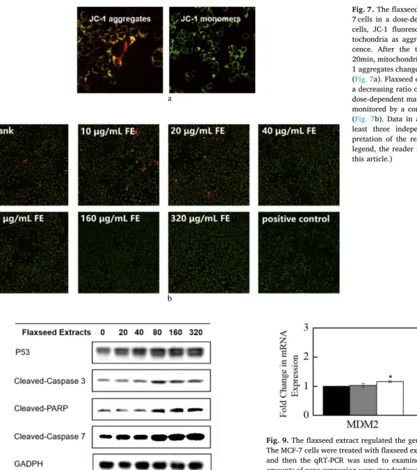
The abnormal mitochondrial structure indicates the dysfunction of mitochondria, accompanied by changes in MMP. The loss of mi-tochondrial transmembrane potential in MCF-7 cells induced by the flaxseed extract was determined using JC-1 fluorescent probe. In healthy cells, the JC-1 fluorescent probes accumulate in mitochondria as aggregates, showing red fluorescence. When apoptosis occurs, the JC-1 aggregates convert to green fluorescent monomers. As a result, the red/green fluorescence ratio detected could be used to evaluate the state of MCF-7 cancer cells (Fig. 7a). The MCF-7 human breast cancer cells were treated with flaxseed extract (0–320 μg/mL) for 24 h before monitoring with a confocal laser scanning microscope. The result
showed that the MCF-7 cells treated with flaxseed extract resulted in a decreasing ratio of the red/green fluorescence in a dose-dependent manner, which suggested that the disruption of MMP after the treat-ment of flaxseed extract to MCF-7 cells (Fig. 7b).
Several gene products have been found to be effective in controlling the apoptotic process. The Western blot analysis of the flaxseed extract on the expression of these gene products in MCF-7 human breast cancer cells was examined. Also, the underlying signaling pathways of flaxseed extract-induced apoptosis, which involved the mitochondrial dysfunc-tion pathway, were explored. The results, summarized inFig. 8, showed that incubation with flaxseed extract for 24 h resulted in an apparent increase in the expression of p53, Cleaved-Caspase 3, Cleaved-PARP, as well as Cleaved-Caspase 7 in a dose-dependent manner, which were all involved in the molecular events of mitochondrial dysfunction pathway. As a result, the decrease of mitochondrial transmembrane potential and the data of western blotting analysis indicated the acti-vation of mitochondrial dysfunction pathway in response to the in-cubation of flaxseed extract to induce apoptosis in MCF-7 cells.
3.7. Expression of genes associated with apoptosis
It was found that the gene of p53 and MDM2 were involved in apoptosis induced by various agents. The qRT-PCR was used to examine whether flaxseed extract had any effect on the expression of these two genes in MCF-7 human breast cancer cells. As shown inFig. 9, after the treatment of flaxseed extract (320 μg/mL) for 24 h, significant increase (p < 0.01) of p53 gene expression was observed with respect to the control group and the experimental group (78% increase compared with the control). For the MDM2 gene expression, only 16% increase was observed for the same treatment with flaxseed extract (320 μg/mL). These data implied the result that the flaxseed extract inhibited the growth and induced apoptosis in MCF-7 human breast cancer cells.
4. Discussion
Apoptosis is a process of regulated cell death, which is an active dedicated molecular process to keep the balance between cell replica-tion and cell death (Curti et al., 2017). The morphological features of apoptosis include nuclear chromatin condensation, DNA fragmentation, blebbing of the plasma membrane, and the presence of apoptotic bodies (Gavrieli et al., 1992). Flaxseed is known to be rich in mammalian lignan precursor secoisolariciresinol diglucoside (Lee and Cho, 2012) and it also has an exceptionally high concentration of α-linolenic acid (Thompson et al., 2005), both of which have protective effects against breast cancer. Therefore, it is hypothesized that flaxseed may have preventive effect on breast cancer. The results of this study revealed the molecular mechanisms underlying flaxseed-induced apoptosis in MCF-7 cells. It was found that the flaxseed extract inhibited the growth of Fig. 3. Flaxseed extract reduced the growth of MCF-7 human breast cancer cells. The MCF-7 cells were treated with flaxseed extract (0–320 μg/mL) for 24, 48, 72 h. Data represent means ± SD of at least three independent experiments. Asterisk (*) indicates that the values significantly differed from the control (*p < 0.05; **p < 0.01; ***p < 0.001).
MCF-7 cells in a dose- and time-dependent manner. It was also found that the flaxseed extract could increase the apoptosis of MCF-7 cells at the early and late stages remarkably, which indicated the flaxseed ex-tract might regulate the growth of MCF-7 cells through the process of apoptosis.
Previous study reported that ROS involved in many cellular pro-cesses, such as cell proliferation, differentiation, and apoptosis (Deng et al., 2010). At the same time, ROS played a dual role in a number of cellular signaling pathways, one of which was that ROS could trigger cellular senescence and function as anti-cancer species (Valko et al.,
2007). Therefore, changes in ROS production was monitored in this study to determine the role of ROS in flaxseed extract-induced apoptosis in MCF-7 cells. Flaxseed extract induced the generation of ROS in a dose-dependent manner after the treatment for 24 h.
As indicated in previous study, the dramatically increased ROS production was associated with the collapse of MMP, which could in-duce a series of mitochondria-related events including apoptosis (Cao et al., 2010). The p53 is a key protein known for keeping the balance between cell survival and cell death by mediating the cell-cycle arrest (Cao et al., 2009). Further, the PARP is a protein helping cells to Fig. 4. Flaxseed extract induced apoptosis of MCF-7 cells. The MCF-7 cells were treated with flaxseed extract (0–320 μg/mL) for 24 h followed by a flow cytometry analysis. Harvested cells were stained with Annexin V and propidium iodide (PI) (Fig. 4a). Results represented viable cells (lower-left quadrant), necrotic cells (upper-left quadrant), early apoptotic cells (lower-right quadrant) and late apoptotic cells (upper-right quadrant). Changes of MCF-7 cells apoptosis percentage after ex-posure to flaxseed extract were shown (Fig. 4b). Data represent means ± SD of at least three independent experiments. Asterisk (*) indicates that the values significantly differed from the control (*p < 0.05; **p < 0.01; ***p < 0.001).
maintain their viability. The cleavage PARP facilitates cellular dis-assembly and is used extensively as a marker during the execution phase of apoptosis (Oliver et al., 1998). Caspases are a family of pro-tease enzymes playing critical role in programmed cell death, especially the executional phase of apoptosis (Cohen, 1997). Caspases are sub-categorized as initiator caspases and executioner caspases. After ap-propriate stimulus, initiator caspases are cleaved and activated, pro-ducing a chain reaction, hence activating several other executioner caspases such as caspase-3 and caspase-7 (Elmore, 2007).
In the present study, the fatty acid profiles in flaxseed were de-termined by gas chromatography mass spectrometry. It was shown in-creased lipid peroxidation in MCF-7 cells at the presence of flaxseed extract, which might be responsible for the observed apoptosis. It was also observed the loss of mitochondrial transmembrane potential in a dose-dependent manner, which was a key event in the induction of apoptosis. In addition, the result revealed the increase of the expression of the p53, cleaved-caspase 3, cleaved-PARP, as well as cleaved-caspase 7 in flaxseed extract-induced apoptosis.
Fig. 5. The flaxseed extract increased the generation of ROS in MCF-7 human breast cancer cells in a dose-dependent manner. The MCF-7 cells were treated with flaxseed extract (0–320 μg/mL) for 24 h. The DCF fluorescence intensity was measured by confocal laser scanning microscope. Data are representative results from at least three independent experiments (Fig. 5a). Flow cytometry analysis of ROS generation in MCF-7 cells was shown inFig. 5b. Data represent means ± SD of at least three independent experiments. Asterisk (*) indicates that the values significantly differed from the control (*p < 0.05; **p < 0.01; ***p < 0.001).
Fig. 6. The flaxseed extract increased the lipid peroxidation in MCF-7 cells in a dose-dependent manner. The MCF-7 cells were treated with flaxseed extract (0–320 μg/mL) for 24 h. The lipid peroxidation in MCF-7 cells was measured by MDA assay kit and the TBAR values were normalized by the total protein content of each sample examined by BCA Protein Assay Kit. The optical density of each sample was measured by fluorescent microscope. Data represent means ± SD of at least three independent experiments. Asterisk (*) indicates that the values significantly differed from the control (*p < 0.05; **p < 0.01; ***p < 0.001).
5. Conclusion
In recent years, advances in therapy and treatment have improved the breast cancer survival rates dramatically, but the cure of breast cancer still has a long way to go. In conclusion, the present study in-vestigated flaxseed extract-induced apoptosis in MCF-7 human breast cancer cells. Our results may clarify potential molecular mechanisms of flaxseed on breast cancer cells. The future work will focus on eluci-dating the precise mechanisms of the flaxseed extract-induced apoptosis in MCF-7 cells.
Conflicts of interest
The authors declare that there are no conflicts of interest.
Acknowledgments
This study was supported by grants from China Agriculture Technology Research System (CARS-16-E-04).
Transparency document
Transparency document related to this article can be found online at https://doi.org/10.1016/j.fct.2019.03.029.
References
Ashkenazi, A., Dixit, V.M., 1998. Death receptors: signaling and modulation. Science 1305–1308.
Cadenas, E., Davies, K.J., 2000. Mitochondrial free radical generation, oxidative stress, and aging1. Free Radical Biol. Med. 29, 222–230.
Call, J.A., Eckhardt, S.G., Camidge, D.R., 2008. Targeted manipulation of apoptosis in cancer treatment. Lancet Oncol. 9, 1002–1011.
Cao, X., Wang, A., Jiao, R., Wang, C., Mao, D., Yan, L., Zeng, B., 2009. Surfactin induces apoptosis and G2/M arrest in human breast cancer MCF-7 cells through cell cycle
Fig. 7. The flaxseed extract decreased MMP in MCF-7 cells in a dose-dependent manner. In the healthy cells, JC-1 fluorescent probes accumulated in mi-tochondria as aggregates and released red fluores-cence. After the treatment of CCCP (10 μM) for 20min, mitochondrial potential collapsed and the JC-1 aggregates changed to green fluorescent monomers (Fig. 7a). Flaxseed extract (0–320 μg/mL) resulted in a decreasing ratio of the red/green fluorescence in a dose-dependent manner after the treatment for 24 h, monitored by a confocal laser scanning microscope (Fig. 7b). Data in are representative result from at least three independent experiments. (For inter-pretation of the references to colour in this figure legend, the reader is referred to the Web version of this article.)
Fig. 8. Effects of flaxseed extract on the apoptosis-related proteins in MCF-7 cells. The MCF-MCF-7 cells were treated with flaxseed extract (0–320 μg/mL) for 24 h and the expression of p53, Cleaved-Caspase 3, Cleaved-PARP, as well as Cleaved-Caspase 7 were measured by western blot analysis. The expression of GADPH was used as the internal control for equivalent loading. The western blotting data presented were representative of that obtained in at least three separated experiments.
Fig. 9. The flaxseed extract regulated the gene expression of p53 and MDM2. The MCF-7 cells were treated with flaxseed extract (0, 20, 320 μg/mL) for 24 h, and then the qRT-PCR was used to examined gene expression. The relative amounts of gene expression were standardized and calculated by the expression of house-keeping gene GADPH. Data represent means ± SD of at least three independent experiments. Asterisk (*) indicates that the values significantly differed from the control (*p < 0.05; **p < 0.01).
factor regulation. Cell Biochem. Biophys. 55, 163.
Cao, X.-h., Wang, A.-h., Wang, C.-l., Mao, D.-z., Lu, M.-f., Cui, Y.-q., Jiao, R.-z., 2010. Surfactin induces apoptosis in human breast cancer MCF-7 cells through a ROS/JNK-mediated mitochondrial/caspase pathway. Chem. Biol. Interact. 183, 357–362.
Cohen, G.M., 1997. Caspases: the executioners of apoptosis. Biochem. J. 326, 1–16.
Curti, V., Di Lorenzo, A., Da Crema, M., Xiao, J.B., Nabavi, S.M., Daglia, M., 2017. In vitro polyphenol effects on apoptosis: an update of literature data. Semin. Canc. Biol. 46, 119–131.
Deng, Y.T., Huang, H.C., Lin, J.K., 2010. Rotenone induces apoptosis in MCF‐7 human breast cancer cell‐mediated ROS through JNK and p38 signaling. Mol. Carcinog. 49, 141–151.
Deshpande, R., Mansara, P., Suryavanshi, S., Kaul-Ghanekar, R., 2013. Alpha-linolenic acid regulates the growth of breast and cervical cancer cell lines through regulation of NO release and induction of lipid peroxidation. J. Mol. Biochem. 2.
Doiron, K.J., Yu, P., 2017. Recent research in flaxseed (oil seed) on molecular structure and metabolic characteristics of protein, heat processing-induced effect and nutrition with advanced synchrotron-based molecular techniques. Crit. Rev. Food Sci. Nutr. 57, 8–17.
El-Sayed, E., Ibrahim, K., 2017. Effect of the types of dietary fats and non-dietary oils on bone metabolism. Crit. Rev. Food Sci. Nutr. 57, 653–658.
Elmore, S., 2007. Apoptosis: a review of programmed cell death. Toxicol. Pathol. 35, 495–516.
Gago-Dominguez, M., Castelao, J.E., Pike, M.C., Sevanian, A., Haile, R.W., 2005. Role of lipid peroxidation in the epidemiology and prevention of breast cancer. Cancer Epidemiol. Biomark. Prev. 14, 2829–2839.
Gavrieli, Y., Sherman, Y., Ben-Sasson, S.A., 1992. Identification of programmed cell death in situ via specific labeling of nuclear DNA fragmentation. J. Cell Biol. 119, 493–501.
Jemal, A., Bray, F., Center, M.M., Ferlay, J., Ward, E., Forman, D., 2011. Global cancer statistics. CA A Cancer J. Clin. 61, 69–90.
Kang, J.X., Wang, J., 2005. A simplified method for analysis of polyunsaturated fatty acids. BMC Biochem. 6, 5.
Kroemer, G., Galluzzi, L., Brenner, C., 2007. Mitochondrial membrane permeabilization in cell death. Physiol. Rev. 87, 99–163.
Lee, J., Cho, K., 2012. Flaxseed sprouts induce apoptosis and inhibit growth in MCF-7 and MDA-MB-231 human breast cancer cells. In Vitro Cell. Dev. Biol. Anim., vol. 48, 244–250.
Mohanan, A., Nickerson, M.T., Ghosh, S., 2018. Oxidative stability of flaxseed oil: effect of hydrophilic, hydrophobic and intermediate polarity antioxidants. Food Chem. 266, 524–533.
Oliver, F.J., de la Rubia, G., Rolli, V., Ruiz-Ruiz, M.C., de Murcia, G., Ménissier-de Murcia, J., 1998. Importance of poly (ADP-ribose) polymerase and its cleavage in apoptosis Lesson from an uncleavable mutant. J. Biol. Chem. 273, 33533–33539.
Oomah, B.D., 2001. Flaxseed as a functional food source. J. Sci. Food Agric. 81, 889–894.
Reed, J.C., 1997. Cytochrome c: can't live with it—can't live without it. Cell 91, 559–562.
Robertson, J.D., Orrenius, S., 2000. Molecular mechanisms of apoptosis induced by cy-totoxic chemicals. Crit. Rev. Toxicol. 30, 609–627.
Simon, H.-U., Haj-Yehia, A., Levi-Schaffer, F., 2000. Role of reactive oxygen species (ROS) in apoptosis induction. Apoptosis 5, 415–418.
Sledge, G.W., Mamounas, E.P., Hortobagyi, G.N., Burstein, H.J., Goodwin, P.J., Wolff, A.C., 2014. Past, present, and future challenges in breast cancer treatment. J. Clin. Oncol. 32, 1979.
Thompson, L.U., Chen, J.M., Li, T., Strasser-Weippl, K., Goss, P.E., 2005. Dietary flaxseed alters tumor biological markers in postmenopausal breast cancer. Clin. Cancer Res. 11, 3828–3835.
Thornberry, N.A., Lazebnik, Y., 1998. Caspases: enemies within. Science 281, 1312–1316.
Valko, M., Leibfritz, D., Moncol, J., Cronin, M.T.D., Mazur, M., Telser, J., 2007. Free radicals and antioxidants in normal physiological functions and human disease. Int. J. Biochem. Cell Biol. 39, 44–84.
Wang, H., Wang, J., Qiu, C., Ye, Y., Guo, X., Chen, G., Li, T., Wang, Y., Fu, X., Liu, R.H., 2017. Comparison of phytochemical profiles and health benefits in fiber and oil flaxseeds (Linum usitatissimum L.). Food Chem. 214, 227–233.
Weinrauch, Y., Zychlinsky, A., 1999. The induction of apoptosis by bacterial pathogens. Annu. Rev. Microbiol. 53, 155–187.
Wiggins, A.K., Mason, J.K., Thompson, L.U., 2015. Growth and gene expression differ over time in alpha-linolenic acid treated breast cancer cells. Exp. Cell Res. 333, 147–154.