SN Applied Sciences (2019) 1:144 | https://doi.org/10.1007/s42452-018-0117-5
Enzyme‑free colorimetric sensing of glucose using
l‑cysteine
functionalized silver nanoparticles
Sumaira Adnan1 · Nazar Hussain Kalwar2,3,4 · Malik Waseem Abbas1 · Razium Ali Soomro1,4,5 · Mumtaz Ali Saand6 ·
Fazli Rabbi Awan1 · Ahmet Avci3 · Erol Pehlivan7 · Sadia Bajwa1 © Springer Nature Switzerland AG 2019
Abstract
The study demonstrates an efficient, simple and on-site viable approach for sensitive determination of blood glucose using colorimetric sensing approach. The devised colorimetric sensor works based on cysteine functionalized silver nanoparticles. The as-synthesized Ag NPs were elaborately characterized using advanced analytical techniques such as ultra-violet visible spectroscopy, Fourier transform infrared spectroscopy, transmission electron microscopy, atomic force microscopy and X-ray diffraction. The detection of glucose was carried under ambient air conditions where change in the optical characteristics of Ag NPs and subsequent interaction between glucose and functional moiety (i.e. surface bind cysteine) of Ag NPs was considered as the signal response. This interaction led to direct detection of glucose in the concentration range between 0.01 to 0.17 µM with limit of detection up to 1 × 10⁻⁴ µM. It is worthwhile mentioning that
success of the assay lies in application of the developed colorimetric sensor in real blood glucose measurements, which also proved its capability for field based analysis. In addition its simple design, low cost, and more reliability signifies the usefulness of colorimetric sensor and it can be extended monitor other biologically important molecules.
Keywords Glucose · Enzyme free · Colorimetric probes · Functionalized Ag NPs
1 Introduction
As diabetes mellitus have become pressing health prob-lem around the globe when dealing with glucose metabo-lism disorders. This chronic disease, associated with insulin deficiency/resistance may lead to heart or kidney failure, high blood pressure, nerve damage and blindness due to continuous variation in blood glucose [1]. Thus, research on portable devices and chemical assays which can allow fast, sensitive and reliable monitoring of blood glucose level is an area of scientific research gaining significant interest of the people. Conventional approaches which include high-performance liquid chromatography (HPLC)
[2], fluorescence spectrometry and chemiluminescence methods [2], despite being sensitive, fail to offer advan-tages like fast analysis, affordability, portability and ease of analysis. In contrast, colorimetric assay based on the use of nano-sized noble metal nanoparticles such as gold or silver have proven efficient, cheap and capable for on-site monitoring or screening of biologically important molecules. Among various metals available, silver and gold are most extensively used for the development of colorimetric probes based on their excellent surface plas-mon resonance (SPR) characteristics. However, the use of such noble metals for probing biologically important molecules for example glucose is normally associated
Received: 29 October 2018 / Accepted: 11 December 2018 / Published online: 3 January 2019
* Nazar Hussain Kalwar, [email protected] | 1National Institute for Biotechnology and Genetic Engineering (NIBGE),
Faisalabad 38000, Pakistan. 2Institute of Chemistry, Shah Abdul Latif University, Khairpur 66020, Pakistan. 3Department of Mechanical Engineering, Faculty of Engineering, University of Selcuk, Campus, 42079 Konya, Turkey. 4Interface Analysis Centre, School of Physics, University of Bristol, Bristol BS8 1TL, UK. 5National Centre of Excellence in Analytical Chemistry, University of Sindh, Jamshoro 76080, Pakistan. 6Department of Botany, Shah Abdul Latif University, Khairpur 66020, Pakistan. 7Department of Chemical Engineering, Faculty of Engineering, University of Selcuk, Campus, 42079 Konya, Turkey.
with the application of enzyme (glucose oxidase, GOx) for the detection [1, 2]. In this connection many reports have already been published likewise Zhang et al. [3] reported the use of Ag NPs for detection of glucose based on the change in SPR response as a consequence of H2O2 pro-duced from the enzymatic reaction between glucose and glucose oxidase previously immobilized on surface of Ag NPs. In a similar scenario, use of gold nanoparticle (Au NPs) coupled with Ag NPs have also been proposed for the detection of glucose [4]. Literature reveals that recently efforts have been carried out for developing fluorescence probe based on GOx loaded nanocomposites of carbon nanodots supported over Ag NPs for gulucose detection. Nanocomplex based on Ag NPs and graphene quantum dots (AgNPs/GQDs) loaded with glucose oxidase have also been used for the development of colorimetric probe for glucose detection [5]. In addition to these, use of bi-metallic composite particles and metal oxides has also been reported for the enzyme based optical probes [6, 7].
Despite the extreme selectivity offered by enzymatic assays, the use of such probes for practical application is restricted by the limitations offered by the enzyme. The temperature depended stability, high cost and immobili-zation complexities of the enzyme marks these assays as unfavorable candidates for diverse application. Recently, enzyme-free approaches have gained substantial atten-tion due to their improved sensitivity and selectivity. An optical probe based on nano-mimicking capability of gold and iron oxide magnetic nanoparticles composite has been proposed by Shin et al. [8]. Despite the success of nano-mimicking assays, efforts for finding newer, simpler and cost-efficient protocols for reliable pre-screening of clinically important biomolecules are still under way. In this context, colorimetric sensor which rely on the interac-tion between the probe (nanoparticle) and target analyte are gaining reasonable consideration [9]. These assays are not only simple in mechanistic consideration but also involve minimum chemicals and complexity which make them suitable to be used practically in low-resource situations [10]. A number of such assays have already been developed for various heavy metal ions and other toxic compounds such as pesticides and drugs [9, 10]. In the present study we have functionalized Ag NPs with
l-cysteine amino acid and carried out direct detection of
glucose (i.e. enzyme free) on their surface via colorimet-ric sensing approach. The assay rely on the competitive interaction between functionalizing agent (l-cysteine)
and glucose molecules leading to particle aggregation and subsequent change in SPR response. The developed assay provides a simplest route towards reliable detection of glucose without the need of any enzyme or any addi-tional chemicals. The practical characteristic of the assay
has been validated by considering it for glucose detection from human serum samples.
2 Materials and methods
2.1 Chemicals
All chemicals were analytical grade and used without further purification. Pure Milli-Q water as the prepara-tory medium. Silver nitrate (AgNO3–97%), was obtained from E. Merck. l-cysteine (C3H7NO2S), sodium hydroxide
(NaOH–98%) and hydrochloric acid (HCl–37%) were pur-chased from Sigma–Aldrich. The stock solutions of metal salts were prepared in pure Milli-Q water and all experi-ments were performed at ambient temperature (25 ± 2 °C).
2.2 Instrumentation
UV–Visible spectroscopy (Lambda 35 of PerkinElmer) was considered for monitoring the variation in SPR signal of Ag NPs within the spectral range of 300–800 nm. Confirma-tion of surface funcConfirma-tionalizaConfirma-tion was achieved using fou-rier transform infrared (FTIR) spectroscopy (Nicolet 5700 of Thermo) with ATIR mode. Further, characterizations of functionalized Ag NPs were obtained using transmission electron microscopy (TEM) (Jeol JEM 1200 EX MKI), atomic force microscopy (AFM) and X-ray diffraction (XRD) (model D-8 of Bruker). The change in color was noted by capturing photographs of the colloidal Ag NPs solutions before and after interaction with glucose using 18 mega pixel digital camera.
2.3 Synthesis and functionalization of silver nanoparticles (Ag NPs)
The Ag NPs were synthesized and functionalized using one-pot wet-chemical reduction approach. In a typical experiment, a 10 ml AgNO3 (1 M) was homogenized with 1 ml of l-cysteine (1 mM) under constant stirring
condi-tion. The clear solution was then introduced into 60 ml of NaBH4 (1 M) until a stable bright yellow colloidal solution was obtained with SPR wavelength of 395 nm. The pH of formed colloidal solution was maintained at 6.5 in accord-ance to pka value of l-cysteine to obtain effective
func-tionalization and smaller size. The use of l-cysteine was
considered based on its capability to act both as efficient surface protecting agent and mild reducing agent. This allowed preserving homogeneity of synthesized Ag NPs with generation of an inert atmosphere, which controlled the use of protective gas and hence tedious strategies to avoid oxidation.
2.4 Colorimetric sensing of glucose using cyst‑Ag NPs
The colorimetric sensing of glucose using cyst-Ag NPs was carried out at room temperature in aqueous solution. For a representative experiment, a certain range of glucose concentration (0.05 mM to 1.5 mM) was introduced within a fixed aliquot of cyst-Ag NPs. The mixtures were allowed to stabilize for few minutes followed by SPR measurement using spectrophotometer. The absorbance was measured at SPR wavelength of 395 nm, in reference to a blank sam-ple (without glucose). The change in absorbance (ΔA) was taken as the analytical signal for the corresponding concentration, while the change in color from bright yel-low to colorless was considered the visual response of the devised sensor. The color change was recorded using an 18 mega pixel digital camera after stabilizing the colloidal mixture for 5 min.
3 Results and discussion
3.1 Characterization of cyst‑Ag NPs
Optical characteristics of nano-sized noble metal parti-cles are a function of geometry and size; thus the optical response of such particles is highly sensitive to change in size of the particles [9]. Owing to the surface plasmon modes of noble metallic NPs which belong to the optical region of electromagnetic spectrum [11–13], one can eas-ily use optical spectroscopy as an efficient tool to observe different molecules based on change in the intra-particle distance of noble metals. In this case, the most stable and small sized cyst-Ag NPs (with blue shift in spectrum) were
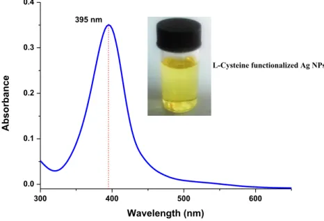
obtained after careful optimization of reaction parameters like concentrations of the precursor salt and functional-izing agent, pH of colloidal solution (not shown). Figure 1
shows the UV–Vis absorbance of a represented sample of the as prepared Ag NPs with SPR wavelength noted at 395 nm.
The morphological characteristics were evaluated using TEM analysis. Figure 2 shows a typical image for as-synthe-sized cyst-Ag NPs. The formed Ag NPs were evident of high surface homogeneity and dispersion indicating the suc-cess of surface functionalization with l-cysteine molecules.
The average size of cyst-Ag NPs were estimated to be in range of 10–35 nm ± 1.4 nm. The EDX spectra provided as the inset of Fig. 2, further confirms the surface purity of the as-synthesized Ag NPs.
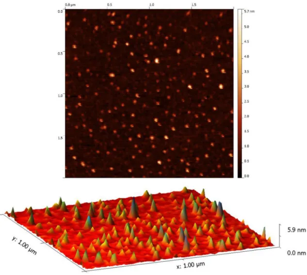
The AFM images for the same Ag NPs are presented in Fig. 3. The findings are apparent of narrow and homog-enous distribution which is less likely in case of biologi-cal methods where use of plant extracts are considered as reducing/capping agents [14]. The topographical map shows significant variation in topography along the sur-face. This reflects the smaller cross-section of the synthe-sized nanoparticles with larger number of active sites available at the surface for reaction participation.
The XRD pattern shown in Fig. 4 consists of peak indexed to (111), (200), (220) and (311) planes of face cen-tered cubic structure (FCC) as referenced against ICCDD (file no: 89-3722). The absence of peaks other than metallic phase silver confirms the compositional purity of the as-synthesized cyst-Ag NPs.
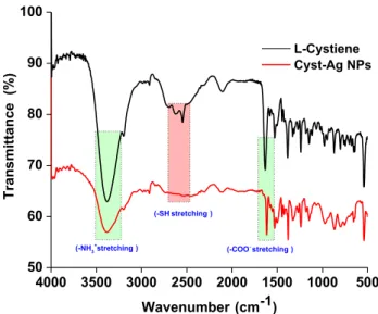
Confirmation for the functionalization was obtained using FTIR analysis. Figure 5 shows the FTIR spectra of both standard l-cysteine and Cyst-Ag NPs. The
charac-teristics band for l-cysteine includes bands at 1630 and
Fig. 1 SPR wavelength for cyst-Ag NPs with inset figure showing the corresponding bright yellow colloidal sol
300 400 500 600 0.0 0.1 0.2 0.3 0.4 Absorbance Wavelength (nm) 395 nm L-Cysteine functionalized Ag NPs
Fig. 2 TEM analysis of a Cyst-Ag NPs with EDX spectra as inset figure and b Cyst-Ag NPs in the absence and presence of glucose (0.01 µM) respectively
1390 cm−1 for asymmetric and symmetric stretching of
(COO−) and 1530 cm−1 for –NH bending vibration.
A broad band near 3000 for –NH3+ stretching and a
band near 2500 cm−1 for –SH [15–17]. In case of cyst-Ag
NPs, the band for –SH was not observed. This confirms the interaction of Ag with cysteine molecules via thiol group. This interaction was further strengthened by interaction of carbonyl from cysteine over Ag NPs sur-face, as indicated by shift in the vibrational frequency of COO− from 1630 to 1620 cm−1. A slight shift in the overall
frequencies was also observed which can be explained on the bases of dipole change as a result of surface func-tionalization of Ag NPs with cysteine molecules [18].
3.2 Enzyme free colorimetric sensing of glucose
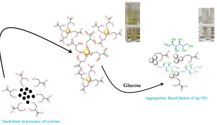
Due to the bright yellow color of colloidal solution of Ag NPs it exhibit a sharp SPR band near 400 nm in UV–Vis spectral window. Therefore a typical narrow SPR peak at 395 nm was evidenced by size homogeneous and crys-talline Cyst-Ag NPs as shown in Fig. 1. This noted optical characteristic can be attributed to the effective capping/ functionalizing agent of Ag NPs with cysteine molecules [19, 20]. The detection mechanism of glucose can be explained on the basis of inter-particle fusion caused by favorable intermolecular forces as a consequence of addi-tion of glucose. This generalized mechanism is illustrated in Scheme 1. It is well-known that SPR of Ag NPs is highly sensitive towards change in the refractive index near the vicinity of metal nanoparticles. In this case when glucose is introduced into colloidal sol of Cyst-Ag NPs, it tends to contribute to the existing inter-ionic forces that prevail between the cysteine functionalized Ag NPs. The glucose based on its hydroxyl functionality can easily facilitates the amine-carbonyl interactions subsequently allowing nanoparticles to fuse together. Unlike the absorption-based colorimetric sensing where change in inter-parti-cle distance is responsible for the shift in SPR wavelength [21], the said process results in complete aggregation of NPs leading to decline in the SPR intensity. The proposed mechanism is further supported by the TEM analysis of cyst-Ag NPs before and after the introduction of glucose molecules. Figure 3a shows that in the absence of glucose, the cyst-Ag NPs tends to be spherical and well-dispersed. In contrast, the TEM image Fig. 3b for cyst-Ag NPs with glucose shows evidence of particle fusion and formation of aggregates with size greater than 100 nm. Quantitative response measurements were carried out by monitor-ing the changes in absorbance (ΔA) for cyst-Ag NPs with glucose in concentration range of 0.01–0.17 µM (Fig. 6). The linear regression analysis was proof of good linearity with LOD (3 × ∂/slope) and LOQ (10 × ∂/slope) estimated to be 1.2 × 10⁻⁴ and 0.03 µM respectively. Table 1 compares
the analytical characteristics of the described sensor with other competitor. It is clear that, the discussed sensor offer greater analytical leverage with additional advantage of working without enzyme and cost-friendly. The selectiv-ity of the assay was evaluated by monitoring the change in SPR response against common co-existing interfering species such as ascorbic acid, sucrose and metal ions like Cu2+, Zn2+ and K+ ions with concentration ten folds higher
than glucose (0.01 µM). The spectral profile shows negligi-ble variation in the SPR intensity of Ag NPs in the presence of interferents (Fig. 7). This negligible variation in the SPR characteristics of Ag NPs, is proof of the high tolerance offered by of cyst-Ag NPs against these interferents.
30 40 50 60 70 80 0 25 50 75 100 125 150 175 ICCDD No. 89-3722 (222) (311) (220) (200) Intensity (a.u.) 2 theta Scale (111)
Fig. 4 XRD pattern recorded for Cyst-Ag NPs with major peaks indexed to cubic unit cell of FCC crystal structure for standard silver
4000 3500 3000 2500 2000 1500 1000 500 50 60 70 80 90 100 (-SH stretching ) (-COO- stretching ) Transmittance (% ) Wavenumber (cm-1) L-Cystiene Cyst-Ag NPs (-NH3+stretching )
It is clear that, the discussed sensor offer greater analyti-cal leverage with additional advantage of working with-out enzyme and cost-friendly. The selectivity of the assay was evaluated by monitoring the change in SPR response against common co-existing interfering species such as ascorbic acid, sucrose and metal ions like Cu2+, Zn2+ and
K+ ions with concentration ten folds higher than glucose
(0.01 µM). The spectral profile shows negligible variation in the SPR intensity of Ag NPs in the presence of interferes (Fig. 7). This negligible variation in the SPR characteristics of Ag NPs, is proof of the high tolerance offered by of Cyst-Ag NPs against these interferes.
Scheme 1 A generalize illustration representing formation and aggregation of Ag NPs in the presence of cysteine and glucose respectively
300 400 500 0.0 0.1 0.2 0.3 0.4 0.17 M 0.16 M 0.14 M 0.12 M 0.10 M 0.08 M 0.02 M 0.04 M Absorbance Wavelength (nm) Blank (without glucose)
0.01 M 0.02 M 0.03 M 0.04 M 0.05 M 0.06 M 0.07 M 0.08 M 0.09 M 0.10 M 0.11 M 0.12 M 0.13 M 0.14 M 0.15 M 0.16 M 0.17 M A Blank
(a)
0.00 0.02 0.04 0.06 0.08 0.10 0.12 0.14 0.16 0.18 0.00 0.05 0.10 0.15 0.20 0.25 0.30 y = 1.5348x - 0.0122 R² = 0.9922 Concentration ( M) A(b)
Fig. 6 a Variation in the noted SPR of Cyst-Ag NPs against succes-sive addition of glucose solution with inset figure representing the corresponding decline in the colour intensity and b Calibration plot
for the devised colorimetric sensor with linear fit analysis for glu-cose in concentration range of 0.01–0.17 µM
3.3 Testing glucose from human serum samples
To validate the practical applicability of the developed col-orimetric assay, the cyst-Ag NPs were subjected to human
serum for glucose analysis taken from lab volunteers with informed consents. Serum was obtained after centrifugation of whole blood followed by two fold dilution for easiness of analysis. The serum samples were then tested for the con-centration of glucose according to the procedure mentioned in Sect. 3.2. The concentration measured using Ag based col-orimetric assay was cross-checked with the results obtained with commercial glucometer. Table 2 shows the obtained data with RSD value for each samples < 10% indicating the reliability of the assay for practical applications.
4 Conclusion
In conclusion, the study demonstrates the use of
l-cysteine functionalized Ag NPs as colorimetric probe
for enzyme free detection of blood glucose. The detec-tion system relied on direct interacdetec-tion between the Table 1 Analytical comparison
of designed sensor with various other competitors
S. no Type of nanoparticles Concentrations (µM) LOD (µM) References
1. Ag Nanoprism 0.2–100 0.2 [21]
2. Ag NPs 868.6 – [22]
3. Ag NPs 215.50–3448 99.13 [23]
4. Au–Ag NPs 5–70 3 [24]
5. Ag NPs/GQDs 500–8000 30 [5]
6. Isotropic silver nanoparticles
(iAg NPs) 0.2–32 0.09 [25]
7. GQDs/Ag NPs 0.5–400 0.17 [26]
8. Ch-Ag NPs 500–200 0.1 [27]
9. C Dots/Ag NPs 2–4000 1.39 [28]
10. L-cyst-Ag NPs 0.01–0.17 1 × 10⁻⁴ This work
Fig. 7 The spectra profile representing the selectivity for cyst-Ag NPs against common interferents with inset figure depicting the corresponding color variation 300 400 500 600 0.0 0.1 0.2 0.3 0.4 Absorbance Wavelength (nm) Sucrose Ascorbic Acid Cu 2+ Zn2+ K+ Glucose
Table 2 Blood glucose determination from real blood serum sam-ple using devised colorimetric probe in reference to measured val-ues obtained using commercial glucometer
a The measured concentration is the mean value for three consecu-tive measurements
Sample Blood glucose (Glucometer) mmol/L Blood glucose (Colorimetric sen-sor) mmol/La Relative standard deviation (RSD %) 1 4.5 4.3 4.0 2 4.8 4.4 9.0 3 5.2 5.0 4.0 4 5.6 5.4 3.7
glucose molecules and functional groups of surface bound cysteine molecules. The assay demonstrated excellent working linearity for enzyme free glucose detection in range of 0.01–5.4 µM. The assay provides a non-enzymatic approach for the detection of glucose avoiding use of toxic chromophores or unstable enzymes like glucose oxidase or HRP. The successful application of the developed sensor in real blood glucose proves its feasibility for practical application and its applicabil-ity as detection strips for enzyme free glucose screen-ing method. Moreover, the preparation of this sensor is very simple, fast, inexpensive and extendable to monitor other biologically important molecules.
Acknowledgements We acknowledge The Scientific and Technologi-cal Research Council of Turkey (TUBITAK) for provision funding for research expenses, traveling and living expenses to conduct the research during post-doctoral fellowship under 2221-Fellowships for Visiting Scientists and Scientists on Sabbatical Leave.
Funding This study was funded by The Scientific and Technological Research Council of Turkey (TUBITAK) under 2221-Fellowships for Visit-ing Scientists and Scientists on Sabbatical Leave, the Grant Number of the project is 1059B211600383.
Compliance with ethical standards
Conflict of interest The authors declare that they have no conflict of interest.
References
1. Akrema, Rahisuddin (2018) Metal nanoparticles as glucose sensor. In: Khan ZH (ed) Nanomaterials and their applications. Springer, Singapore, pp 143–168
2. Rauf S, Nawaz MAH, Badea M, Marty JL, Hayat A (2016) Nano-engineered biomimetic optical sensors for glucose monitoring in diabetes. Sensors 16(11):1–17
3. Zhang Y, Zhang Y-J, Xia X-D, Hou X-Q, Feng C-T, Wang J-X, Deng L (2013) A quantitative colorimetric assay of H2O2 and glucose using silver nanoparticles induced by H2O2 and UV. Chin Chem Lett 24(12):1053–1058
4. Gao Y, Wu YT, Di JW (2017) Colorimetric detection of glucose based on gold nanoparticles coupled with silver nanoparticles. Spectrochimica Acta A Mol Biomol Spectrosc 173:207–212 5. Nguyen ND, Nguyen TV, Chu AD, Tran HV, Tran LT, Huynh CD
(2018) A label-free colorimetric sensor based on silver nanopar-ticles directed to hydrogen peroxide and glucose. Arab J Chem 11(7):1134–1143
6. Zhang X, Wei M, Lv B, Liu Y, Liu X, Wei W (2016) Sensitive colori-metric detection of glucose and cholesterol by using Au@Ag core-shell nanoparticles. RSC Adv 6(41):35001–35007
7. Li Z, Zhang Y, Ye JY, Guo MQ, Chen J, Chen WY (2016) Nonenzy-matic glucose biosensors based on silver nanoparticles depos-ited on TiO2 nanotubes. J Nanotechnol 2016:1–7
8. Shin HY, Cho S, Kim MI (2017) Enzyme-free colorimetric detec-tion of glucose using a composite entrapping gold and mag-netic nanoparticles within an agarose gel matrix. J Nanosci Nanotechnol 17(11):7971–7977
9. Soomro RA, Nafady A, Memon N, Sherazi TH, Kalwar NH (2014) l-cysteine protected copper nanoparticles as colorimetric sen-sor for mercuric ions. Talanta 130:415–422
10. Hussain M, Nafady A, Sirajuddin, Hussain-Sherazi ST, Shah MR, Alsalme A, Kalhoro MS, Mahesar SA, Siddiqui S (2016) Cefuro-xime derived copper nanoparticles and their application as a colorimetric sensor for trace level detection of picric acid. RSC Adv 6(86):82882–82889
11. Chen F, Alemu N, Johnston RL (2011) Collective plasmon modes in a compositionally asymmetric nanoparticle dimer. AIP Adv 1(3):032134-16
12. Ghosh SK, Pal T (2007) Interparticle coupling effect on the sur-face plasmon resonance of gold nanoparticles: from theory to applications. Chem Rev 107(11):4797–4862
13. Sherazi STH, Soomro RA, Uddin S, Memon N (2014) Synthesis and characterizations of highly efficient copper nanoparticles and their use in ultra fast catalytic degradation of organic dyes. Adv Mater Res 829:93–99
14. Ahmed S, Ahmad M, Swami BL, Ikram S (2016) A review on plants extract mediated synthesis of silver nanoparticles for antimicro-bial applications: a green expertise. J Adv Res 7(1):17–28 15. Barth A (2007) Infrared spectroscopy of proteins. Biochem
Bio-phys Acta 1767(9):1073–1101
16. Panigrahi S, Kundu S, Basu S, Praharaj S, Jana S, Pande S, Ghosh SK, Pal A, Pal T (2006) Cysteine functionalized copper organosol: synthesis, characterization and catalytic application. Nanotech-nology 17(21):5461
17. Sirajuddin, Nafady A, Afridi HI, Sara S, Shah A, Niaz A (2011) Direct synthesis and stabilization of Bi-sized cysteine-derived gold nanoparticles: reduction catalyst for methylene blue. J Iran Chem Soc 8(1):S34–S43
18. Cai Z-X, Yang H, Zhang Y, Yan X-P (2006) Preparation, characteri-zation and evaluation of water-soluble l-cysteine-capped-CdS nanoparticles as fluorescence probe for detection of Hg(II) in aqueous solution. Anal Chim Acta 559(2):234–239
19. Kalwar NH, Nafady A, Sherazi STH, Soomro RA, Hallam KR, Khaskheli AR, Jamali AA (2015) Catalytic degradation of imi-dacloprid using l-serine capped nickel nanoparticles. Mater Express 5(2):121–128
20. Kalwar NH, Sirajuddin, Soomro RA, Sherazi STH, Hallam KR, Khaskheli AR (2014) Synthesis and characterization of highly efficient nickel nanocatalysts and their use in degradation of organic dyes. Int J Metals. https ://doi.org/10.1155/2014/12610 3
21. Jazayeri MH, Aghaie T, Avan A, Vatankhah A, Ghaffari MRS (2018) Colorimetric detection based on gold nano particles (GNPs): an easy, fast, inexpensive, low-cost and short time method in detection of analytes (protein, DNA, and ion). Sens Bio-Sensing Res 20:1–8
22. Xia Y, Ye J, Tan K, Wang J, Yang G (2013) Colorimetric visualization of glucose at the submicromole level in serum by a homog-enous silver nanoprism-glucose oxidase system. Anal Chem 85(13):6241–6247
23. Serra A, Filippo E, Re M, Palmisano M, Vittori-Antisari M, Bucco-lieri A, Manno D (2009) Non-functionalized silver nanoparticles for a localized surface plasmon resonance-based glucose sen-sor. Nanotechnology 20(16):165501
24. Gao Y, Wu Y, Di J (2017) Colorimetric detection of glucose based on gold nanoparticles coupled with silver nanoparticles. Spec-trochim Acta Part A Mol Biomol Spectrosc 173:207–212 25. Zong C, Li B, Wang J, Liu X, Zhao W, Zhang Q, Nie X, Yu Y (2018)
Visual and colorimetric determination of H2O2 and glucose based on citrate-promoted H2O2 sculpturing of silver nanopar-ticles. Mikrochim Acta 185(3):199
26. Chen S, Hai X, Chen X-W, Wang J-H (2014) In situ growth of silver nanoparticles on graphene quantum dots for
ultrasensitive colorimetric detection of H2O2 and Glucose. Anal Chem 86(13):6689–6694
27. Jiang H, Chen Z, Cao H, Huang Y (2012) Peroxidase-like activity of chitosan stabilized silver nanoparticles for visual and colori-metric detection of glucose. Analyst 137(23):5560–5564
28. Ma J-L, Yin B-C, Wu X, Ye B-C (2017) Simple and cost-effective glucose detection based on carbon nanodots supported on silver nanoparticles. Anal Chem 89(2):1323–1328