R E S E A R C H A R T I C L E
Open Access
Cancer-testis gene expression is associated with
the methylenetetrahydrofolate reductase 677 C>T
polymorphism in non-small cell lung carcinoma
Kerem M Senses
1, Mithat Gonen
2, Ahmet R Barutcu
1,6, Zeynep Kalaylioglu
3, Murat Isbilen
1, Ozlen Konu
1,
Yao T Chen
4, Nasser K Altorki
5and Ali O Gure
1*Abstract
Background: Tumor-specific, coordinate expression of cancer-testis (CT) genes, mapping to the X chromosome, is observed in more than 60% of non-small cell lung cancer (NSCLC) patients. Although CT gene expression has been unequivocally related to DNA demethylation of promoter regions, the underlying mechanism leading to loss of promoter methylation remains elusive. Polymorphisms of enzymes within the 1-carbon pathway have been shown to affect S-adenosyl methionine (SAM) production, which is the sole methyl donor in the cell. Allelic variants of several enzymes within this pathway have been associated with altered SAM levels either directly, or indirectly as reflected by altered levels of SAH and Homocysteine levels, and altered levels of DNA methylation. We, therefore, asked whether the five most commonly occurring polymorphisms in four of the enzymes in the 1-carbon pathway associated with CT gene expression status in patients with NSCLC.
Methods: Fifty patients among a cohort of 763 with NSCLC were selected based on CT gene expression status and typed for five polymorphisms in four genes known to affect SAM generation by allele specific q-PCR and RFLP. Results: We identified a significant association between CT gene expression and the MTHFR 677 CC genotype, as well as the C allele of the SNP, in this cohort of patients. Multivariate analysis revealed that the genotype and allele strongly associate with CT gene expression, independent of potential confounders.
Conclusions: Although CT gene expression is associated with DNA demethylation, in NSCLC, our data suggests this is unlikely to be the result of decreased MTHFR function.
Keywords: Cancer-testis genes, 1-carbon pathway, Adomet, DNA methylation Background
Cancer-testis (CT), or cancer-germline genes, currently with more than 100 members, are distinctly expressed in cancer, germline and trophoblast cells but not in other normal tissues in the adult. Most CT genes constitute multigene families organized in clusters along the X chromosome. Members within a family are highly hom-ologous, however, no conservation of sequence exists be-tween families [1]. Despite the lack of sequence similarity (including promoters), re-expression of almost all CT genes in tumors correlates with the demethylation of their
promoters that occurs in parallel to a genome-wide de-methylation event, primarily affecting repeat regions [2]. The mechanisms leading to CT gene promoter demethyla-tion in cancer are unknown. Increased BORIS expression has been associated with upregulated CT gene expression [3,4], but the protein is likely not the sole responsible fac-tor in this event. Histone acetylation has also been shown to facilitate CT gene expression, primarily when it associ-ates with DNA demethylation [5].
As most CT gene products are highly antigenic they have been utilized in clinical trials based on immunothera-peutic approaches targeting these antigens [6]. Since pa-tient eligibility for CT targeting immunotherapy requires that the tumor express CT genes, it is important to know whether CT gene expression can be induced. It is expected
* Correspondence:[email protected]
1
Department of Molecular Biology and Genetics, Bilkent University, Ankara, Turkey
Full list of author information is available at the end of the article
© 2013 Senses et al.; licensee BioMed Central Ltd. This is an Open Access article distributed under the terms of the Creative Commons Attribution License (http://creativecommons.org/licenses/by/2.0), which permits unrestricted use, distribution, and reproduction in any medium, provided the original work is properly cited.
that any approach leading to CT gene expression should also result in the demethylation of their promoters.
Production of the sole methyl donor in the cell, S-adenosylmethionine (SAM), depends on the efficient utilization of folate, by the 1-carbon pathway. Several enzymes in this pathway contain common polymorphic variants that reduce the efficiency of the enzyme and thus, the rate of SAM production. Hypomorphic alleles of four of these enzymes (methylenetetrahydrofolate reductase (MTHFR), methionine synthase reductase (MTRR), methionine synthase (MTR), and reduced fol-ate carrier (RFC)), have been associfol-ated with cellular under-utilization of folate and homocysteine, increased DNA hypomethylation, and decreased CpG methylation [7-11]. More recently, the hypomorphic 677 T allele of MTHFR, has been associataed with the expression of MAGE-A1, a CT gene, in glioblastoma multiforme [12]. Others, however, could not reproduce these findings in ovarian carcinoma [13]. In the present study we asked if polymorphisms of the 1-carbon pathway enzymes asso-ciate with CT gene expression in non-small cell lung cancer (NSCLC) patients. Our results show a strong
as-sociation between theMTHFR677 CC genotype as well
as theMTHFR 677 C allele and CT gene expression
in-dependent of age, sex, histology, and tumor stage. Methods
Patients and tumor material
Tumor samples obtained from patients undergoing cura-tive surgical resection for primary NSCLC at the Depart-ment of Cardio-Thoracic Surgery, Weill Medical College of Cornell University, from 1991 to July 2005 were analyzed in this study. Informed consent was obtained from all pa-tients. The study was approved by the Institutional Review Board of Weill Medical College of Cornell University. Fifty tumor samples were selected solely based on CT gene ex-pression from 763 samples that had been evaluated for the presence of transcripts from up to 9 CT genes (NY-ESO-1, LAGE-1, MAGE-A1, MAGE-A3, MAGE-A4, MAGE-A10, CT-7, SSX2, and SSX4), by semi-quantitative PCR, as de-scribed previously [14]. Twenty one samples with CT ex-pression in at least 4 of the 9 CT genes tested, with strong expression in at least one gene, constituted the CT (+) group. Twenty-nine samples with no CT expression in any of the CT genes tested (with a minimum of 5 CT genes tested) were selected as CT (-) tumors for this study. CT gene expression was determined as strong (+++), inter-mediate (++), weak (+ or +/-), or none (-) as previously described [14], and is shown in Additional file 1: Table S1.
DNA analysis
Genomic DNA extracted from tumor tissues were typed using pre-designed 5’-nuclease TaqMan SNP geno-typing assays (Applied Biosystems, Foster City, CA) using
a Stratagene Mx3005P instrument according to the manu-facturer’s instructions. The SNPs typed and their reference
IDs were: MTHFR 677 C> T (rs1801133), MTHFR 1298
A>C (rs1801131), MTR 2756 A>G (rs1805087), and
MTRR 66 A>G (rs1801394). Nested PCR-RFLP was used to type the RFC 80 G>A (rs1051266) polymorphism for which the first round PCR conditions were previously de-scribed [10]. Nested PCR primers were: 5’- AGCCGTAGA AGCAAAGGTAGC-3’ and 5’-AGCGTCACCTTCGTCC CCTC-3’. PCR was performed using DyNAzyme™ II Hot Start DNA Polymerase (Finnzymes, Keilaranta, Finland). PCR conditions were: 10’ activation at 94°C, followed by 35 cycles of 94°C, 62°C and 72°C; 30” each, with a final 72°C, 7’ extension. HinP1I (New England Biolabs, Hert-fordshire, UK) digested PCR products were analyzed as described previously [10]. All analyses were repeated at least twice.
Genotypes for all polymorphisms were determined successfully in all cases (Additional file 2: Table S2). Genotype distributions did not deviate from Hardy-Weinberg equilibrium (Additional file 3: Table S3). Minor allele frequencies for individual loci were: 40%
for MTHFR 677 C > T, 26% for MTHFR 1298 A > C,
14% for MTR 2756 A > G, 54% for MTRR 66, and 42%
for RFC 80 G > A. MTHFR genotypes were not
inde-pendently distributed across the 2 loci. The major 677C allele was in linkage disequilibrium with the minor 1298C allele (D’ = 0.99, r2
= 0.23) [15].
In silico association analysis
Paired datasets, GSE14471 and GSE15714, containing gene expression and SNP genotyping data, respectively, from 111 pediatric acute myeloid leukemia samples (of which 109 were typed successfully), were analyzed for
an association between CT gene expression andMTHFR
677 genotype distribution [16]. A principal component analysis using 44 probesets corresponding to 9 CT gene families was performed for the expression dataset. The first principal component, explaining 0.48 of variance for CT gene expression was used to generate groups representing samples with low, intermediate, and high CT gene expression by K means clustering using a cus-tomized R code [17]. Optimum number of clusters according to Elbow criterion was determined as five. Therefore, five initial cluster centers were placed equally distant from each other where the first and last centers represented the minimum and maximum values of PC1, respectively. Centers were iteratively updated based on the median value of the reassigned cluster members until no change in cluster membership took place. The five clusters were regrouped into three representing low (clusters 1 & 2), intermediate (cluster 3), and high CT gene expression (clusters 4 & 5).
Statistical analysis
To analyze the association between 1-carbon pathway en-zyme polymorphisms and CT gene expression, the genotype distributions were compared in CT (+) and CT (-) tumors by Pearson’s Chi-Square (2 degrees of freedom) or Fisher’s exact tests. Odds ratios (OR) were estimated by multivariate logistic regression. To evaluate whether CT gene expression was related to sex, smoking status, tumor size, and disease stage, Fisher's exact test or Chi-square tests were used. Race information was available for only 29 patients of which 25 were non-Hispanic white, one was a non-Hispanic black, and 3 were of mixed race, and was not included in statistical analyses. All statistical tests were two-sided with a 5% type I error rate, unless indicated otherwise, and were carried out using SAS (version 9.3) software (SAS Institute, Cary, NC). P < 0.05 was considered statistically significant.
Results
Demographics and clinical characteristics of patients and their distribution within CT (+) and (-) groups are shown in Table 1 and Additional file 1: Table S1. Tumors with non-squamous cell carcinoma histology and earlier tumor stage (T stage) showed lower CT gene expression, similar to what has been reported previously [14]. Distribution of individual genotypes among CT (+) and (-) tumors are shown in Table 2 and Additional file 2: Table S2. A
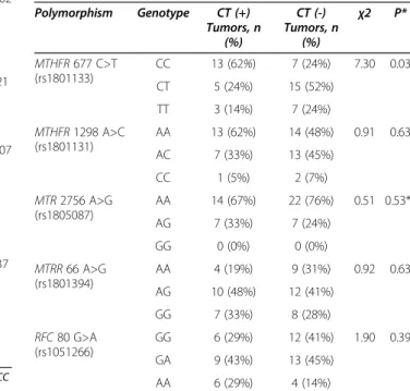
signifi-cant association between the MTHFR 677CC genotype
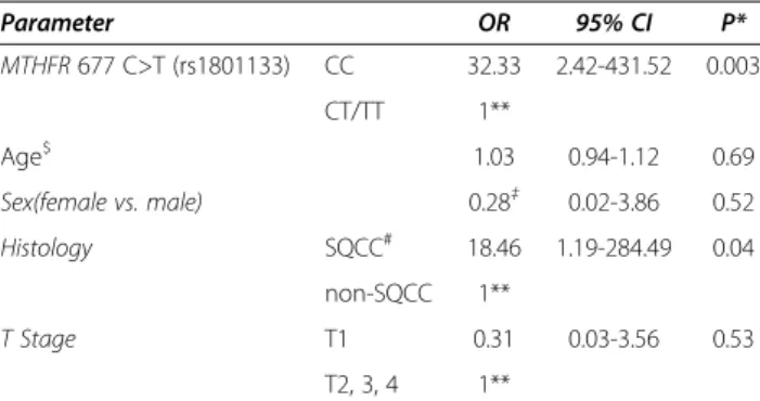
and CT expression was observed (P = 0.03). CT expres-sion was not related to any other genotype tested. A multi-variate logistic regression analysis (MVA) of CT gene expression that included theMTHFR 677 genotype distri-bution, age, sex, histology and T stage revealed that the MTHFR 677 genotype and histology were independent predictors of CT gene expression in this cohort (Table 3).
The MTHFR 677 SNP was found to be associated with
CT gene expression when analyzed on a per allele basis, controlling for confounding factors, while other markers were not (Table 4). We performed anin silico association
analysis for CT expression and theMTHFR 677 genotype
using two datasets derived from childhood acute myeloid leukemia (AML) where both gene expression and SNP genotyping data were available [16]. This analysis, how-ever, did not reveal a statistically significant association be-tween these two parameters (Table 5 and Additional file 4: Figure S1).
Table 1 Demographics and clinical characteristics
CT (+) patients (n=21) CT (-) patients (n=29) P* Age >60 12 19 0.74 <=60 6 7 Unknown$ 3 3 Sex Male 10 12 0.76 Female 8 14 Unknown 3 3 Smoking history No 1 5 0.37 Yes 15 18 Unknown 5 6 Histology SQCC# 8 2 0.002 non-SQCC 7 24 Unknown 6 3
Pathological tumor size >3 cm 10 8 0.21
<=3 cm 8 17 Unknown 3 4 T stage 1 5 14 0.007 2 8 12 3 3 0 4 2 0 Unknown 3 3 TNM stage (Pathologic stage of primary tumor)
I 9 18 0.37
II 5 3
III 4 5
IV 0 0
Unknown 3 3
* Chi-square (Fisher's exact test, two sided) or chi-square test for trend;#
SQCC Squamous cell carcinoma;$
patients with missing clinical data were not included in statistical analyses.
Table 2 Distribution of individual genotypes among CT (+) and CT (-) tumors Polymorphism Genotype CT (+) Tumors, n (%) CT (-) Tumors, n (%) χ2 P* MTHFR 677 C>T (rs1801133) CC 13 (62%) 7 (24%) 7.30 0.03 CT 5 (24%) 15 (52%) TT 3 (14%) 7 (24%) MTHFR 1298 A>C (rs1801131) AA 13 (62%) 14 (48%) 0.91 0.63 AC 7 (33%) 13 (45%) CC 1 (5%) 2 (7%) MTR 2756 A>G (rs1805087) AA 14 (67%) 22 (76%) 0.51 0.53** AG 7 (33%) 7 (24%) GG 0 (0%) 0 (0%) MTRR 66 A>G (rs1801394) AA 4 (19%) 9 (31%) 0.92 0.63 AG 10 (48%) 12 (41%) GG 7 (33%) 8 (28%) RFC 80 G>A (rs1051266) GG 6 (29%) 12 (41%) 1.90 0.39 GA 9 (43%) 13 (45%) AA 6 (29%) 4 (14%)
Discussion
Among the five markers analyzed in this study, we find a
strong association between the major MTHFR 677 CC
genotype, as well as theMTHFR 677 C allele and CT gene expression in lung cancer. This contrasts with earlier stud-ies where the minor allele of this SNP was associated with decreased SAM production, decreased methylation levels
and decreased MAGE-A1 expression [12]. Although our
analysis included only 7% of patients within a large cohort with the highest and lowest amount of CT gene expression, we don’t think this is a reason for bias, as the distribution of the 1-carbon pathway genotypes of our samples are similar to those where much larger lung cancer patient co-horts were evaluated [18-20]. Tumors of squamous cell histology were previously identified as showing more fre-quent and stronger CT gene expression; however, MVA
shows that the association betweenMTHFR 677 CC
geno-type or the C allele of the same polymorphism and CT gene expression is independent of histology. On the other hand, tumor type is known to affect CT gene expression rates, as some blood-derived tumors and cancers originat-ing from the kidney rarely express CT genes [21]. In this line, one reason for our inability to replicate our q-PCR based results in silico might be related to the fact that AML is not a tumor with strong CT expression and thus,
the K-means based classification of this tumor is some-what artificial. Therefore, a similar analysis with datasets ideally derived from lung cancer might reveal associations not identified in this study.
We calculated the sample size that would give us 80% power to detect a significant association between
poly-morphisms other thanMTHFR 677 and CT gene
expres-sion using the observed effect sizes in this study as true values. We found that at least 250 patients would be required to find one more polymorphism significant. Therefore, analysis of larger cohorts might reveal add-itional associations as well as compound effects of SNPs within the 1-carbon pathway enzymes on CT gene ex-pression. Models to test for such effects were not com-puted in this study due to the limited sample size.
Although decreased SAM levels might be expected to result in DNA demethylation, the exact SAM concentra-tion threshold required for gene re-expression might be affected by various other parameters not tested in this study. A candidate is thymidylate synthase (TS) whose levels are known to fluctuate widely in cancer and which can inhibit MTHFR activity [22]. CT gene expression is as-sociated with larger tumors and advanced stage [14]. If this is to be taken as a sign of increased proliferation, it would imply increased TS activity, and thus, possibly suppressed MTHFR, which in turn could affect CT gene expression. On the other hand, increased SAM production might indirectly inhibit methylation reactions via methyl-thioadenosine (MTA), a nucleoside produced from SAM through the polyamine biosynthetic pathway. MTA can strongly inhibit H3K4 methylation, possibly by inhibiting Set1 methyltransferase, which could in turn result in repressed CT gene expression [23-25]. Future studies are necessary to explain which of these primarily affect methy-lation rates and thus CT gene expression in cancer. Conclusion
Why some NSCLC cells express CT genes when others don’t, remains an interesting and unanswered question. We show a strong association between the normoactive
allele of MTHFR 677 and CT gene expression in this
study. This argues against the hypothesis of low level MTHFR activity leading to DNA hypomethylation, which in turn could lead to genome-wide hypomethylation and
Table 3 Multivariate analysis of CT gene expression with MTHFR 677 genotypes Parameter OR 95% CI P* MTHFR 677 C>T (rs1801133) CC 32.33 2.42-431.52 0.003 CT/TT 1** Age$ 1.03 0.94-1.12 0.69 Sex(female vs. male) 0.28‡ 0.02-3.86 0.52 Histology SQCC# 18.46 1.19-284.49 0.04 non-SQCC 1** T Stage T1 0.31 0.03-3.56 0.53 T2, 3, 4 1**
* Computed from a logistic regression model using the EXACT option of PROC LOGISTIC in SAS to account for the small data set;$
continuous variable.
#
SQCC Squamous cell carcinoma;‡reference = male; **reference group.
Table 4 Multivariate logistic regression modeling of association between 1-carbon enzyme alleles and CT gene expression Polymorphism OR$ 95% CI P* MTHFR 677 (rs1801133) 13.18 1.96-88.5 0.004 MTHFR 1298 (rs1801131) 0.56 0.15-2.09 0.53 MTR 2756 (rs1805087) 0.81 0.14-4.75 1 MTRR 66 (rs1801394) 0.67 0.22-2.00 0.52 RFC 80 (rs1051266) 0.7 0.24-2.07 0.62 $
Based on number of major alleles, adjusted for age, sex, histology and t stage; * computed using the EXACT option of PROC LOGISTIC in SAS to account for the small data set.
Table 5In silico correlation of CT gene expression with MTHFR 677 genotypes in acute Myeloid Leukemia
CT gene expression clusters MTHFR 677 C>T (rs1801133) P (chi-square) CC CT/TT Untyped* High 15 16 0 0.17 Intermediate 20 13 0 Low 29 16 2
CT gene expression. However, due to the limited power of this study, we might have missed individual or cumulative effects of SNPs within other enzymes of the 1-carbon pathway on CT gene expression. SAM/SAH ratios for the tissues analyzed here were also unknown. Hence, we only contributed to, but did not resolve this interesting story, and hope future studies reveal the intricacies of the rela-tion between CT gene expression and genetic variants of the 1-carbon pathway enzyme genes.
Additional files
Additional file 1: Table S1. CT Gene Expression and Distribution of Clinical Parameters within NSCLC Patients.
Additional file 2: Table S2. Genotypes of NSCLC Patients. Additional file 3: Table S3. Hardy-Weinberg Distributions of Single Nucleotide Polymorphisms in NSCLC Patients.
Additional file 4: Figure S1. Principal component analysis based in silico clustering of AML. Tumor samples are shown ordered from the lowest to the highest first principal component (PC1) value. The 5 clusters generated by K-means clustering are indicated. Tumors with low, intermediate and high CT gene expression correspond to clusters 1-2, 3, and 4-5, respectively.
Competing interests
The authors declare that they have no competing interest. Authors’ contributions
Overall study design: KMS, MG, ZK, YTC, AOG; patient recruitment and sample collection: NKA and YTC; genotyping experiments: KMS and ARB; principal component analysis and other in silico analyses: MI, OK and KMS; statistical analyses: MG, ZK, OK and KMS. All authors read and approved the final manuscript.
Acknowledgements
We thank Lloyd J. Old, for years of continued support and encouragement; David Kelly and Jay Lee for excellent technical assistance.
Author details
1Department of Molecular Biology and Genetics, Bilkent University, Ankara,
Turkey.2Department of Epidemiology and Biostatistics, Memorial Sloan
Kettering Cancer Center, New York, NY, USA.3Department of Statistics,
Middle East Technical University, Ankara, Turkey.4Department of Pathology,
Weill Medical College of Cornell University, New York, NY, USA.5Department
of Cardiothoracic Surgery, Weill Medical College of Cornell University, New York, NY, USA.6Department of Cell and Developmental Biology, University of
Massachusetts Medical School, Worcester, MA, USA.
Received: 17 April 2013 Accepted: 20 September 2013 Published: 24 September 2013
References
1. Caballero OL, Chen YT: Cancer/testis (CT) antigens: potential targets for immunotherapy. Cancer science 2009, 100(11):2014–2021.
2. Loriot A, Reister S, Parvizi GK, Lysy PA, De Smet C: DNA methylation-associated repression of cancer-germline genes in human embryonic and adult stem cells. Stem cells (Dayton, Ohio) 2009, 27(4):822–824.
3. Vatolin S, Abdullaev Z, Pack SD, Flanagan PT, Custer M, Loukinov DI, Pugacheva E, Hong JA, Morse H 3rd, Schrump DS, et al: Conditional expression of the CTCF-paralogous transcriptional factor BORIS in normal cells results in demethylation and derepression of MAGE-A1 and reactivation of other cancer-testis genes. Cancer Res 2005, 65(17):7751–7762. 4. Hong JA, Kang Y, Abdullaev Z, Flanagan PT, Pack SD, Fischette MR, Adnani MT, Loukinov DI, Vatolin S, Risinger JI, et al: Reciprocal binding of CTCF and BORIS to the NY-ESO-1 promoter coincides with derepression of this cancer-testis gene in lung cancer cells. Cancer Res 2005, 65(17):7763–7774.
5. Gure AO, Wei IJ, Old LJ, Chen YT: The SSX gene family: characterization of 9 complete genes. International journal of cancer 2002, 101(5):448–453. 6. Jager E, Knuth A: The discovery of cancer/testis antigens by autologous
typing with T cell clones and the evolution of cancer vaccines. Cancer Immun 2012, 12:6.
7. Castro R, Rivera I, Ravasco P, Camilo ME, Jakobs C, Blom HJ, de Almeida IT: 5,10-methylenetetrahydrofolate reductase (MTHFR) 677C–>T and 1298A–>C mutations are associated with DNA hypomethylation. J Med Genet 2004, 41(6):454–458.
8. Friso S, Girelli D, Trabetti E, Olivieri O, Guarini P, Pignatti PF, Corrocher R, Choi SW: The MTHFR 1298A>C polymorphism and genomic DNA methylation in human lymphocytes. Cancer Epidemiol Biomarkers Prev 2005, 14(4):938–943.
9. Olteanu H, Munson T, Banerjee R: Differences in the efficiency of reductive activation of methionine synthase and exogenous electron acceptors between the common polymorphic variants of human methionine synthase reductase. Biochemistry 2002,
41(45):13378–13385.
10. Chango A, Emery-Fillon N, de Courcy GP, Lambert D, Pfister M, Rosenblatt DS, Nicolas JP: A polymorphism (80G->A) in the reduced folate carrier gene and its associations with folate status and homocysteinemia. Molecular genetics and metabolism2000, 70(4):310–315.
11. Paz MF, Avila S, Fraga MF, Pollan M, Capella G, Peinado MA, Sanchez-Cespedes M, Herman JG, Esteller M: Germ-line variants in methyl-group metabolism genes and susceptibility to DNA methylation in normal tissues and human primary tumors. Cancer Res 2002, 62(15):4519–4524.
12. Cadieux B, Ching TT, Vandenberg SR, Costello JF: Genome-wide hypomethylation in human glioblastomas associated with specific copy number alteration, methylenetetrahydrofolate reductase allele status, and increased proliferation. Cancer Res 2006,
66(17):8469–8476.
13. Woloszynska-Read A, Zhang W, Yu J, Link PA, Mhawech-Fauceglia P, Collamat G, Akers SN, Ostler KR, Godley LA, Odunsi K, et al: Coordinated cancer germline antigen promoter and global DNA hypomethylation in ovarian cancer: association with the BORIS/CTCF expression ratio and advanced stage. Clin Cancer Res 2011, 17(8):2170–2180. 14. Gure AO, Chua R, Williamson B, Gonen M, Ferrera CA, Gnjatic S, Ritter G,
Simpson AJ, Chen YT, Old LJ, et al: Cancer-testis genes are coordinately expressed and are markers of poor outcome in non-small cell lung cancer. Clin Cancer Res 2005, 11(22):8055–8062.
15. Gaunt TR, Rodriguez S, Day IN: Cubic exact solutions for the estimation of pairwise haplotype frequencies: implications for linkage
disequilibrium analyses and a web tool 'CubeX'. BMC bioinformatics 2007, 8:428.
16. Radtke I, Mullighan CG, Ishii M, Su X, Cheng J, Ma J, Ganti R, Cai Z, Goorha S, Pounds SB, et al: Genomic analysis reveals few genetic alterations in pediatric acute myeloid leukemia. Proc Natl Acad Sci USA 2009, 106(31):12944–12949.
17. R Core Team: R: A language and environment for statistical computing. Vienna, Austria: R Foundation for Statistical Computing; 2013. http://www.R-project.org/. ISBN 3-900051-07-0.
18. Shen H, Spitz MR, Wang LE, Hong WK, Wei Q: Polymorphisms of methylene-tetrahydrofolate reductase and risk of lung cancer: a case-control study. Cancer Epidemiol Biomarkers Prev 2001, 10(4):397–401.
19. Shi Q, Zhang Z, Li G, Pillow PC, Hernandez LM, Spitz MR, Wei Q: Polymorphisms of methionine synthase and methionine synthase reductase and risk of lung cancer: a case-control analysis. Pharmacogenetics and genomics 2005, 15(8):547–555.
20. Suzuki T, Matsuo K, Hiraki A, Saito T, Sato S, Yatabe Y, Mitsudomi T, Hida T, Ueda R, Tajima K: Impact of one-carbon metabolism-related gene polymorphisms on risk of lung cancer in Japan: a case control study. Carcinogenesis2007, 28(8):1718–1725.
21. Hofmann O, Caballero OL, Stevenson BJ, Chen YT, Cohen T, Chua R, Maher CA, Panji S, Schaefer U, Kruger A, et al: Genome-wide analysis of cancer/testis gene expression. Proc Natl Acad Sci USA 2008, 105(51):20422–20427. 22. Matthews RG, Daubner SC: Modulation of methylenetetrahydrofolate
reductase activity by S-adenosylmethionine and by dihydrofolate and its polyglutamate analogues. Advances in enzyme regulation
23. Song MR, Ghosh A: FGF2-induced chromatin remodeling regulates CNTF-mediated gene expression and astrocyte differentiation. Nat Neurosci 2004, 7(3):229–235.
24. Chau CM, Lieberman PM: Dynamic chromatin boundaries delineate a latency control region of Epstein-Barr virus. J Virol 2004, 78(22):12308–12319. 25. Huang J, Kent JR, Placek B, Whelan KA, Hollow CM, Zeng PY, Fraser NW,
Berger SL: Trimethylation of histone H3 lysine 4 by Set1 in the lytic infection of human herpes simplex virus 1. J Virol 2006, 80(12):5740–5746.
doi:10.1186/1471-2350-14-97
Cite this article as: Senses et al.: Cancer-testis gene expression is associated with the methylenetetrahydrofolate reductase 677 C>T polymorphism in non-small cell lung carcinoma. BMC Medical Genetics 2013 14:97.
Submit your next manuscript to BioMed Central and take full advantage of:
• Convenient online submission
• Thorough peer review
• No space constraints or color figure charges
• Immediate publication on acceptance
• Inclusion in PubMed, CAS, Scopus and Google Scholar
• Research which is freely available for redistribution
Submit your manuscript at www.biomedcentral.com/submit