This article has been accepted for publication and has undergone full peer review but has not been through the copyediting, typesetting, pagination, and proofreading processes, which may
lead to differences between this version and the Version of Record.
FULL TITLE: Ultrasound guided rhomboid intercostal block provides effective pain control after
video-assisted thoracoscopic surgery: a brief report of three cases
RUNNING HEAD TITLE: RIB for video-assisted thoracoscopic surgery The authors declare that they do not have any conflict of interest. The authors did not receive any funding to produce this manuscript. Any necessary ethical approval.
1. Author
• Title: Bahadir Ciftci, MD, Assist. Prof. (Corresponding Author)
• Affiliation: Istanbul Medipol University, School of Medicine, Mega Medipol University Hospital, Department of Anesthesiology and Reanimation, Istanbul, TURKEY
• Email: [email protected] 2. Author
• Title: Mursel Ekinci, MD, Assist. Prof.
• Affiliation: Istanbul Medipol University, School of Medicine, Mega Medipol University Hospital, Department of Anesthesiology and Reanimation, Istanbul, TURKEY
• Email: [email protected]
3. Author
• Title: Yunus Oktay Atalay, MD, Assoc. Prof.
• Affiliation: Istanbul Medipol University, School of Medicine, Mega Medipol University Hospital, Department of Anesthesiology and Reanimation, Istanbul, TURKEY
• Email: [email protected]
Corresponding Author:
Ciftci, Bahadir, MD
Assist. Prof. Istanbul Medipol University School of Medicine, Department of Anesthesiology and Reanimation
Address: Istanbul Medipol University, School of Medicine, Mega Medipol University Hospital, Department of Anesthesiology and Reanimation, Istanbul, TURKEY
34040, Bagcilar/Istanbul/ TURKEY Phone: + 90 532 503 44 28
[email protected], [email protected]
Acknowledgment
The authors declare that they have no conflict of interest.
Patient/Parent consent
Ultrasound guided rhomboid intercostal block provides effective pain control after video-assisted thoracoscopic surgery: a brief report of three cases
- Letter to the Editor -
Dear editor;
Rhomboid intercostal block (RIB) is a novel block and it may be used for several indications [1,2]. It may be performed for open thoracotomy [3], however more information is needed for other
thoracic procedures. Video-assisted thoracic surgery (VATS) has the advantages of reduced pain
over open thoracotomy. However patients may still feel moderate to severe pain after VATS [4]. Herein, we report RIB in 3 patients for analgesia management after VATS. Written informed consent for the procedure and future publishing were obtained from the patients.
Patient 1 was a 45-year-old, 170 cm height and 75 kg weight male patient. He had no extra co-morbidity and was ASA I. He used no extra medications. He was diagnosed with lung carcinoma in right lung. His laboratory parameters were in normal ranges and vital signs were stable. He underwent thoracoscopic surgery. There was no complication during the surgery. The operation lasted 130 mins. Before extubation RIB was performed. After RIB he was extubated. After observing sufficient spontaneous respiration he was transferred to the intensive care unit (ICU) for further monitoring. Intravenous ibuprofen (400 mg) was ordered for postoperative pain control at
every 8 hours postoperatively. The VAS was evaluated at 1st, 6th, 12th, and 24th hours. The static
and dynamic (on deep breathing) VAS were 0/1, 1/2, 0/2, 1/1 respectively. He needed no extra analgesic drug. After 24h period, he shifted to the ward. After 36h period, the chest drain was
removed. On the 4th day after surgery, he was discharged.
Patient 2 was a 53-year-old, 165 cm height and 72 kg weight male patient. He had no extra co-morbidity and was ASA I. He used no extra medications. He was diagnosed with lung carcinoma
in left lung. His laboratory parameters were in normal ranges and vital signs were stable. He underwent thoracoscopic surgery. There was no complication during the operation. The operation lasted 125 mins. Before extubation RIB was performed. After RIB he was extubated. After observing sufficient spontaneous respiration he was transferred to the intensive care unit (ICU) for further monitoring. Intravenous ibuprofen (400 mg) was ordered for postoperative pain control at
every 8 hours postoperatively. The VAS was evaluated at 1st, 6th, 12th, and 24th hours. The static
and dynamic VAS were 1/2, 2/3, 1/3, 0/1 respectively. He needed no extra analgesic drug. After
24h period, he shifted to the ward. After 48h period, the chest drain was removed. On the 5th day
after surgery, he was discharged.
Patient 3 was a 58-year-old, 178 cm height and 86 kg weight male patient. He had no extra co-morbidity and was ASA I. He used no extra medications. He was diagnosed with lung carcinoma in right lung. His laboratory parameters were in normal ranges and vital signs were stable. He underwent thoracoscopic surgery. There was no complication during the procedure. The operation lasted 110 mins. Before extubation RIB was performed. After RIB he was extubated. After observing sufficient spontaneous respiration he was transferred to the intensive care unit (ICU) for further monitoring. Intravenous ibuprofen (400 mg) was ordered for postoperative pain control at
every 8 hours postoperatively. The VAS was evaluated at 1st, 6th, 12th, and 24th hours. The static
and dynamic VAS were 0/2, 1/2, 1/1, 0/1 respectively. He needed no extra analgesic drug. After
24h period, he shifted to the ward. After 36h period, the chest drain was removed. On the 3rd day
after surgery, he was discharged.
All of the patients underwent unilateral thoracoscopic lobectomy due to lung carcinoma. A
standard three-port VATS approach (at 5th, 8th, and 9th intercostal space) was performed. A 24 F
chest tube was placed at 8thintercostal space in midaxillary line. General anesthesia induction was
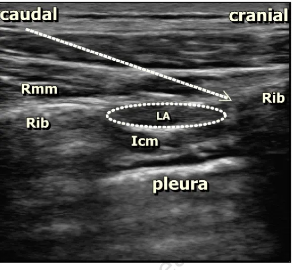
Double-lumen tube was used for orotracheal intubation. The position of the double lumen tube was confirmed via a fiberoptic bronchoscopy. The patients were placed in a lateral decubitus position for the surgery. The mechanical ventilation was performed using a one-lung mechanical ventilation model. Perioperative analgesia was provided with a remifentanil infusion at a rate of 0.01–0.1 µg/kg/min. A dose of 400 mg ibuprofen and tramadol 100 mg was administered IV before 20 minutes the end of the surgery for multimodal postoperative pain management. At the end of the surgery, RIB was performed in lateral decubitus position, with using a Vivid q US system (GE Healthcare, Wauwatosa, WI, USA) in these three of patients. Same regional block technique was administered and same dosages of drugs were applied to the patients. After placing linear high-frequency probe (12 MHz) in a sagittal plane medially on the medial border of the scapula at the T5-6 level; 22 G, 80 mm needle (Braun Stimuplex Ultra, 360 30°) was inserted into the fascial plane between the rhomboid major and intercostal muscles in a caudal cranial direction. A dose of 20 ml 0.25% bupivacaine was injected into the fascial plane (Figure 1).
As a new technique, Rhomboid intercostal block (RIB) defined by Elsharkawy et al. in 2016 may also be good alternative for chest wall analgesia according to a cadaveric examination [5]. This cadaveric examination using methylene blue contrast dye of the RIB showed the spread of dye from caudad to cephalad including T2-T8 tissue plane, as far as lateral branches of the intercostal nerves T3-T8, the posterior primary rami near the midline, and clavipectoral fascia within the axilla. The authors concluded that RIB may be effective for managing the anterior and posterior hemithorax pain [5]. The analgesic efficacy of RIB after thoracotomy has been reported by Altiparmak et al. In their report the authors performed RIB in two patients for thoracotomy and demonstrated a good postoperative pain management [3]. We also demonstrated that RIB has provided effective analgesia for VATS in our patients.
As conclusion, RIB may be an alternative technique for pain management after VATS. There is a requirement of a larger comparative study as to decide and advice regarding the practical applications in routine practice.
References
1. Elsharkawy H, Hamadnalla H, Altinpulluk EY, Gabriel RA. Rhomboid intercostal and subserratus plane block: a case series. Korean J Anesthesiol. 2020 Feb 12. doi: 10.4097/kja.19479. Epub ahead of print. PMID: 32046475.
2. Ekinci M, Ciftci B, Alici HA, Ahiskalioglu A. Ultrasound-guided rhomboid intercostal block effectively manages myofascial pain. Korean J Anesthesiol. 2020 May 12. doi: 10.4097/kja.20211. Epub ahead of print. PMID: 32392670.
3. Altıparmak B, Korkmaz Toker M, Uysal Aİ, Turan M, Gümüş Demirbilek S. The usage of
single-shot ultrasound guided rhomboid intercostal block for analgesia after thoracotomy: Clinical experience in two patients. J Clin Anesth. 2019; 56: 98–9.
4. Steinthorsdottir KJ, Wildgaard L, Hansen HJ, Petersen RH, Wildgaard K. Regional
analgesia for video-assisted thoracic surgery: a systematic review. Eur J Cardiothorac Surg. 2014; 45: 959-66.
5. Elsharkawy H, Saifullah T, Kolli S, Drake R. Rhomboid intercostal block. Anaesthesia. 2016; 71: 856–7.
Figure 1. Sonographic anatomy of block, needle direction, and spread of local anesthetic (LA). The
rhomboid major muscle (Rmm), intercostal muscle (Icm), and rib are seen. The white arrow indicates the direction of needle.