http://dx.doi.org/10.1080/01694243.2016.1168340
© 2016 informa uK limited, trading as Taylor & francis group
Load-bearing capacity and failure types of premolars restored
with sonic activated bulk-fill-, nano-hybrid and
silorane-based resin restorative materials
Funda Öztürk-Bozkurta, Tugba Toz-Akalina, Burcu Gözeticia, Mahmut Kusdemira, Alev Özsoya, Harika Gozukara-Bağb and Mutlu Özcanc
adepartment of restorative dentistry, school of dentistry, istanbul Medipol university, istanbul, Turkey; bdepartment of Biostatistics, school of Medicine, inonu university, Malatya, Turkey; cdental Materials unit, center for dental and oral Medicine, clinic for fixed and removable Prosthodontics and dental Materials science, university of Zurich, Zurich, switzerland
Introduction
More than five hundred million direct dental restorations are placed every year worldwide that represents one of the most prevalent medical interventions in the human body.[1] The
ABSTRACT
This study evaluated the load-bearing capacity of premolars restored with three types of resin composite materials in mesio-occluso-distal (MOD) cavities. Extracted human premolars (N = 30, n = 10 per group) were randomly divided into three groups; MOD cavities were prepared and restored with one of the following resin materials: Group BFC: Bulk-fill composite (SonicFill, Kerr Corporation), Group NC: Nanohybrid resin composite (Filtek Z550, 3M ESPE), Group SC: Silorane-based composite (Filtek Silorane, 3M ESPE). For NC Clearfil SE Bond (Kuraray, Japan) and for SC, Silorane adhesive resin (Filtek Silorane System Adhesive, 3M ESPE) were used. The cavities in NC and SC groups were restored incrementally, while those in BFC groups were restored in bulk. After water storage (24 h, 37 °C), each tooth was subjected to compressive loading with a stainless steel ball (diameter: 4 mm) perpendicular to the occlusal surface (1 mm/ min). Failure types were categorized according to being repairable or irrepairable. Data were statistically analysed using Kruskal-Wallis test (α = 0.05). Two-parameter Weibull distribution values including the Weibull modulus, scale (m) and shape (0) values were calculated. Fracture resistance of premolars restored with BFC (829.84), NC (701.35) and SC (807.73) did not show significant difference (p = 0.72). Weibull distribution presented lower shape (0) for SC (m = 2.77) and NC (m = 3.09) compared to BFC (m = 5.01). The incidence of repairable failures was more common in BFC (80%) compared to NC (60%) and SC (50%). Adhesive failures were more often observed in NC (80%) and SC (70%) groups.
KEYWORDS
adhesion; bulk-fill resin; fracture resistance; nanohybrid; resin composite; silorane; sonicfill
ARTICLE HISTORY
received 1 March 2016 revised 14 March 2016 accepted 16 March 2016
traditional restorative material for posterior dental restorations, amalgam, is increasingly being replaced with resin composite materials applied directly. The general recognition of resin composites could be attributed to the paradigm shift from G.V. Black’s ‘extension for prevention’ to minimal invasive dentistry established by the advances in adhesive technolo-gies.[2] Clinical studies on the survival rate of resin composite posterior restorations report mainly marginal leakage caused by polymerization shrinkage, voids, wear, sensitivity after placement, insufficient proximal contact and contour as failure types.[3–8] Over the years, new filling techniques and resin composites have been suggested in order to diminish such limitations of resin-based restorative materials.
Low polymerization shrinkage of oxirane monomers and increased hydrophobicity due to the presence of siloxane monomers in its composition; silorane-based resin materials are claimed to have advantages over methacrylate-based ones.[9,10] The silorane composite, classified as microhybrid resin composite, contains fine quartz particles and radiopaque yttrium fluoride.[11] One recent other development is the introduction of bulk-fill resin composites that could polymerize in 4-mm-deep cavities when applied even in bulk.[12] Such restorative materials, with their chemical composition being similar to nanohybrid and microhybrid resin composites,[13] are being increasingly used, as they require sim-plified procedures without the necessity of layering technique.[1] While some bulk-fill composites require a final 2-mm increment of a conventional composite material, other bulk-fill composites (i.e. SonicFill, Kerr Corporation, Orange, CA, USA) could be placed in the cavity without such final layer. Sonic Fill is a sonic-activated material that is indicated for posterior restorations up to 5 mm depth in one step unlike the other bulk-fill flowable composites. This highly filled resin has special modifiers that react to sonic energy and rapidly flow into the cavity under sonic activation. The high depth cure is not dictated by the increase in translucency of the material like in other bulk-fill composites, yielding to better optical results.[14]
Endodontically treated teeth are typically subjected to significant loss of tooth structure as a result of restorative and endodontic procedures, and considered more susceptible to fracture than those of intact teeth.[15] Thus, such teeth with moderate remaining tooth structure require more conservative approach where direct resin restorations could preserve the remaining dental tissues with minimal cavity preparation and intracoronal reinforce-ment.[16] On the other hand, maxillary premolar restorations may undergo palatal and buccal strains as a result of chewing loads that generate high stress at the tooth-restoration interface. When the strain is higher than the maximum resistance of the tooth, gap forma-tion, nanoleakage, crack or fracture may also yield to failure of the restoration.[17]
Recent studies claimed weakening of the teeth following mesio-occluso-distal (MOD) cavity preparations but possible strengthening mechanisms of restorative materials to the remaining tissues.[11,18] It has been, however, generally accepted that the strength of a tooth decreases with the increased amount of tooth tissue removed, particularly in relation to the width of the occlusal preparation.[11]
The objectives of this study therefore were to evaluate the load-bearing capacity of pre-molars restored with sonic-activated bulk-fill-, nanohybrid and silorane-based resin restor-ative materials on MOD cavities and to evaluate the failure modes after fracture. The null hypothesis tested was that there would be no significant difference on the fracture resistance premolars restored with different materials.
Materials and methods
The types, brands, main chemical compositions and manufacturers of materials used for the experiments are listed in Table 1.
Tooth specimens
Recently extracted maxillary premolars (N = 30) due to orthodontic reasons with simi-lar dimensions were selected from a pool of extracted teeth for this study at the Istanbul Medipol University, Dental Clinics. Calculus and soft-tissue remnants were removed using a periodontal scaler and they were cleaned using a rubber cup using fine pumice-water slurry. The teeth were examined under stereomicroscope (Olympus SZ61, Olympus Corporation, Tokyo, Japan) at ×10 for cracks, defects and caries. Teeth of similar size (buccolingual width: 6.43–7.25 mm) were chosen measured using a digital micrometer gauge (Series 480-505, resolution 1 μm; SHAN TM Precision Measuring Instruments, Guilin, China) and allowing a maximum deviation of 10% from the determined mean width value. Finally, the selected teeth were stored in distilled water at 37 °C that was changed every 5 days until the experiments.
Cavity preparations
One operator made all cavity preparations and restorations. Prior to the preparation, cavity outlines were drawn with a pencil, MOD cavities were prepared with 1-mm gingival cavo-surface margin coronal to the cement-enamel junction with a cylindrical bur (835-012-4, Diatech, Geneve, Switzerland) under water coolant. After every four cavity preparation, a new bur was used in order to ensure high cutting efficacy. The cavities had flat cavity floors, with a width of one-third of the intercuspal distance of the occlusal surface and proximal boxes had width of one-third of the total bucco-lingual width. The cavosurface margins were prepared at 90° and all internal angles were rounded.
Table 1. The brands, abbreviations, chemical compositions and manufacturers of the restorative mate-rials tested in the study.
Brand Type Chemical Composition Manufacturer
sonicfill (Bfc) Bulk-fill resin
composite glass, oxide, chemicals, 3-trimethoxysilylpropyl methacrylate, silicon dioxide, ethoxylated bisphenol-adimethacrylate bis-phenol-a-bis-(2-hydroxy-3-mehacryloxypropyl) ether, triethyleneglycoldimethacrylate
Kerr corporation, orange, ca, usa
filtek Z550 (nc) nanohybrid
resin composite silane-treated ceramic, bis-phenol a polyethyl-ene glycol diether dimethacrylate, bis-phenol a diglycidyl ether dimethacrylate (bis-gMa), silane-treated silica, diurethane dimethacrylate (udMa)
3M esPe, st. Paul, Minn, usa
silorane (sc) silorane-based
resin composite 3,4 epoxycyclohexylethylcyclopolymethyl-siloxane, bis-3,4-epoxycyclohexylethylphenyl-methylsilane, silanized glass, yttrium fluoride, camphorquinone
Endodontic treatment
After cavity preparations, a conservative, endodontic access was performed on the pulp chamber wall. The canals were prepared with Pro-Taper Ni–Ti Rotary System (Dentsply Malleiffer, DeTrey, Konstanz, Germany), obturated with an AH 26 sealer (Dentsply) and gutta-percha was used for lateral compaction. After removal of gutta-percha, excess sealer was retrieved using cotton pellet with alcohol.
Restorative procedures
Following the endodontic treatments, the teeth were randomly divided into three groups to be restored with one of the following restorative materials: Group BFC: Bulk-fill com-posite (SonicFill, Kerr Corporation, CA, USA), Group NC: Nanohybrid resin comcom-posite (Filtek Z550, 3M ESPE, St. Paul, MN, USA), Group SC: Silorane-based composite (Filtek Silorane, 3M ESPE).
Group BFC
Primer of the adhesive system (Clearfil SE Primer) was scrubbed to the walls of the prepa-ration, waited for 20 s and gently air-thinned. Then, adhesive resin (Clearfil SE Bond) was applied with a brush to the entire cavity walls, air-thinned and photo-polymerized (Elipar Free Light, 3M ESPE) for 10 s. After the adhesive resin application, BFC was applied to the bonded cavities under the sonic energy and photo-polymerized (Elipar Free Light) for 40 s according to the manufacturer’s instruction. After polymerization, the restorations were polished with abrasive disks (Sof-Lex Finishing and Polishing Systems, 3M ESPE) and rubber cups (Jiffy Polishers, Ultradent, South Jordan, USA).
Group NC
Primer (Silorane Primer, 3M ESPE) was scrubbed to the walls of the preparation for 15, gently air-thinned. Then, adhesive resin (Silorane Bond, 3M ESPE) was applied with a brush to the entire walls, air-thinned and photo-polymerized (Elipar Free Light, 3M ESPE, >400 mW/cm2) for 10 s. Thereafter, a metal matrix band (Adapt SuperCap Matrices, Kerr, Bioggio, Switzerland) was placed around the tooth. A thin layer of NC was applied towards the metallic matrix contacting the cavosurface of the proximal box up to the half of the occlusal-cervical extension. Thus, the cavity turned into a class I through the centripetal technique. After polymerization of this layer for 20 s, the second layer was applied over the previous increment contacting the cavosurface margin of the proximal box and forming the marginal ridge. This procedure was applied for both the mesial and distal margins. The cavities were restored with nanohybrid resin composite (Filtek Z550, 3M ESPE) incremen-tally, photo-polymerized for 40 s according to the manufacturer’s instruction and polished as described in Group BFC.
Group FS
All adhesive procedures were similar as in Group NC. The cavities were restored with silorane (Filtek Silorane, 3M ESPE) and polished as described in Group BFC.
The teeth were subjected to 500 thermal cycles (Salubristechnica, Salubris A.S., Istanbul, Turkey) between 5 and 55 °C with a dwell time of 20 s at each bath.
Fracture resistance and failure-type analysis
A thin coat of wax (0.3 mm) was applied on the external root surface of all teeth. The teeth were vertically mounted to a level of 1.0 mm apical to the CEJ. The apical parts of the restored teeth were embedded vertically in metal moulds using auto-polymerized acrylic resin (Pancyril Self Cure Acrylic, Zeist, The Netherlands) up to 1 mm below the CEJ, with the long axis of the tooth parallel to that of the mould. The wax on the root surfaces was removed with boiling water, and this space was filled with silicone (Oranwash vl, Zhermark, Marl, Germany) 1 mm apical to the CEJ to simulate periodontal ligament. The specimens were stored in 100% humidity for 24 h prior to fracture testing.
The specimens were placed in the jig of the Universal Testing machine (Autograph AG-IS, Shimadzu Co, Kyoto, Japan) with the long axis of the roots parallel to the direction of loading. A stainless steel spherical ball (diameter: 4 mm) was used to load the specimens until fracture at a cross-head speed of 1 mm/min. The loading site was the central fissure where the spherical ball contacted the buccal and palatinal cusps of the teeth on the occlusal surface of the restorations.
After fracture loading, the failure modes (Adhesive: Failure between the restoration and the cavity wall; Cohesive: Cohesive failure of the tooth including the root; Mixed: Cohesive failure of the restorative material and adhesive failure between the restoration and the cavity wall) were evaluated under stereomicroscope at a magnification of ×20 and further classified as ‘repairable’ when they were limited to the coronal portion and ‘no-repairable’ when the failure included the root.[19]
Statistical analysis
All statistical procedures were performed using the Statistical Package for Medical Science (SPSS 21.00 for Windows software, SPSS Inc., Chicago, IL, USA). The data were expressed as median, minimum and maximum values. Descriptive statistics were computed, and test of normality was performed using Kolmogorov–Smirmov and Shapiro–Wilk’s tests. Mean fracture resistance data were analysed using Kruskal–Wallis test. Maximum likelihood estimation without a correction factor was used for two-parameter Weibull distribution, including the Weibull modulus, scale (m) and shape (°), to interpret predictability and reliability of fracture resistance (Minitab Software V.16, State College, PA, USA). P values less than 0.05 were considered to be statistically significant in all tests.
Results
Fracture resistance of premolars restored with BFC (829.84), NC (701.35) and SC (807.73) did not show significant difference (p = 0.72) (see Tables 1 and 2).
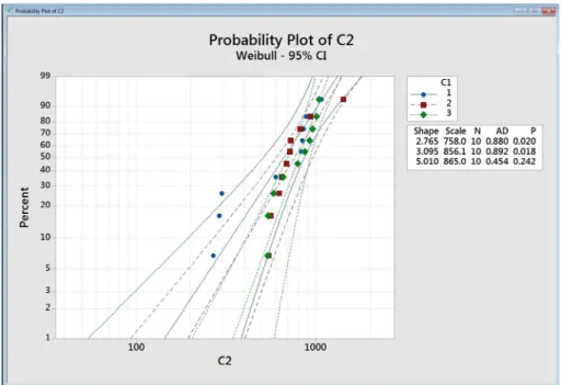
Weibull distribution presented lower shape (0) for SC (m = 2.77) and NC (m = 3.09) compared to BFC (m = 5.01) (Figure 1).
The incidence of repairable failures was more common in BFC (80%) compared to NC (60%) and SC (50%). Adhesive failures were more often observed in NC (80%) and SC (70%) groups (Table 3, Figure 2).
Discussion
This study aimed to evaluate the load-bearing capacity of endodontically treated premolars restored with sonic-activated bulk-fill-, nanohybrid and silorane-based resin restorative materials on MOD cavities and evaluated the failure types after fracture. Based on the results of this study, since fracture resistance did not show significant difference between the experimental groups, the null hypothesis could be accepted (see Figure 2).
Table 2. The median, minimum and maximum and median forces of experimental groups (N).
Note. same lowercase superscript letters in each column indicate no significant difference (p > 0.05). see Table 1 for group abbreviations.
Experimental groups Median Minimum Maximum
Bfc 829.84a 539.69 1044.25
nc 701.35a 544.34 1422.75
sc 807.73a 267.59 1076.50
Figure 1. Probability plot with Weibull curves (95% ci) using maximum likelihood estimation, scale and shape values for all groups. 1: sc; 2: nc; 3: Bfc. see Table 1 for group abbreviations.
Table 3. distribution of failure types in each experimental group after fracture loading. repairable: frac-ture limited to the coronal portion; non-repairable: failure included the root fracfrac-ture; adhesive: failure between the restoration and the cavity wall; cohesive: cohesive failure of the tooth including the root; Mixed: cohesive failure of the restorative material and adhesive failure between the restoration and the cavity wall. see Table 1 for group abbreviations.
Experimental
groups repairable (n, %) Non-repairable (n, %) Adhesive (n, %) Cohesive (n, %) Mixed (n, %)
Bfc 8 (80) 2 (20) 4 (40) 0 (0) 6 (60)
nc 6 (60) 4 (40) 8 (80) 0 (0) 2 (20)
Although the use of resin composites has increased dramatically for the restoration of teeth in a minimal invasive fashion, average lifetime of posterior resin restorations was reported as only six years.[20] Current advances in resin technologies aim at modifying resin and/or filler component of the composite in order to achieve more durable restorations especially with reduced polymerization stress. This effort is a direct response to the sensitive and somewhat complex placement techniques required for the application of such materials. Secondary caries has consistently been reported as the primary reason for replacement of composite restorations in daily clinical practice followed by partial or complete fracture of the restoration.[21–23] In that respect, MOD cavity preparation results in significant reduction in tooth strength due to the loss of marginal ridges and formation of possible micro-fractures caused during chewing forces [24,25] or endodontic treatment.[17,26] For this reason, in this study, large MOD cavities were prepared in maxillary premolars with endodontic access, representing caries affected and subsequently endodontically treated teeth.
Figure 2. (a)–(c) representative photos of (a) adhesive failure between the restorative material Bfc and cavity wall; (b) mixed failure including cohesive failure of the material sc and adhesive failure between the restoration and the cavity wall; (c) cohesive failure of the tooth including the root in group sc. ((a), (b): repairable, (c): non-repairable failure).
The clinical performance of the newer dental composites has been significantly improved over the past decade providing adequate strength and resistance in order to withstand the chewing forces with less polymerization shrinkage and improved polymerization in deep cavities. Nevertheless, the relatively high brittleness and low fracture toughness of current dental composites still remain a problem in stress-bearing posterior restorations.[1] A restored tooth tends to transfer stresses differently than an intact tooth [2] where the filling technique and cavity size affects the bond strength of composite in the preparation.[6,7] Moreover, adhesive restorations transmit and distribute functional stresses across the bond-ing interface more favourable and have the potential to reinforce weakened tooth structure. [8–10] However, polymerization of composites may cause deformation in the surrounding tooth structure resulting in microcracks that eventually predispose the tooth to fracture.[11] Although reduced polymerization shrinkage composites has been reported for silorane-based resin resulting in significant reduction in polymerization shrinkage stresses,[15] and reduced cuspal deflection compared to conventional methacrylate-based resin com-posites,[27] the results of this study did not present higher fracture resistance compared to other materials tested. Other previous studies also revealed comparable fracture resistance of silorane and methacrylate-based resin composites.[11,15,28] In fact, the reduction in shrinkage per unit volume of silorane-based resin composites is not the only factor that determines the fracture resistance. The complexity of the interaction between polymeri-zation shrinkage stress, elastic modulus of the composite, the tooth, cavity design and the extent to which the restoration is bonded, are all responsible for the fracture resistance.[29]
Bulk-fill technique is highly desired in daily dental practice due to decreased number of restorative incremental steps.[30] The reported properties of bulk-fill composites such as decreased polymerization shrinkage,[31] improved self-levelling ability,[32] reduced cus-pal deflection [33] and optimal bond strength [30] are certain advantages of this material. Manufacturers eliminated the disadvantages such as shrinkage stress by modifying the composition through polymerization kinetics. Slower polymerization is employed in bulk-fill materials as a basic strategy to decrease the polymerization shrinkage.[34] In an earlier study, teeth restored with bulk-fill composites presented higher fracture resistance where no sonic activation was practised.[1] In this study, a sonic-activated bulk-fill material was used presenting similar fracture resistance results compared to those of other materials but with higher Weibull modulus indicating more reliable fracture resistance.
The nanohybrid (NC) material was based on bis-GMA, UDMA, bis-EMA, PEGDMA and TEGDMA ( http://multimedia.3m.com/mws/media/619598O/filtek-ultimate-techni-cal-product-profilecee.pdf?fn=Filtek_Ultimate_TPP_CEE.pdf). Higher amounts of flexible UDMA in addition to TEGDMA may result in an extensive deformation before fracture. [35] On the other hand, in UDMA, the long aliphatic segment in repeated units results in more flexibility and coiling of the chain. Despite the ability of ‘O–CO–NH’ groups in UDMA, forming hydrogen bonds, easy damage of these bonds with water yields to low elastic modulus and increased flexibility of chains as a result of moderate change in temper-ature or stress.[35] The other monomer bis-GMA has two aromatic rings that cause a lower cycling of pendant groups and thereby, a higher cross-linking of the polymer.[36] However, this has no effect on the improvement of mechanical properties of the resin composite. Unlike bis-GMA, bis-EMA does not contain any hydroxyl groups. Thus, hydrogen bonds among the molecules of this monomer do not exist. This leads to an increased movement of the molecules resulting in a higher degree of conversion.[37] In turn, while BFC consists
of 3-trimethoxysilylpropyl methacrylate, ethoxylated bisphenol-Adimethacrylate bisphe-nol-A-bis-(2-hydroxy-3-mehacryloxypropyl) ether and TEGDMA, SC is based on silox-ane molecule that makes the material hydrophobic, and oxirsilox-ane with which a volumetric shrinkage of 0.99 volume% is achieved.[38]
Although fracture resistance results were similar, the Weibull modulus was more favoura-ble for BFC and those of SC and NC. Similarly, repairafavoura-ble and adhesive types of failures were more commonly observed with the BFC. This could be attributed to better adhesion of the BFC to the dentin walls and less stress formation in the surrounding dental tissues in this material. Higher incidence of adhesive failure types in NC and SC indicates a weaker link between the tooth-restoration interfaces with the adhesive resin used.[39] Likewise, the use of silorane-based resin composite demonstrated similar bond strength values compared to methacrylate-based ones in cavities with high C-factor with the majority of failures being mainly mixed failure types with the BFC.[40] In this study, the adhesive resin used for both NC and BFC groups was the same. Thus, the failure types of the restorative materials could not be related solely to the adhesive performance of the MC and BFC.
Mechanical properties express the response of materials to external loading, manifested by their ability to sustain reversible or irreversible deformation or to fracture.[41] The high percentage of repairable failures in Group BFC could be related to its low elasticity modulus. Bulk-fill materials were generally classified between hybrid and flowable resin composites with regard to hardness and modulus of elasticity properties.[42] Nevertheless, the results of this study should be verified in a larger sample with and without artificial ageing before such materials could be used for clinical trials.
Conclusions
From this study, the following could be concluded:
(1) Restoring endodontically treated premolars with MOD cavities using sonic-ac-tivated bulk-fill, nanohybrid or silorane-based resin materials did not affect the fracture resistance of the teeth.
(2) Weibull parameters presented more reliable mechanical durability for bulk-fill resin composite compared to nanohybrid and silorane-based materials.
(3) Sonic-activated bulk-fill nanohybrid resin composite presented more repairable failures after maximum loading.
Clinical relevance
Endodontically treated premolars with MOD cavities could be restored with either nano-hybrid, silorane or bulk-fill nanohybrid resin composites, providing that bulk-fill resin showed more reliable Weibull modulus for fracture resistance and also presented more repairable failures.
Acknowledgements
Conflict of interest
The authors did not have any commercial interest in any of the materials used in this study.
References
[1] Rosatto CM, Bicalho AA, Veríssimo C, et al. Mechanical properties, shrinkage stress, cuspal strain and fracture resistance of molars restored with bulk-fill composites and incremental filling technique. J. Dent. 2015;43:1519–1528.
[2] Keulemans F, Palav P, Aboushelib MM, et al. Fracture strength and fatigue resistance of dental resin-based composites. Dent. Mater. 2009;25:1433–1441.
[3] Knobloch LA, Kerby RE, Seghi R, et al. Fracture toughness of packable and conventional composite materials. J. Prosthet. Dent. 2002;88:307–313.
[4] Tantbirojn D, Versluis A, Cheng YS, et al. Fracture toughness and microhardness of a composite: do they correlate? J. Dent. 2003;31:89–95.
[5] Bicalho AA, Valdívia AD, Barreto BC, et al. Incremental filling technique and composite material-Part II: shrinkage and shrinkage stresses. Oper. Dent. 2014;39:e83–e92.
[6] Kramer N, Reinelt C, Frankenberger R. Ten-year clinical performance of posterior resin composite restorations. J. Adhes. Dent. 2015;17:433–441.
[7] Pallesen U, van Dijken JW. A randomized controlled 27 years follow up of three resin composites in Class II restorations. J. Dent. 2015;43:1547–1558.
[8] Pallesen U, van Dijken JW. A randomized controlled 30 years follow up of three conventional resin composites in Class II restorations. Dent. Mater. 2015;31:1232–1244.
[9] Relhan N, Ponnappa KC, Relhan A, et al. An in vitro comparison of micro leakage between two posterior composites restored with different layering techniques using two different led modes. J. Clin. Diag. Res. 2015;9:78–81.
[10] Sadat HashemiKamangar S, Ghavam M, Mirkhezri Z, et al. Comparison of the effects of two different drinks on microhardness of a silorane-based composite resin. J. Dent. (Shiraz)
2015;16:260–266.
[11] Kikuti WY, Chaves FO, Di Hipolito V, et al. Fracture resistance of teeth restored with different resin-based restorative systems. Braz. Oral Res. 2012;26:275–281.
[12] Do T, Church B, Verissimo C, et al. Cuspal flexure, depth-of-cure, and bond integrity of bulk-fill composites. Pediat. Dent. 2014;36:468–473.
[13] Ilie N, Rencz A, Hickel R. Investigations towards nano-hybrid resin-based composites. Clin. Oral Invest. 2013;17:185–193.
[14] Jackon RD. Placing posterior composites: increasing efficiency. Dent. Today. 2011;30:30–31. [15] Taha NA, Maghaireh GA, Bagheri R, et al. Fracture strength of root filled premolar teeth
restored with silorane and methacrylate-based resin composite. J. Dent. 2015;43:735–741. [16] Soares PV, Santos-Filho PC, Queiroz EC, et al. Fracture resistance and stress distribution in
endodontically treated maxillary premolars restored with composite resin. J. Prosthodont.
2008;17:114–119.
[17] Toz T, Öztürk Bozkurt F, Kara Tuncer A, et al. The effect of bulk-fill flowable composites on the fracture resistance and cuspal deflection of endodontically treated premolars. J. Adhes. Sci. Technol. 2015;29:1581–1592.
[18] Dalpino PH, Francischone CE, Ishikiriama A, et al. Fracture resistance of teeth directly and indirectly restored with composite resin and indirectly restored with ceramic materials. Am. J. Dent. 2002;15:389–394.
[19] Soares PV, Santos-Filho PC, Martins LR, et al. Influence of restorative technique on the biomechanical behavior of endodontically treated maxillary premolars. Part I: Fracture resistance and fracture mode. J. Prosthet. Dent. 2008;99:30–37.
[20] Downer MC, Azli NA, Bedi R, et al. Dental restorations: how long do routine dental restorations last? A systematic review Br. Dent. J. 1999;187:432–439.
[21] Bernardo M, Luis H, Martin MD, et al. Survival and reasons for failure of amalgam versus composite posterior restorations placed in a randomized clinical trial. J. Am. Dent. Assoc.
2007;138:775–783.
[22] Soncini JA, Maserejian NN, Trachtenberg F, et al. The longevity of amalgam versus compomer/ composite restorations in posterior primary and permanent teeth. J. Am. Dent. Assoc.
2007;138:763–772.
[23] Ferracane JL. Resin-based composite performance: are there some things we can’t predict? Dent. Mater. 2013;29:51–58.
[24] Bichacho N. The centripetal build-up for composite resin posterior restorations. Pract. Periodont. Aesthet. Dent. 1994;6:17–23.
[25] Moosavi H, Zeynali M, Pour ZH. Fracture resistance of premolars restored by various types and placement techniques of resin composites. Int. J. Dent. 2012;2012:973641.
[26] Kemaloglu H, Emin Kaval M, Turkun M, et al. Effect of novel restoration techniques on the fracture resistance of teeth treated endodontically: an in vitro study. Dent. Mater. J.
2015;34:618–622.
[27] Bouillaguet S, Gamba J, Forchelet J, et al. Dynamics of composite polymerization mediates the development of cuspal strain. Dent. Mater. 2006;22:896–902.
[28] Akbarian G, Ameri H, Chasteen JE, et al. Fracture resistance of premolar teeth restored with silorane-based or dimethacrylate-based composite resins. J. Esthet. Restorative Dent.
2014;26:200–207.
[29] Marchesi G, Breschi L, Antoniolli F, et al. Contraction stress of low-shrinkage composite materials assessed with different testing systems. Dent. Mater. 2010;26:947–953.
[30] Van Ende A, De Munck J, Van Landuyt KL, et al. Bulk-filling of high C-factor posterior cavities: effect on adhesion to cavity-bottom dentin. Dent. Mater. 2013;29:269–277.
[31] El-Damanhoury H, Platt J. Polymerization shrinkage stress kinetics and related properties of bulk-fill resin composites. Oper. Dent. 2014;39:374–382.
[32] Petrovic LM, Zorica DM, Stojanac I, et al. A model of the viscoelastic behavior of flowable resin composites prior to setting. Dent. Mater. 2013;29:929–934.
[33] Moorthy A, Hogg CH, Dowling AH, et al. Cuspal deflection and microleakage in premolar teeth restored with bulk-fill flowable resin-based composite base materials. J. Dent. 2012;40:500–505. [34] Par M, Gamulin O, Marovic D, et al. Effect of temperature on post-cure polymerization of
bulk-fill composites. J. Dent. 2014;42:1255–1260.
[35] Asmussen E, Peutzfeldt A. Influence of UEDMA BisGMA and TEGDMA on selected mechanical properties of experimental resin composites. Dent. Mater. 1998;14:51–56. [36] Elliott JE, Lovell LG, Bowman CN. Primary cyclization in the polymerization of bis-GMA
and TEGDMA: a modeling approach to understanding the cure of dental resins. Dent. Mater.
2001;17:221–229.
[37] Obici AC, Sinhoreti MA, de Goes MF, et al. Effect of the photo-activation method on polymerization shrinkage of restorative composites. Oper. Dent. 2002;27:192–198.
[38] Eick JD, Smith RE, Pinzino CS, et al. Stability of silorane dental monomers in aqueous systems. J. Dent. 2006;34:405–410.
[39] Van Ende A, De Munck J, Mine A, et al. Does a low-shrinking composite induce less stress at the adhesive interface? Dent. Mater. 2010;26:215–222.
[40] Isaac SZ, Bergamin AC, Turssi CP, et al. Evaluation of bond strength of silorane and methacrylate based restorative systems to dentin using different cavity models. J. Appl. Oral Sci. 2013;21:452–429.
[41] Ornaghi BP, Meier MM, Lohbauer U, et al. Fracture toughness and cyclic fatigue resistance of resin composites with different filler size distributions. Dent. Mater. 2014;30:742–751. [42] Ilie N, Bucuta S, Draenert M. Bulk-fill resin-based composites: an in vitro assessment of their