Summary
The effect of amniotic membrane on biochemical parameters was investigated in response to experimentally induced non-sterile clean wound inflammation. In addition, the effort was made to find out the biochemical compounds of human placenta extract. Thirty male Wistar-Albino rats weighing 250-300 g were used and randomly divided into three groups of 10. The control group, subjected to no operation; sham-operated group, subjected to experimental clean wound inflammation; and amniotic membrane group, subjected to experimental clean wound inflammation and received Human Amniotic Membrane (HAM) treatment. All rats had free access to standard laboratory diet and water. In the sham-operated and amniotic membrane groups, a 4 cm-incision was made on the median line in order to create a wound. The abdominal fascia of the amniotic membrane group was then covered with amniotic membrane whilst the abdominal fascia of the sham-operated group was uncovered with a material. At the end of 14 days, the animals were sacrificed and blood samples were obtained and analyzed for biochemical parameters. Amniotic membrane with whole placenta extracts that were rich in enzymes as lactate dehydrogenase (LDH), alanine aminotransferase (ALP) and aspartate aminotransferase (AST), and elements including Fe, Na and Cl. The amniotic membrane group had significantly lower in the mean values of serum C-reactive protein (CRP), LDH and AST whilst this group had significantly higher in the mean concentration of serum albumin and iron compared to sham-operated values (P<0.05). Similarly, the mean values of serum albumin, iron, AST were significantly lower in amniotic membrane group whereas the activity of GGT was higher in this group compared to control values. These results may indicate that amniotic membrane exerts anti-inflammatory effect and may decrease severity of tissue damage and accelerate healing process in rats with experimentally induced non-sterile clean wound inflammation.
Keywords: Amniotic membrane, Inflammation, Biochemical parameters, Wound
Amniyotik Membranın Deneysel Olarak İndüklenmiş Steril
Olmayan Temiz Yara İnflamasyonunda Serum Biyokimyasal
Parametreler Üzerine Etkisi
Özet
Deneysel olarak indüklenmiş steril olmayan temiz yara inflammasyonuna cevap olarak, amniyotik membranın, biyokimyasal parametreler üzerine etkisi incelendi. Ayrıca insan plasenta ekstraktının biyokimyasal bileşenleri de araştırıldı. Ağırlıkları 250-300 g arasında değişen, 30 adet, erkek Wistar-Albino sıçanları kullanıldı ve rastgele 10’arlı 3 grup oluşturuldu. Kontrol grubu hiçbir operasyon geçirmezken, sham-operasyonlu grup, deneysel temiz yara inflammasyonuna maruz kaldı ve amniyotik membran grubu da deneysel temiz yara inflammasyonuna maruz kalıp HAM sağaltımı aldı. Sıçanların hepsi, standart laboratuvar diyeti ve suya serbest ulaşıma sahiptiler. Sham-operasyonlu ve amniyotik membran gruplarında, yara oluşturmak için median hat boyunca 4 cm’lik bir ensizyon yapıldı. Sonrasında, amniyotik membran grubunun abdominal fasiyası, amniyotik membran ile kapatılırken, sham-operasyonlu grubun abdominal fasiyası amniyotik membran eklenmeden kapatıldı. Ondördüncü günün sonunda, hayvanlar öldürüldü ve kan numuneleri alınarak biyokimyasal parametreler yönünden analiz edildi. Tüm plasenta ekstraktlı amniyotik membran; laktat dehidrojenaz (LDH), alanin aminotransferaz (ALP) ve aspartate aminotransferaz; (AST) enzimleri ile Fe, Na ve Cl elementleri yönünden zengindi. Amniyotik membran grubunun C-reactive protein (CRP), LDH ve AST serum ortalama değerleri, belirgin olarak düşük iken, yine bu grubun albumin ve demir serum ortalama konsantrasyonları, sham-operasyonlu grubun değerleriyle karşılaştırıldığında, belirgin olarak daha yüksekti (P<0.05). Benzer şekilde, amniyotik membran grubunun albumin, demir ve AST serum ortalama değerleri belirgin olarak düşük iken, bu grubun GGT aktivitesi, kontrol grubunun değerleriyle karşılaştırıldığında, daha yüksekti. Bu sonuçlar, amniyotik membranın anti-inflammatuvar etkiye sahip olduğunu ve deneysel olarak indüklenmiş steril olmayan temiz yara inflammasyonuna sahip sıçanlarda, doku hasarının ciddiyetini azaltabileceğini ve iyileşmeyi hızlandırabileceğini göstermektedir.
Anahtar sözcükler: Amniyotik membran, İnflammasyon, Biyokimyasal parametreler, Yara
The Effect of Amniotic Membrane on Serum Biochemical
Parameters in Experimentally Induced Non-Sterile Clean
Wound Inflammation
Şahver Ege HİŞMİOĞULLARI * Adnan Adil HİŞMİOĞULLARI *
İsmail YAMAN ***
Özlem YAVUZ ** Kamil SEYREK ** Ömür KARACA ****Cemal KARA ***** Armağan HAYIRLI ******
* ** *** **** ***** ******
Department of Pharmacology and Toxicology, School of Veterinary Medicine, Balıkesir University, TR-10145 Balıkesir - TURKEY
Department of Biochemistry, School of Medicine, Balıkesir University, TR-10145 Balıkesir - TURKEY Department of General Surgery, School of Medicine, Balıkesir University, TR-10145 Balıkesir - TURKEY Department of Anatomy, School of Medicine, Balıkesir University, TR-10145 Balıkesir - TURKEY Surgical Clinic of Karsıyaka Hospital TR-35500 İzmir - TURKEY
Department of Animal Nutrition, School of Veterinary Medicine, Atatürk University, TR-25240 Erzurum - TURKEY
Makale Kodu (Article Code): KVFD-2011-5559
İletişim (Correspondence) +90 266 6121455
INTRODUCTION
In response to major tissue injury due to surgical trauma, a highly complex inflammation and healing process take places, which are modulated by numerous cells and their products such as cytokines, especially tumor necrosis factor-α (TNF- α), interleukin (IL)-1β, and IL-1b 1,2.
Wound healing is a complex insult-initiated biologic process that involves inflammation, new tissue formation and remodeling 3,4. Inflammation, constituting part of the acute
response, results in a coordinated influx of neutrophils at the wound site. These cells, through their characteristic “respiratory burst” activity, produce oxidant, which is well known criterion for defense against bacteria and other pathogens 5.
The amniotic membrane (AM) is a thin avascular membrane composed of an epithelial layer and an inner mesodermal tissue. Amniotic membrane with whole placenta is rich in enzymes, vitamins, amino acids, steroids, fatty acids and elements including Na, K, Ca, Mg, Cu, Fe, P, and Si 6. All these components may possess multiple
biological activities. It has reported that the AM mezoderm can suppress the expression of potent proinflammatory cytokines though the mechanism of action by which AM inhibits inflammation is not clear 7,8. The potent
anti-inflammatory effects 9 of HAM in regards to regeneration
and new tissue formation 3 have also been demonstrated.
It has been suggested that human amniotic membrane (HAM) releases of soluble factors such as (IL-10, IL-6) and angiogenic factors by cells and molecules bound to the collagenous stromal matrix of the HAM patch. This, in turn, exerts paracrine mechanisms to support survival, differentiation and proliferation of host cells 8.
Human amniotic membrane (HAM) has important clinical application, such as a material to accelerate wound healing 10, reconstruct damaged organs 11-13,treat burn
lesion 14, cover surgical wounds to avoid collusion 15, and
induce keratocyte expression 16. This experiment was
therefore conducted to investigate the biochemical profile in response to HAM treatment in a state of inflammation induced experimentally by the non-sterile clean wound technique.
MATERIAL and METHODS
Animals
Upon the approval of the experimental protocol by the Animal Ethics Committee of the Balikesir University, animals were cared in accordance with National Institutes of Health Guide for the Care and Use of Laboratory Animals. Thirty male Wistar-Albino rats at age of five months and weighing 250-300 g were housed in separate cages at 25°C and subjected to a 12:12-h light:dark cycle. The rats
were randomly divided into three groups: Control group (Group 1), subjected to no operation; sham-operated group (Group 2), subjected to experimental clean wound inflammation; and amniotic membrane group (Group 3), subjected to experimental clean wound inflammation and received HAM treatment. All animals fed ad libitum consumption a standard laboratory diet and had free access to water during the experimental period.
Preparation of the Amniotic Membrane
Human amniotic membranes (38 weeks old) were obtained from caesarean deliveries of patients (n = 10) with negative test results for HBsAg, HCV, HIV, syphilis and no histories of premature membrane ruptures, endo-metritis or meconium ileus. Placentas were transferred in a container under sterile conditions at 4°C to the laboratory. At the processing site, the amnion was separated from the rest of the chorion by blunt dissection, then rinsed and soaked in saline and Dakin’s solutions (0.25% sodium hypochloride solution) for 10 min in order to remove blood and other contaminants. The amnions were then stored in saline solution containing 50 μg/mL penicillin, 50 μg/ mL streptomycin, 100 μg/mL neomycin, and 2.5 μg/mL amphotericin B for 10 min. Finally, amnions were carefully flattened onto sterile nitrocellulose paper with the epithelium facing up. The nitrocellulose paper containing adherent membranes was then cut into 20-mm wide segments.
Operation Procedure
The rats were anaesthetized with intramuscular injection of 60 mg/kg of ketamin hydrochloride (Ketalar, Eczacibasi, Warner-Lambert Laboratories, Istanbul, Turkey) and 10 mg/ kg of xylazine hydrochloride (Rompun, Bayer Laboratories, Istanbul, Turkey). All procedures were performed under clean but non-sterile conditions and the animals were allowed to breath spontaneously during the surgery. The body temperature was maintained around 37°C by the use of a heating lamp. After shaving and scrubbing the abdominal skin with a povidone-iodine, a 4 cm-long mid-line incision was made. Immediately, the abdominal fascia was closed by a continuous suture (silk 3/0) in the HAM and sham-operated groups. Following that the fascia of the study group was covered by HAM while the fascia of the sham-operated group was covered no material. Finally, skin of these groups were closed by a continuous suture (silk 3/0). All animals were sacrificed 14 days after surgery administering an overdose of sodium pentobarbital (300 mg/kg, intraperitoneal). Prior to sacrifice, cardiac blood samples were collected into vacutainers.
Preparation of the Placenta Extract
Placenta samples (1 g) was transferred to 9 vol. (w/v) of ice-cold buffered sucrose (0.25 M containing 1 mM HEPES pH 7.4.). The placenta was cut into several large pieces and swirled around in the buffer to remove blood as much as possible. The placenta was minced finely with a sharp
scissors and transferred to ice-cold homogenising vessel and were finally homogenised with about six strokes of the pestle at full speed. The homogenate was made up to 10 vol. (w/v) with sucrose buffer solution. A sample of homogenate (3-4 ml) was centrifuged in a fixed angle rotor at 4°C for 10 min at 6.000 хg to obtain supernatant. The supernatant was used for biochemical analysis.
Biochemical Analysis
Serum was separated by centrifuging at 825 xg for 10 min for analyses of urea, creatinine, cholesterol, tri-glyceride, high density lipoprotein-cholesterol (HDL-C), total protein and C-reactive protein (CRP) as well as alanine aminotransferase (ALT), aspartate aminotransferase (AST), lactate dehydrogenase (LDH), and alkaline phosphatase (ALP) activities using commercially available kits in an auto- analyzer (Cobas Integra 800; Roche Diagnostics GmbH; Mannheim, Germany). Serum levels of tumor necrosis factor-alpha (TNF-α) were determined by enzyme-linked immunosorbent assay (ELISA) using commercially available kits (eBioscience, Rat TNF-α Platinum ELISA, Austria) in a diagnostic instrument (BioTek, ELx 800, USA).
Statistical Analysis
After determining normality, data were subjected to one-way ANOVA (SPSS, version 11.0, Chicago, IL). Group mean differences were attained using the Bonferoni post- hoc test option. The data were expressed as mean ± SE and differences were considered significant when the p value was less than 0.05.
RESULTS
Human Placenta Extracts
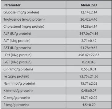
The biochemical parameters were expressed as biochemical values per gram placenta extract protein for standardization and accuracy. The placental LDH, ALP, and AST activities were more pronounceable as compared to the placental ALT and GGT activities (Table 1). Moreover, the placental extract were relatively rich in Fe, Na, Cl, and P, whereas poor in K (Table 1). The other biochemical para-meters which were present in placenta extracts were glucose (12.14 mg/g protein), cholesterol (14.25 mg/g protein), and triglycerides (26.42 mg/g protein/g protein) as well as CRP (0.55 mg/g protein) (Table 1).
Amniotic Membrane Treatment
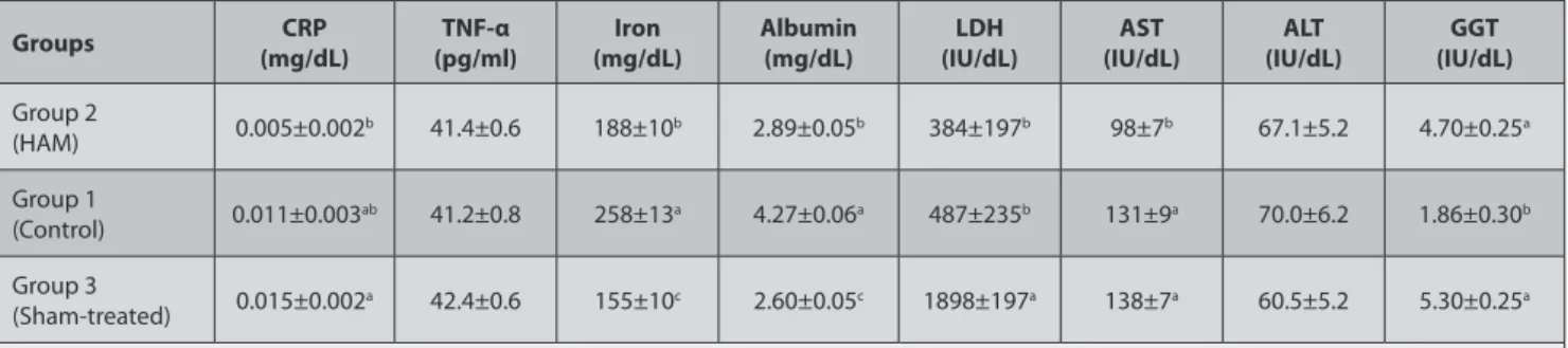
Table 2 summarizes biochemical parameters in response to the (HAM) treatment in rats induced experimentally non-sterile clean wound inflammation. Induction of non- sterile clean wound inflammation causes a 36% elevation in CRP level, which was reduced by the HAM treatment (Group 3) to the control level (Group 1). However, there were no differences in TNF-α across the experimental groups. As compared with the control group (Group 1),
sham operation (Group 2) decreased serum Fe concentration by 40%. Although the HAM treatment (Group 3) increased serum Fe concentration by 21% as compared to the sham-operated group (Group 2), it did not reach the control group (Group 1) level. Alterations in serum albumin concentration in response to experimental groups were similar to those in serum Fe concentrations. The activities of enzymes in response to the experimental groups were variable. The serum LDH activity increased by 4.1-fold in the shame operated group and this elevation was depressed by the HAM treatment (Group 3). The sham-operation (Group 2 did not affect the serum AST activity, whereas the HAM treatment decreased the serum AST activity as compared with the control and sham-operated groups. The serum ALT activity did not differ by the experimental groups. The sham-operation caused a 186% elevation in the serum GGT activities in comparison with the control group and the HAM treatment failed to reduce this elevation.
DISCUSSION
Placenta extract with HAM has been used for a long time as a wound healer and a cosmetic in many countries 17.
It is rich in enzymes such as AST and ALT; nucleic acids,
Table 1. Biochemical profile of the human placenta extracts (n = 10) Tablo 1. İnsan plasenta ekstraktlarının biyokimyasal profili (n = 10)
Parameter Mean±SD
Glucose (mg/g protein) 12.14±2.14
Triglyceride (mg/g protein) 26.42±4.46
Cholesterol (mg/g protein) 14.28±4.14
ALP (IU/g protein) 347.0±74.16
ALT (IU/g protein) 2.71±0.42
AST (IU/g protein) 53.78±9.67
LDH (IU/g protein) 498.42±77.67 GGT (IU/g protein) 8.20±0.8 CRP (mg/g protein) 0.55±0.01 Fe (µg/g protein) 92.75±21.36 Na (mmol/g protein) 15.71±2.02 K (mmol/g protein) 0.48±0.07 Cl (mg/g protein) 15.71±2.02 P (mg/g protein) 4.5±0.70
The placenta extracts were analysed for glucose, triglyceride, cholesterol, ALP, ALT, AST, LDH, GGT, CRP, Fe, Na, K, Cl and P
ALT: alanine aminotransferase; AST: aspartate aminotransferase; LDH:
lactate dehydrogenase; CRP: C-reactive protein; GGT: gamma-glutamyl transferase; ALP: alkaline phosphatase; SD: standard deviation
Plasenta ekstraktından glukoz, triglyiserid, kolesterol, ALP, ALT, AST, LDH, GGT, CRP, Fe, Na, K, Cl ve P analizleri gerçekleştirildi
ALT: alanin aminotransferaz; AST: aspartat aminotransferaz; LDH: laktat dehidrojenaz; CRP: C-reaktif protein; GGT: gamma-glutamil transferaz;
vitamins, amino acids, steroids, fatty acids and minerals (Table 1). These biochemical molecules may have multiple biological effects, some of which are anti-inflammatory, immunotrophic and anti-oxidative 18. To our knowledge,
very few studies have dealt with the effects of AM in long term. Therefore, the attempt was to investigate effects of HAM on metabolism on 14th day 14,19. In the
current experiment, serum biochemistry tests were used to assess the effects of HAM on inflammation induced experimentally by the non-sterile clean wound technique.
The human amniotic membrane enhances epitheliza-tion 20-23, through secreting anti-inflammatory cytokines
and suppressing TGF-β signaling at the transcriptional level, leading to down-regulation of several downstream genes that are responsible for scar formation 24,25. In general,
HAM causes release of potent immune-modulatory and anti-inflammatory cytokines (IL-10, IL-6) 26through a direct
suppressive effect of amniotic membrane matrix on the expression of two of the most potent proinflammatory cytokines, IL-1α and IL-1β, at both protein and mRNA levels 13,27
indicated that HAM possesses an immunosuppressor effect on human peripheral blood mononuclear cells, including apoptosis, and inhibiting proliferation in response to a polyclonal stimulus, as well as inhibiting the synthesis and secretion of pro-inflammatory cytokines on exposure to lypopolysaccharide. Similarly, it has been shown that HAM’s anti-inflammatory actions may be mediated in part by its secretion of anti-inflammatory cytokines such as IL-10, inhibin, activin, and IL-1 receptor antagonist as well as anti-inflammatory protease inhibitors such as a1-anti-trysin inhibitor and inter-a-trysin inhibitor 28. Moreover, HAM
suppresses innate immunity by trapping both
mono-nuclear and polymorphomono-nuclear granulocytes within its stromal matrix and inducing them to undergo apoptosis 29.
Tseng 30 has reported that HAM modulates acquired
immunity by suppressing alloreactive responses and down regulate production of Th1 and Th2 cytokines. C-reactive protein is a well-known acute-phase protein and a marker of systemic inflammation in the body 31. In the present study,
a lower level of CRP occurred in the HAM-treated group when compared with the sham-operated group (Table 2). The reduction in CRP level may indicate that HAM could facilitate wound healing by inhibiting inflammation. How-ever, lacking difference in serum TNF-α levels (Table 2) may suggest that TNF-α acts locally rather than systematically.
To our knowledge, the HAM effects on the activity of enzymes have not been studied in detail. Aspartate amino-transferase, one of the transaminase enzymes, is regarded as markers of muscular dystrophy and cell damage 32.
The decreased activity of AST in the serum of the HAM-treated group may be due to restoration of muscular and cell damages induced by the non-sterile clean wound inflammation. Lactate dehydrogenase, an intracellular enzyme, increases in serum in case of cell damage and/ or death 33. The HAM treatment (Group 3) reduced the
serum LDH activity which increased drastically in response to the sham operation (Table 2). Overall, enzyme activity responses may suggest that HAM may benefit to alleviate tissue damage. Additionally, decreased serum Fe and albumin concentration seem directly to be related to trauma induction. The partial restoration of these two parameters in the HAM-treated group (Group 3) could be related to having HAM served as a reservoir for these compounds (Table 2).
Table 2. Serum biochemical parameters in response to the human amniotic membrane (HAM) treatment in rats induced experimentally non-sterile clean
wound inflammation (n = 10)*
Tablo 2. Deneysel steril olmayan temiz yara inflammasyonuyla indüklenmiş sıçanlarda, insan amniyotik membran (HAM) sağaltımına cevap olarak serum
biyokimyasal parametreler (n =10)*
Groups (mg/dL)CRP (pg/ml)TNF-α (mg/dL)Iron Albumin (mg/dL) (IU/dL)LDH (IU/dL)AST (IU/dL)ALT (IU/dL)GGT
Group 2 (HAM) 0.005±0.002b 41.4±0.6 188±10b 2.89±0.05b 384±197b 98±7b 67.1±5.2 4.70±0.25a Group 1 (Control) 0.011±0.003ab 41.2±0.8 258±13a 4.27±0.06a 487±235b 131±9a 70.0±6.2 1.86±0.30b Group 3 (Sham-treated) 0.015±0.002a 42.4±0.6 155±10c 2.60±0.05c 1898±197a 138±7a 60.5±5.2 5.30±0.25a
The sera were analysed for CRP, TNF-α , iron, albumin, LDH, AST, ALT and GGT
* The data are expressed as mean ± SE
1 Control: rats that were not operated; Sham-operated: rats that were subjected to experimental clean wound inflammation; HAM-treated: rats that were
subjected to experimental clean wound inflammation and received HAM treatment
Different superscripts within the same rows differ (P<0.05). CRP: C-reactive protein; TNF-α: tumor necrosis factor-alpha; LDH: lactate dehydrogenase;
AST: aspartate aminotransferase; ALT: alanine aminotransferase; GGT: gamma-glutamyl transferase
Serumlardan CRP, TNF-α , demir, albumin, LDH, AST, ALT ve GGT analizleri gerçekleştirildi
* Veriler, ortalama ± SE olarak gösterildi
1 Kontrol: sıçanları, operasyon geçirmediler; Sham-operasyonlu: sıçanlar, deneysel temiz yara inflammasyonuna maruz kaldılar; HAM-maruziyetli sıçanlar,
deneysel temiz yara inflammasyonuna maruz kaldılar ve HAM sağaltımı aldılar
Aynı satırdaki farklı üst-simgeler, farklıdır (P<0.05). CRP: C-reaktif protein; TNF-α: tümör nekroz faktör-alfa; LDH: laktat dehidrojenaz; AST: aspartat aminotransferaz; ALT: alanin aminotransferaz; GGT: gamma-glutamil transferaz
In conclusion, sham-operation resulted in tissue damage. The HAM treatment alleviated tissue damage as reflected by decreases in CRP and the serum LDH and AST activities. It also served as metabolite supplement for tissue restoration. Namely, the AM attenuates indicators of inflammation and tissue injury in experimental induced non-sterile clean wound inflammation.
A
cknowledgementsThe authors are grateful to Eren KIRDAR, Zeynep ATALAY and Selim YIRIK for their technical assistance.
REFERENCES
1. Aller MA, Arias JI, Alonso-Poza A, Arias J: A review of metabolic
staging in severely injured patients. Scan J Trauma Resusc Emerg Med, 18, 27-39, 2011.
2. Yücel O, Güler A, Gamsızkan M, Ersöz N, Eken A, Şirin YS, Balkan M, Genç O: Protective effects of proanthocyanidin on allograft renal
oxidative stress: An experimental study. Kafkas Univ Vet Fak Derg, 16 (1):
7-12, 2010.
3. Singer AJ, Clark RA: Cutaneous wound healing. New Engl J Med, 341,
738-746, 1999.
4. Atalay G, Demirkan İ, Yaman H, Cihan M, Önder F, Sözmen M: Effect
of topical kefir application on open wound healing: An in vivo study.
Kafkas Univ Vet Fak Derg, 9 (1): 43-47, 2003.
5. Babior BM: Oxygen-dependent microbial killing by phagocytes
(second of two parts). N Engl J Med, 298, 721-725, 1978.
6. Ansari KU, Gupta N, Bapat SK: An experimental and clinical evaluation
of immunomodulating potential of human placental extracts. Indian J
Pharmacol, 26, 130-132, 1994.
7. Solomon A, Rosenblatt M, Monray D, Ji Z, Pflugfelder SC, Tseng SC: Suppression of interleukin 1a and interleukin 1β in human limbal
epithelial cells cultured on the amniotic membrane stromal matrix. Br J
Ophthalmol, 85, 444-449, 2001.
8. Manuelpillai U, Moodley Y, Borlongan CV, Parolini O: Amniotic
membrane and amniotic cell: Potential therapeutic tools to combat tissue inflammation and fibrosis? Placenta, S320-S325, 2011.
9. Garfias Y, Zago-Clavellina V, Vadillo-Ortega F, Osorio M, Jimenez-Martinez MC: Amniotic membrane is an immunosuppressor of peripheral
blood mononuclear cells. Immunol Invest, 40, 183-196, 2011.
10. Gomes MF, Dos Anjos MJ, Nogueira, TO, Guiaraes SA: Histological
evaluation of the osteoinductive property of autogenous demineralized dentin matrix on surgical bone defects in rabbit skull using human amniotic membrane for guided bone regeneration. Int J Oral Maxillofac
Implant, 16, 563-571, 2001.
11. Kim JS, Kim JC, Na BK, Jeong JM, Song CY: Amniotic membrane
patching promotes healing and inhibits proteinase activity on wound healing following acute corneal alkali burn. Exp Eye Res, 70, 329-337, 2000.
12. Lee SH, Tseng SC: Amniotic membrane transplantation for persistent
epithelial defect with ulceration. Am J Ophthalmol, 123, 303-312, 1997.
13. Shimazaki J, Yang HY, Tsubota K: Amniotic membrane
trans-plantation for ocular surface reconstruction in patients with chemical and thermal burns. Ophthalmology, 104, 2068-2076, 1997.
14. Choi JA, Choi JS, Joo CK: Effects of amniotic membrane suspension
in the rat alkali burn model. Molecular Vision, 17, 404-412, 2011.
15. Toda A, Okabe M, Yoshida T, Nikaido T: The potential of amniotic
membrane/amnion-derived cells for regeneration of various tissues.
J Pharmacol Sci, 105, 215-228, 2007.
16. Espana EM, He H, Kawakita T, Di Pascuale MA, Raju VK, Liu CY, Tseng SC: Human keratocytes cultured on amniotic membrane stroma
preserve morphology and Express keratocan. Invest. Ophthalmol Vis Sci, 44, 5136-5141, 2003.
17. Tonello G, Daglio M, Zaccarelli N, Sottofattori E, Mazzei M, Balbi A: Characterization and quantitation of the active polynucleotide fraction
(PDRN) from human placenta, a tissue repair stimulating agent. J Pharm
Biomed Anal, 14, 1555-1560, 1996.
18. Watanabe S, Togashi S, Takahashi N, Fukui T: L-tryptophan as an
antioxidant in human placenta extract. J Nutr Sci Vitaminol (Tokyo), 48, 36-39, 2002.
19. Rinastiti M, Harijadi, Santoso ALS, Sosroseno W: Histological
evaluation of rabbit gingival wound healing transplanted with human amniotic membrane. Int J Oral Maxillofac Surg, 35, 247-251.
20. Deprest JA, Van Ballaer PP, Evrard VA, Peers KH, Spitz B, Steegers EA, Vandenberghe K: Experience with fetoscopic cord ligation. Eur J Obstet Gynecol Reprod Biol, 81, 157-164, 1998.
21. Devlieger R, Deprest JA, Gratacos E, Pijnenborg R, Leask R, Riley SC: Matrix metalloproteinases-2 and -9 and their endogenous tissue
inhibitors in fetal membrane repair following fetoscopy in a rabbit model.
Mol Hum Reprod, 6, 479-485, 2000.
22. Fortunato SJ, Menon R: Distinct molecular events suggest different
pathways for preterm labor and premature rupture of membranes. Am J
Obstet Gynecol, 184, 1399-1405, 2001.
23. Choi, JA, Jin HJ, Jung S, Yang E, Choi JS, Chung SH, Joo CK: Effects
of amniotic membrane suspension in human corneal wound healing in
vitro. Mol Vis, 15, 2230-2238, 2009.
24. Letko E, Papaliodis DN, Papaliodis GN, Daoud YJ, Ahmed AR, Foster CS: Stevens-Johnson syndrome and toxic epidermal necrolysis: A
review of the literature. Ann Allergy Asthma Immunol, 94, 419-436, 2005.
25. Tseng SC, Li DQ, Ma X: Suppression of transforming growth factor
isoform, TGF-b receptor II, and myofibroblast differentiation in cultured human corneal and limbal fibroblasts by amniotic membrane matrix. J
Cell Physiol, 179, 325-335, 1999.
26. Paradowska E, Blach-Olszewska Z, Gejdel E: Constitutive and induced
cytokine production by human placenta and amniotic membrane at term.
Placenta, 18, 441-6, 1997.
27. Chang YJ, Hwang SM, Tseng CP, Cheng FC, Huang SH, Hsu LF, Hsu LW, Tsai MS: Isolation of mesenchymal stem cells with neurogenic
potential from the mesoderm of the amniotic membrane. Cells Tissues
Organs, 192, 93-105, 2010.
28. Shimmura S, Shimazaki J, Ohashi Y, Tsubota K: Anti-inflammatory
effects of amniotic membrane transplantation in ocular surface disorders.
Cornea, 20, 408-413, 2001.
29. Solomon A, Wajngarten M, Alviano F, Anteby, I, Elchalal U, Pe’er, J, Levi-Schaffer F: Suppression of inflammatory and fibrotic responses in
allergic inflammation by the amniotic membrane stromal matrix. Clin Exp
Allergy, 35, 941-948, 2005.
30. Tseng SC: Amniotic membrane transplantation for ocular surface
reconstruction. Biosci Rep, 21, 481-489, 2001.
31. Gabay C, Kushner I: Acute-phase proteins and other systemic
responses to inflammation. N Engl J Med, 340, 448-454, 1999.
32. Kaplan A, Szabo LL, Opheim KE: Clinical Chemistry: Interpretation
and Techniques, 3rd ed., pp. 226-228, Lea and Febiger, Philadelphia, USA, 1988.
33. Prabhu R, Balasubramanian KA: Effect of oxidants on small intestinal
brush border membranes and colonic apical membrane- a comparative study. Comp Biochem Physiol C Toxicol Pharmacol, 134, 329-339, 2003.