Olgu Sunumu
© 2009
DEÜ
TIP FAKÜLTESİ DERGİSİ CİLT 23, SAYI 3 , (EYLÜL) 2009, S: 135 - 138135
A Case Of Apert Syndrome Presented With
Ventricular Septal Defect
VENTRİKÜLER SEPTAL DEFEKTLİ BİR APERT SENDROMU OLGUSU
Savaş DEMİRPENÇE
1, Vedide TAVLI
1, Derya ERÇAL
2, Timur MEŞE
11
Dr. Behçet Uz Çocuk Hastanesi Çocuk Kardiyolojisi Bölümü
2
Dokuz Eylül Üniversitesi Tıp Fakültesi Çocuk Sağlığı ve Hastalıkları Anabilim Dalı Genetik Bilim Dalı
Savaş DEMİRPENÇE Dr. Behçet Uz Çocuk Hastanesi Çocuk Kardiyolojisi Bölümü Alsancak, İZMİR Tel. Tel. Tel. Tel. (232) 4895656 (6186) GSM. GSM. GSM. GSM. (505) 5252849 SUMMARY
Apert syndrome is a congenital malformation syndrome which is associated with craniosynostosis, craniofacial anomalies, syndactyly and congenital heart defects. Several cardiovascular abnormalities including atrial septal defect, ventricular septal defect or patent ductus arteriosus were reported in 10% of the patients with Apert syndrome. Herein, we report a case of Apert syndrome with ventricular septal defect diagnosed at 2-month-old age and aim to emphasize the clinical and laboratory features of Apert syndrome in the light of this case. We also aim to attract the attention of the pediatricians to the careful cardiologic examination in every newly diagnosed case of Apert syndrome for early detection of possible heart defects.
Key words: Apert syndrome, ventricular septal defect ÖZET
Apert sendromu, kraniyosinostoz, kraniyofasyal anomaliler, sindaktili ve konjenital kalp defektleri ile giden bir konjenital malformasyon sendromudur. Apert sendromlu hastaların %10’unda atriyal septal defekt, ventriküler septal defekt veya patent duktus arteriozus gibi çeşitli kardiyovasküler anormallikler bildirilmiştir. Bu yazıda 2 aylıkken tanı alan ventriküler septal defektli bir olgu sunuyor ve bu olgunun ışığında Apert sendromunun klinik ve laboratuar özelliklerini vurgulamayı amaçlıyoruz. Ayrıca yeni tanı alan her Apert semdromlu olguda, olası kardiyak defektlerin erken teşhisi için dikkatlı bir kardiyak muayene yapılmasının gerekliliğine çocuk hekimlerinin dikkatini çekmeyi amaçlıyoruz.
Anahtar sözcükler: Apert sendromu, ventriküler septal defekt
Apert syndrome is a rare congenital malformation syndrome which was first described by Wheaton in 1894 and named by Apert in 1906. It is associated with craniosynostosis, craniofacial anomalies including maxil-lary hypoplasia, low-set ears, hypertelorism, low posterior hairline, frontal bossing symmetrical syndactly of the
hands (1). Although autosomal dominant inheritance and germinal mosaicism may occur, it is usually a sporadic condition (2,3). More than 98% of cases arise by new mutation. Its prevalance is estimated at 1 in 65,000 live births (4). The clinical features of Apert syndrome are caused by allelic mutations in the fibroblast growth factor
A case of Apert syndrome presented with ventricular septal defect
136
receptor 2 (FGFR2) gene. In most cases, the disorder results from a mutation in the father. Intracranial anomalies include hypoplastic white matter, progressive hydrocephalus and agenesis of the corpus callosum leading to mental retardation. Several cardiovascular abnormalities including Atrial Septal Defects (ASD) and Ventricular Septal Defects (VSD) were reported in 10% of the patients with Apert syndrome (5).
Here we report a case of Apert syndrome with VSD diagnosed at 2 months of age.
CASE REPORT
The 2-month-old male patient was born at term by caeserean section. His birthweight was 3200 g. The mother had no infectious, physical, psychological or phar-macological trauma during the pregnancy. The perinatal period was also uneventful. The parents were not con-sanguineous. No similar cases were present in the family.
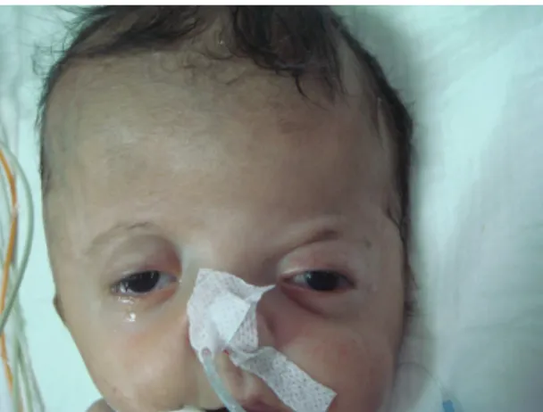
At birth the patient manifested several craniofacial abnormalities including a short, wide head (brachy-cephaly), midfacial hypoplasia, flat forehead, hypertelo-rism, ocular proptosis, depression of the lateral palpebral fissures, low-set ears and a low posterior hairline. Intraoral examination showed complete cleft palate in the midline (Figures 1-3). On cardiac examination a 2/6 holosystolic murmur at the left second intercostal space was heard.
Figure 1. Facial appearence of the patient (midfacial hypo-plasia, flat forehead, hypertelorism, ocular proptosis, depression of the lateral palpebral fissures, low-set ears)
Examination of the extremities showed symmetrical syndactyly leading to the fusion of the fingers and toes (Figure 4).
Figure 2. Low posterior hairline of the patient
Figure 3. Complete cleft palate in the midline
A craniofacial X-ray and X-rays of the extremities were obtained. The cranial X-ray demonstrated hypoplasia of the midline third part of the face, decreased anteroposterior size of the cranium and brachycephaly (Figure 5). Hand and feet radiographs showed the pathognomonic syndactyly of the syndrome and the absence of the distal phalanx (Figure 4). Transfontanel
A case of Apert syndrome presented with ventricular septal defect 137
ultrasonographic examination showed dilatation of the right lateral ventricle. Electrocardiogram was normal. Echocardiography demonstrated a perimembraneous restrictive VSD.
Figure 4. Syndactyly leading to the fusion of the fingers and toes
Plastic surgery, pediatric neurology and genetics consultations for genetic counselling were planned. But the patient died because of severe pulmonary infection which was resistent to antibiotics.
DISCUSSION
Apert syndrome can be detected in the newborn period with craniosynostosis and associated features including syndactyly and craniofacial anomalies. The cranial malformations are the most apparent features of Apert syndrome. Brachycephaly due to the fusion of the coronal suture is the common pattern of growth (1). Our patient also had brachycephaly.
Figure 5. Cranial X-ray demonstrated hypoplasia of the midline third part of the face, decreased anteroposterior size of the cranium and brachycephaly
A high, prominent forehead with a flat posterior skull is another common characteristic cranial finding. A flat face and prognatism develop as a result of deficient growth in the midfacial bones. The patient also had midfacial hypoplasia and hypertelorism. Dental anomalies were also described in Apert syndrome (3). Because our patient was 2-months old, it was not possible to determine dental anomalies.
Syndactyly of the hands and feet is a major association in of Apert syndrome. There is symmetrical fusion of fingers or toes with an equal number on both sides of the body. Syndactyly of the second, third and fourth fingers is common, which may be joined to the thumb and the fifth finger in the hands and feet. The thumb may be broad (5,6). Our patient had syndactyly of the hands, feet and the thumbs were broad.
Congenital heart defects such as VSD, ASD and PDA were reported in 10% of the patients with Apert syndrome. Apert syndrome patients with congenital heart defects are frequently associated with early death (7). Our patient also died at 2-month of age. Cohen et al. reported 136 cases of Apert syndrome. It was emphasized that complex and multiple cardiac anomalies were frequently
A case of Apert syndrome presented with ventricular septal defect
138
associated with early death (8). The case presented here is in accordance with the literature.
The patient presented here had VSD. In literature, it was reported that, most of the cases presented with VSD. However, small ASD defects could also occur (9). Therefore, a careful cardiologic examination should be done in every new diagnosed case of Apert syndrome for early detection of possible heart defects.
In conclusion, we emphasize that each case of Apert syndrome has to be classified and investigated indi-vidually and carefully in regard of possible heart defects. REFERENCES
1. Hukki J, Saarinen P, Kangusniemi M. Single suture craniosynostosis: diagnosis and imaging. Front Oral Biolog 2008; 12: 179-190.
2. Allanson JE. Germinal mosaicism in Apert syndrome. Clin Genet 1986; 29: 429-433.
3. Hollway GE, Sutters GK, Haan EA et al. Mutation detection in FGF-2 craniosynostosis syndromes. Human
Genet 1997; 99: 251-255.
4. Cohen MM Jr, Kreiberg S, Lammer EJ et al. Birth prevelance study of the Apert Syndrome. Am J Med Genet 1992; 42: 655-659.
5. Breugem CC, Fitzpatrick DF, Verchere C. Monozygotic twins with Apert syndrome. Cleft palate craniofac J 2008;45:101-104.
6. Holten IW, Smith AW, Bourne AJ, David DJ. The Apert syndrome hand: pathologic anatomy and clinical manifestations. Plas Reconstr Surg 1997;99:1681-1687.
7. Quinteno-Rivera F, Robson CD, Reis RE, Levine D, Benson C, Müllihan JB, Kinanis VE. Apert syndrome: what prenatal radiographic findings should prompt its consideration? Prenat Diagn 2006;26: 966-972.
8. Cohen MM Jr, Kreiborg S. Visceral anomalies in the Apert syndrome. Am J Med Genet. 1993 Mar 15;45:758-760.
9. Alp E, Alp H, Koç H, Uçar C, Çimen D. Apert Sendromu. Türkiye Klinikleri Pediatri Dergisi 2007;16: 264-268.