18
The Effect of Energy Deficiency on Plasma İnsulin and Cortisol Concentrations During Late Pregnancy in Chios Ewes
Mehmet Hanifi Durak*, Ayşen Altıner**
*
Department of Biochemistry, Faculty of Veterinary Medicine, Dicle University, 21280 Diyarbakır-TURKEY **Department of Biochemistry, Faculty of Veterinary Medicine, Istanbul University, 34320 İstanbul-TURKEY
Summary
The aim of this study was to investigate the effect on plasma insulin and cortisol concentrations by feeding
Chios ewes with a low energy ration in the late period of gestation. 38 Chios ewes were divided into three groups
as pregnant normal energy (PNE), pregnant low energy (PLE) and non pregnant normal energy (N-PNE). Feeding with the treatment rations started on day 105 of the gestation and continued until lambing. Blood samples were obtained on days 120. 127. 134. 141. and 148. during gestation. Plasma insulin concentrations of ewes in the pregnant normal energy were significantly higher than those of the pregnant low energy on days 134 and 141. No significant difference was observed in plasma insulin concentration between pregnant normal energy ewes and non pregnant ewes. Plasma cortisol concentration of ewes in the pregnant low energy was lower on day 120 but higher on day 134 than those of ewes in the pregnant normal energy. As a conclusion an energy intake of greater than 10 MJ/kg ME should be assured for pregnant ewes in the late pregnancy, although energy level of 8.0 MJ/kg ME does not cause a serious energy deficiency.
Keywords: cortisol, energy deficiency, ewe, insulin, pregnancy
Sakız Irkı Koyunlarda İleri Gebelikte Enerji Noksanlığının Plazma İnsülin ve Kortizol Konsantrasyonları Üzerine Etkisi
Özet
Araştırmanın amacı gebeliğin son dönemindeki enerji yetersizliğinin Sakız ırkı koyunlarda plazma insülin ve kortizol konsantrasyonlarını incelemektir. 38 adet Sakız ırkı koyun normal enerjili gebe grup, yetersiz enerjili gebe grup ve normal enerjili gebe olmayan grup olacak şekilde üç gruba ayrılmışlardır. Gebeliğin 105. gününden doğuma kadar bulundukları gruplara uygun bir şekilde beslenmeye başlanmışlardır. Kan numuneleri gebeliğin 120. 127. 134. 141. ve 148. günlerinde alınmıştır. Normal enerjili gebe koyunların 134. ve 141. günlerindeki plazma insülin konsantrasyonları, yetersiz enerjili gebe gruptakilere göre önemli oranda yüksek çıkmıştır. Normal enerjili gebe gruptaki koyunlar ile normal enerjili gebe olmayan gruptaki koyunların plazma insülin konsantrasyonları arasında bir fark bulunmamıştır. Yetersiz enerjili beslenen gebe koyunlar, normal enerjili beslenen gebelerden gebeliğin 120. gününde daha düşük, 134. gününde ise daha yüksek plazma kortizol konsantrasyonlarına sahip olmuşlardır. Sonuç olarak koyunlar gebeliğin son dönemlerinde 10 MJ/kg ME’den daha yüksek enerji içeren yemlerle beslenmelidir ve gebeliğin son dönemlerinde koyunların 8.0 MJ/kg ME ile beslenmesi ciddi bir enerji eksikliğine yol açmamıştır.
Anahtar sözcükler: gebelik, insülin, kortizol, koyun, yetersiz enerji
ARAŞTIRMA
Elektronik:ISSN: 1308-0679
http://www.dicle.edu.tr/bolum/Muh/veteriner/dergi/
19
Introduction
Pregnancy and lactation periods are known as the most critical periods in ovine nutrition (1). Energy requirements of pregnant ewes significantly increase in the last period of the pregnancy (2). Sufficient growth and development of fetus depend on feeding of the dam with a balanced ration in the last 6 weeks of gestation in which 70-80% of fetal growth occurs (1). In addition, placenta and uterus rapidly develop, and mammary glands are prepared for lactation. If the dam enters to the last 6 weeks of gestation with poor body conditions, this may result in low survival rate of the fetus. Furthermore, development of mammary glands slows down, and the ewe can not produce milk despite the high genetic capacity (3).
It has been reported that nutrition in the last period of gestation affects lambing performance, milk yield, birth and growth weights of lambs, and survival rates. It should be taken into account that nutrition of ewes becomes more important in the last period if multiple fetuses and unwilling of ewe to hay due to pressure of large uterus to rumen are present. In the last 6 weeks, sufficient nutrients should be provided to the ewes (1). Deficiency in any of the main nutrients including water, lipids, carbohydrates, proteins, minerals and vitamins affects growth and reproductive performance of sheep (3). Many studies have indicated (1, 3)revealed that nutrition deficiency resulted in low yield conditions. Nutrient deficiency during gestation of ewes causes underdevelopment of the fetus. Development of hypertension was reported in lambs when they reached 3-months of age if they were born from ewes suffering from malnutrition in the last period of gestation (4).
Feeding ewes with energy deficient ration results in remarkable decreases in the insulin secretion (5). Increase of ovulation
rate by well feeding regime is related to increase in the concentration of insulin (6). Insulin enhances the growth of fetus during the early embryonic development (7). Cortisol is secreted as a response to stress conditions (8). Gestation and environmental stress conditions such as deficient nutrition cause increases in the plasma cortisol levels which may lead to abortion. Furthermore, prepartum increased cortisol level may suppress the immunity (9).
The aim of this study is to investigate the changes in plasma insulin and cortisol levels of ewes fed low energy during the late period of gestation.
Material and Methods
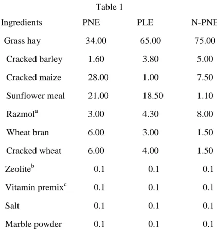
Thirty-eight Chios ewes at 4 to 6 years of age were used. They were housed in 3 separate boxes (3x4x5m). A 15-day flushing was applied to the ewes before estrus synchronization to increase the pregnancy rate. The ewes were synchronized for estrus to increase the twin rate and simultaneous pregnancy. For synchronization, sponges containing 60 mg of medoxyprogesterone acetate were applied to the ewes intravaginally using special applicators. After 15 days, the sponges were removed and 600 IU of PMSG per ewe was administered intramuscularly. Forty-eight hours after the injection, the ewes were placed in boxes with 1 Chios ram per 10 ewes for mating. On day 105 after random mating, the ewes were subjected to ultrasound examination to determine pregnancy. The ultrasound examination revealed that 24 of the 38 ewes were pregnant. The animals were divided into 3 groups: pregnant normal energy (PNE), pregnant low energy (PLE), and non-pregnant normal energy (N-PNE). On day 105 of gestation, the animals were begun to be fed with the treatment rations (Table 1).
20
Table 1. Composition of the diets (%). Table 1
Ingredients PNE PLE N-PNE
Grass hay 34.00 65.00 75.00 Cracked barley 1.60 3.80 5.00 Cracked maize 28.00 1.00 7.50 Sunflower meal 21.00 18.50 1.10 Razmola 3.00 4.30 8.00 Wheat bran 6.00 3.00 1.50 Cracked wheat 6.00 4.00 1.50 Zeoliteb 0.1 0.1 0.1 Vitamin premixc 0.1 0.1 0.1 Salt 0.1 0.1 0.1 Marble powder 0.1 0.1 0.1 a
Fine bran, flour and embryo of wheat.
b
1 kg zeolite: SiO2 48.58g, Al2O3 14.72g, CaO 11.11g, MgO 11.65g, Fl2O3 9.19g, LiO 2.5g,
Na2O 1.28g, K2O 0.44g, TiO2 0.38g, MnO 0.16 g, Cr2O3 0.06g, P2O5 0.03g. c
1 kg vitamin premix: vitamin A 10.000.000 IU, vitamin D3 1.500.000 IU, vitamin E 25g, niacin
20g, d-pantothenic acid 7g, vitamin B2 2.5g, vitamin B1 1.5g, vitamin B6 1.5g, vitamin B12
15mg.
PNE= Pregnant normal energy. PLE= pregnant low energy. N-PNE= Non-pregnant normal energy.
The feeding regime continued until lambing. All ewes were fed 700g of the corresponding rations in the morning (08:00) and evening (16:00). Water was provided ad libitum. Two ewes died in the PNE group during the final periods of pregnancy. It was determined that 2 ewes had acute pneumonia in the PLE group and 4 ewes had metritis in the N-PNE group. In total 8 ewes were removed from the 3 groups in this study.
21
Feeding between days 0 and 105 of gestation: All ewes (n=38) in all groups were fed a ration with 11% crude protein (CP) and 8.8 MJ/kg ME (1400 g/day) (Table 1, ration for N-PNE). Feeding between day 106 of gestation and birth: The ewes in PNE were fed a ration containing 13% CP and 10 MJ/kg ME, while those in PLE were fed a ration containing 13% CP and 8.0 MJ/kg ME.
The ration for the ewes in N-PNE contained 11% CP and 8.8 MJ/kg ME. Blood samples (10 ml) were taken from the jugular vein into tubes with and without anticoagulant before feding in the morning on days 120. 127. 134. 141. and 148. during gestation. The samples were centrifuged at 3000 rpm for 10 min for separating plasma and serum. The plasma and serum samples were stored at -20ºC until analyzed. Plasma insulin and cortisol concentrations were analyzed by commercial test kits (DSL-1600 insulin RIA, Diagnostic Systems Lab. USA.; DSL-2100 active cortisol RIA, Diagnostic Systems Lab. USA. respectively) and radioimmunoassay.
Statistical Analyses: Parameters including insulin and cortisol were statistically compared between PNE and PLE ewes and PNE and N-PNE ewes using Independent-sample t-test. In addition, concentrations of insulin and cortisol were compared between sampling days using One-Way ANOVA (Duncan’s test). SPSS was used for all statistical analysis. (SPSS for Windows. Standard version 10.00 SPSS Inc. Headquarters, Chicago, USA.1999). All results were expressed as mean ± SD. A significance level of P<0.05 was employed in the analysis of data from treatment groups.
Results
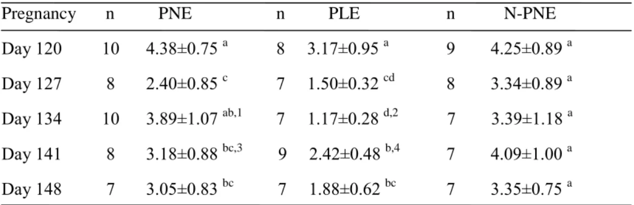
Mean plasma insulin concentrations of pregnant ewes fed with PNE and PLE rations and those of N-PNE ewes are presented in Table 2. Results indicated that the insulin concentration of PLE was always lower during the study period compared to the other two treatment groups. Plasma insulin concentrations of PNE were significantly higher than those of PLE on days 134 and 141 (P<0.001 and P<0.05, respectively). No significant difference was observed in plasma insulin concentrations
between PNE ewes and N-PNE ewes. The plasma insulin concentration of PNE exhibited a significant decrease on day 127 followed by an increase on day 134 and then gradually decreased. The highest plasma insulin concentration in PLE animals was observed on day 120 and then significantly (P<0.05) decreased during the subsequent days. In N-PNE ewes, there were no differences in insulin concentrations between sampling days.
22
Table 2 Plasma insulin concentrations during late pregnancy of Chios ewes fed different (µU/ml, x±SD).
Pregnancy n PNE n PLE n N-PNE Day 120 10 4.38±0.75 a 8 3.17±0.95 a 9 4.25±0.89 a Day 127 8 2.40±0.85 c 7 1.50±0.32 cd 8 3.34±0.89 a Day 134 10 3.89±1.07 ab,1 7 1.17±0.28 d,2 7 3.39±1.18 a Day 141 8 3.18±0.88 bc,3 9 2.42±0.48 b,4 7 4.09±1.00 a Day 148 7 3.05±0.83 bc 7 1.88±0.62 bc 7 3.35±0.75 a
abcd Different superscripts indicate significant differences in same column (P<0.05).
1-2
Statistical difference is significant between two groups within lines (P<0.001). 3-4 Statistical difference is significant between two groups within lines (P<0.05).
PNE= Pregnant normal energy. PLE= pregnant low energy. N-PNE= Non-pregnant normal energy.
Mean plasma cortisol concentrations of pregnant ewes fed with PNE and PLE rations and those of N-PNE ewes are presented in Table 3. Plasma cortisol concentration of PLE was lower on day 120 but higher on day 134 than those of PNE. PNE had significantly (P<0.05) higher plasma cortisol concentration than those of N-PNE ewes on day 120 whereas no significant difference was found between the two groups on the subsequent sampling days. Plasma cortisol level in PNE showed a significant increase only on day 148. On the other hand, plasma cortisol concentration continuously increased in PLE until day 134 and then slightly decreased followed by a significant (P<0.05) increase on day 148. In N-PNE animals, plasma cortisol concentration did not change significantly between sampling days.
Table 3 Plasma cortisol concentrations during late pregnancy of Chios ewes fed different (ng/ml, x±SD).
Pregnancy n PNE n PLE n N-PNE Day 120 9 60.9±21.5 b,1 8 54.3±12.8 c,2 7 41.8±12.4 a,2 Day 127 8 66.7±16.2 b 6 67.3±4.7 bc 8 47.2±13.4 a Day 134 7 60.6±15.9 b,1 7 89.6±6.7 ab,2 6 39.3±9.0 a Day 141 9 57.9±14.2 b 9 67.2±19.8 bc 7 43.6±12.9 a Day 148 8 87.9±23.6 a 10 105.5±17.0 a 7 53.8±11.6 a abc
Different superscripts indicate significant differences in same column (P<0.05).
1-2
Statistical difference is significant between two groups within lines (P<0.05).
PNE= Pregnant normal energy. PLE= pregnant low energy. N-PNE= Non-pregnant normal energy.
23
Discussion
In sheep as in other mammals, the blood insulin level is regulated by the blood glucose concentration, while the cellular metabolism of glucose is regulated by this hormone (5, 10). Bergman et al. (11) reported that insulin resistance developed in sheep resulted from unbalanced alimentation that caused fattening. Faulkner and Martin (12) determined that insulin level increased twice 2-6 h after feeding. The same researchers reported that plasma insulin level of sheep under negative energy balance was low. Gonda et al. (10) reported that increasing energy level in the rations of rams resulted in an increase in plasma insulin level. The plasma insulin concentrations of PLE ration were found
lower than those of PNE or N-PNE ewes (Table 2). This finding is consistent with those reported by Faulkner and Martin (12) and by Gonda et al. (10) El-Sherif and Assad (13) reported that the insulin response is decreased by gestation. However, the absence of a significant difference between insulin concentrations of PNE ewes and N-PNE ewes (Table 2) indicates that pregnancy does not affect insulin secretion.
As in other mammals, cortisol in sheep increases permeability of muscle cells to aminoacids and glucose, acts as antagonist to insulin (14), is the chief corticosteroid released as response to stress conditions such as
pregnancy and environmental temperature (8, 15) Green (16) reported that plasma cortisol level increased in ewes fed with deficient ration on the days 100 and 125 of gestation period. Henze et al. (17) found that cortisol levels increased in ewes with pregnancy toxemia. Similarly with findings of other researchers (15, 16, 17), we observed lower cortisol concentrations in N-PNE ewes than in pregnant and higher concentrations of this hormone in PLE ewes than in PNE ewes (Table 3).
Cortisol increases fetal glucogenesis at the end of pregnancy (13). Maternal cortisol reaches fetus by passing placenta resulting in an increase in the level of fetal cortisol. Cortisol also triggers the parturition mechanism (18). Chen et al. (19) and Roche et al. (20) reported that cortisol levels of maternal plasma in cow and goat continuously increased. In agreement with the results of those researchers, significant increases in cortisol levels of pregnant ewes were observed in the last week of pregnancy (day 148) in the present study. There are various approaches about the reason for this increase. Some researchers (18, 21) thought
that increased cortisol in the last 10-15 days of gestation plays an important role for maturation of lungs, digestive system, brain and kidneys of fetus as well as facilitating the enlargement of the uterus. Fowden et al. (22) reported that cortisol stopped growth of axial skeleton and fetal growth in general. Some other researchers (19, 20) believed that increased cortisol before the delivery prepares the fetus for potential trauma during birth and for adapting the postparturition environment.
In our study, plasma insulin concentrations of all pregnant animals decreased towards the end of pregnancy, the fetus’s growth rate may be declining this period. Beaceuse insulin is a hormone that encourages the growth of the fetus. Cortisol increase in the levels of both groups of pregnant ewes as a cause, 8.0 MJ/kg ME and 10 MJ/kg ME diet with sources of stress in sheep in late pregnancy is tought to be created.
As a result of the current study, it is concluded that an energy intake greater than 10 MJ/kg ME should be ensured for ewes in the last period of pregnancy, although an
24
energy level of 8.0 MJ/kg ME kg does not
cause a serious energy deficiency. References
1. Demirel M, Aygün T, Altın T, Bingol M. (2000). Hamdani ve Karakaş koyunlarında gebeliğin son döneminde farklı düzeylerde beslemenin koyunlarda canlı ağırlık, kuzularda doğum ağırlığı ve büyüme üzerine etkileri. Turk J Vet Anim Sci, 24: 243-249. 2. Ozpinar A. (1994). Ruminantlarda
ketozisin olusum mekanizmasi. Çiftlik. 4:20-26.
3. Johnson KA. (1997). Nutritional Management of the Sheep Flock. Washington State Cooperative Extension, Washington, USA. 3–7. 4. Dodic M, Pers A, Coghlan JP, Wintour
M. (1999). Can excess glucocorticoid, in utero, predispose to cardiovascular and metabolic disease in middle age? Trends Endocrin Met, 10: 86–91.
5. Elmahdi B, Sallmann HP, Fuhrmann H, Engelhardt WV, Kaske M. (1997). Comparative aspects of glucose tolerance in camels, sheep, and ponies. Comp Biochem Phys A, 118A: 147-51. 6. Al-Haboby AH, Salman AD, Abdul
Karem TA. (1999). Influence of protein supplementation on reproductive traits of Awassi sheep grazing cereal stubble. Small Rum Res, 34: 33-40.
7. Forbes CD, Fernandez JM, Bunting LD, Southern LL, Thompson DL. Gentry LR, Chapa AM. (1998). Growth and metabolic characteristics of Suffolk and Gulf coast native yearling ewes supplemented with chromium tripicolinate. Small Rum Res, 28: 149– 160.
8. Fleming MW. (1997). Cortisol as an indicator of severity of parasitic infections of Haemonchus contortus in lambs (Ovis aries). Comp Biochem Phys B, 116B: 41-44.
9. Symonds ME, Bryant MJ, Lomax MA. (1990). Lipid metabolism in shorn and
unshorn pregnant sheep. Brıt j nutr, 63: 397–400.
10. Gonda HL, Lindberg JE, Holtenius K. (1997). Plasma levels of energy metabolites and pancreatic hormones in relation to the level of intake and intraruminal infusions of volatile fatty acids in fed wether sheep. Comp Biochem Phys A, 116A: 65–73.
11. Bergman EN, Reulein SS, Corlett RE. (1989). Effects of obesity on insulin sensitivity and responsiveness in sheep. Am J Physiol Endocrinol Metab, 257: E772-E781.
12. Faulkner A, Martin PA. (1997). The concentrations of some gut polypeptides are elevated during lactation in ruminants. Comp Biochem Phys B, 118B: 563-568.
13. El-Sherif MMA, Assad F. (2001). Changes in some blood constituents of Barki ewes during pregnancy and lactation under semi arid conditions. Small Rum Res, 40: 269–277.
14. Andrews AH, Holland-Howes VE, Wilkinson JID. (1996). Naturally occuring pregnancy toxaemia in the ewe and treatment with recombinant bovine somatotropin. Small Rum Res, 23: 191– 197.
15. Silanikove N. (2000). Effects of heat stress on the welfare of extensively managed domestic ruminants. Livest Prod Sci, 67: 1–18.
16. Green LR. (2001). Programming of endocrine mechanisms of cardiovascular control and growth. Reprod Sci, 8(2): 57–68.
17. Henze P, Bickhardt K, Fuhrmann H. (1994). The influences of insulin, cortisol, growth hormone and total oestrogen on the pathogenesis of ketosis in sheep. Dtsch Tierarztl Wochenschr, 101: 61-65.
18. Challis JRG, Sloboda D, Matthews SG, Holloway A, Alfaidy N, Patel FA, Whittle W, Fraser M, Moss TJM, Newnham J. (2001). The fetal placental hypothalamic-pituitary-adrenal (HPA)
25
axis, parturition and post natal health. Mol cell endocrinol, 185(1): 135-144. 19. Chen JC, Chang CJ, Peh HC, Lee SL.
(1999). Perinatal adrenocortical function in relation to the growth rate and immunoglobulin acquisition of goat kids. Small Rum Res, 33: 255-262. 20. Roche JF, Mackey D, Diskin MD.
(2000). Reproductive management of postpartum cows. Anim Reprod Sci, 60-61(1): 703-712.
21. Edwards LJ, Bryce AE, Coulter CL, McMillen IC. (2002). Maternal undernutrition throughout pregnancy increases adrenocorticotrophin receptor and steroidogenic acute regulatory protein gene expression in the adrenal gland of twin fetal sheep during late gestation. Mol cell endocrinol, 196: 1-10.
22. Fowden AL, Szemere J, Hughes P, Gilmour RS, Forhead AJ. (1996). The effects of cortisol on the growth rate of the sheep fetus during late gestation. J endocrınol, 151: 97-105.