1 Dicle University Faculty of Medicine, Department of Pediatric, Diyarbakir, Turkey, 2 Yüzüncü Yıl University Faculty of Medicine, Department of Pediatric, Van, Turkey 3 Department of Pediatric Neurology, Yüzüncü Yıl University Faculty of Medicine, Van, Turkey
Yazışma Adresi /Correspondence: Fesih Aktar,
Dicle University Medical Faculty, Department of Pediatric, 21280, Diyarbakır, Turkey Email: [email protected] Geliş Tarihi / Received: 10.03.2016, Kabul Tarihi / Accepted: 21.04.2016
Copyright © Dicle Tıp Dergisi 2016, Her hakkı saklıdır / All rights reserved
Dicle Tıp Dergisi / 2016; 43 (2): 356-359
Dicle Medical Journal doi: 10.5798/diclemedj.0921.2016.02.0695
356
CASE REPORT / OLGU SUNUMU
Magnetic Resonance Spectroscopy in Sjögren-Larsson Syndrome
Sjögren-Larsson Sendromunda Manyetik Rezonans Spektroskopi
Fesih Aktar1, Kamuran Karaman2, Berfin Ö. Özmen2, Muhammed Akıl2,Gökmen Taşkın2, Hüseyin Çaksen3 ABSTRACT
Sjögren-Larsson syndrome (SLS) is a rare neurocutane-ous disease showing an autosomal recessive transmis-sion due to a lack of fatty acid aldehyde dehydrogenase. Spastic diplegia or triplegia, mental retardation and con-genital lamellar ichthyosis are the major findings of the disease. The syndrome may be accompanied by various eye and teeth features, skeletal system anomaly, speak-ing defects, hypertelorism and epilepsy. A 9-month male patient has been hospitalized for convulsion and flaking on body. The patient history showed that flaking skin thickening and peeling was started at the birth, and he suffered a right-side focused seizure when he was three month-old and he was treated with phenobarbital and car-bamazepine upon the epilepsy diagnosis. Wide ichthyo-sis, hypertelorism and bilateral simian line were observed in the physical examination. Bilateral punctuate lesions in cornea, pigment epithelial atrophy in the right eye and esotropia in the left eye have been determined during the eye examination. An epiteliform anomaly has been ob-served in the left hemisphere by electroencephalography. In brain magnetic resonance imaging (MRI), an increase in cerebral-cerebellar brain parenchyma and T1-T2 relax-ation time and in the signal in corpus callosum (delayed myelination) have been determined. With the observa-tion of the white matter in centrum semi oval using brain MRI spectroscopy, signs of a sphingolipid peak at 1.3 ppm have been observed. An SLS diagnosis has been proposed upon clinical and laboratory observations. We want to emphasize on the fact that in epilepsy cases with ichthyosis, SLS should be considered.
Key words: Magnetic resonance spectroscopy, Sjögren-Larsson syndrome, epilepsy, ichthyosis, child
ÖZET
Sjögren-Larsson sendromu (SLS), yağ asidi aldehid de-hidrogenaz enzim eksikliği sonucu gelişen, otozomal resesif geçiş gösteren nadir bir nörokutanöz hastalık-tır. Spastik dipleji veya tetrapleji, mental retardasyon ve konjenital lameller ihtiyozis hastalığın major bulgularıdır. Sendroma çeşitli göz ve diş bulguları, iskelet sistemi ano-malileri, konuşma defektleri, hipertelorizm ve epilepsi de eşlik edebilmektedir. Dokuz aylık erkek hasta havale ge-çirme ve vücudunda pullanma şikâyetleri ile getirildi. Öz-geçmişinde, ciltteki pullanmalarının doğumundan itibaren olduğu ve zamanla deride kalınlaşma ve soyulmaların başladığı, ilk kez üç aylıkken sağ tarafa lokalize havale geçirdiği ve epilepsi tanısı ile fenobarbital ve karbamaze-pin kullandığı öğrenildi. Fizik muayenesinde ciltte yaygın ihtiyozis, hipertelorizm ve bilateral simian çizgisi vardı. Göz muayenesinde korneada bilateral punktat lezyonlar, sağ gözde pigment epitel atrofisi ve sol gözde ezotropia tespit edildi. Elektroensefalografide sol hemisferde epi-leptiform anomali izlendi. Beyin Manyetik Rezonans (MR) incelemesinde serebral-serebellar beyin parankiminde ve T1-T2 relaksasyon süresinde artış ve korpus kallozum-da sinyal artışı (gecikmiş miyelinizasyon) saptandı. Beyin MR spektroskopide sentrum semiovaledeki beyaz cevhe-re yönelik yapılan incelemede 1,3 ppm’de sfingolipid piki-ni içeren bulgular izlendi. Klipiki-nik ve laboratuar bulgularıyla SLS tanısı kondu. Bu vaka dolayısıyla epilepsi ile birlikte ihtiyozisi olan vakalarda SLS’nin de düşünülmesi gerekti-ğini vurgulamak istedik.
Anahtar kelimeler: Manyetik rezonans spektroskopi, Sjögren-Larsson sendromu, epilepsi, ihtiyozis, çocuk
F. Aktar ve ark. MR Spectroscopy in Sjögren-Larsson Syndrome 357
Dicle Tıp Derg / Dicle Med J www.diclemedj.org Cilt / Vol 43, No 2, 356-359
INTRODUCTION
Sjögren-Larsson syndrome (SLS), is a rare neu-rocutaneous disease showing an autosomal reces-sive transmission due to a lack of fatty acid alde-hyde dehydrogenase [1]. Approximately 200 cases have been declared from the definition of the syn-drome. Most of the cases are from Sweden, and the incidence in the North of that country is 2.7-10.2/100.000 [1,2].
With the study of this case, we aimed to em-phasize on the fact that in rare epilepsy cases with ichthyosis, SLS cases shall be considered.
CASE REPORT
A 9-month male patient has been hospitalized in our clinic for convulsion and flaking on body. The pa-tient went through flaking skin thickening and peel-ing at the birth and the patient history showed that lamellar ichthyosis diagnosis was established from the dermatology department they applied. He suf-fered a right-side focused seizure when he was three month-old and he was treated with phenobarbital and carbamazepine upon the epilepsy diagnosis.
The patient history also showed that he was born by caesarian section at twenty eight weeks; with a weight of 1800 grams and that he was un-der phototherapy due to jaundice. The mental and motor development was normal. There was no par-ticularity in the family history. In physical examina-tion, his height was 66 cm (10 percentile), his body weight was 7.6 kg (10-25 percentile), and his head circumference was 44 cm (10 percentile). Wide ichthyosis has been observed in scalp, nape, arms, chest, shoulders, legs, face, and abdomen, palm and sole. Hypertelorism and bilateral simian line have been observed (Figure 1). Hemogram, biochemical and urinary analyses, coagulation tests and levels of thyroid hormone, B12 vitamin and folate were nor-mal in laboratory analyses. The tandem mass study result was normal. Bilateral punctuate lesions in cornea, pigment epithelial atrophy in the right eye and esotropia in the left eye have been determined during the eye examination. No anomalies except of a slight kyphoscoliosis have been observed in the skeletal survey. An epiteliform anomaly has been observed in the left hemisphere by electroenceph-alography. In brain magnetic resonance imaging
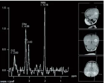
(MRI), an increase in cerebral-cerebellar brain pa-renchyma and T1-T2 relaxation time and in the sig-nal in corpus callosum (delayed myelination) have been determined (Figure 2). With the observation of the white matter in centrum semi oval using brain MRI spectroscopy, signs of a sphingolipid peak at 1.3 ppm have been observed (Figure 3). Sjögren-Larsson Syndrome diagnosis has been proposed upon clinical and laboratory observations for the patient. He is still under observation, squams are present in some area of his skin and the convulsions are kept under control using a double antiepileptic treatment.
Figure 1. Wide ichthyosis and squams
Figure 2. Brain MRI images showing increase in
cerebral-cerebellar brain parenchyma and T1-T2 relaxation time and in the signal in corpus callosum (delayed myelina-tion)
F. Aktar ve ark. MR Spectroscopy in Sjögren-Larsson Syndrome 358
Dicle Tıp Derg / Dicle Med J www.diclemedj.org Cilt / Vol 43, No 2, 356-359
Figure 3. MRI spectroscopy image of white matter in
centrum semi oval showing sphingolipid peak at 1.3 ppm
DISCUSSION
The biochemical and metabolic defect observed in this syndrome is not well known. However, a lack of NAD+ oxidoreductase enzyme of the fatty alcohol responsible for the oxidation of the fatty alcohol to the fatty acid in skin fibroblast cultures and periph-eral blood leucocytes has been determined. The ac-cumulating fatty alcohols or its metabolic products are supposed to modify the epidermal lipid com-position that has a water retainer property and thus may play a role in the pathogenesis [2]. Enzymatic studies have not been performed on our patient due to technical difficulties. However, the diagnosis has been established as SLS considering the clinical and radiological observations of the patient.
Spastic diplegia or triplegia, mental retardation and congenital lamellar ichthyosis are major find-ings of the disease. In addition to this, punctuate epithelial erosions in the surface, white glistening dots (pathognomonic for this syndrome), pigment degeneration in the retina, some observations in the eye such as blepharitis, conjunctivitis, presence of simian line, short height, teeth anomalies, kypho-sis, scoliosis and skeletal system anomalies such as metaphysial dysplasia with small irregular epiphy-sis, speaking defects, hypertelorism, aminoaciduria and epilepsy may appear with this syndrome [1-3]. In our case; congenital lamellar ichthyosis, epilep-sy, hypertelorism, bilateral simian line and slight kyphoscoliosis were observed. Bilateral punctuate lesions in cornea, pigment epithelial atrophy in the
right eye and esotropia in the left eye have been de-termined during the detailed eye examination but no glistening dots have been observed in the macula.
Ichthyosis is present everywhere in the body but is more marked in flexural regions of the body and in lower abdomen [4]. In most patients, ichthy-osis appears at birth and immediately after birth and looks like lamellar ichthyosis [2]. The skin lesions of our patient correspond to this particularity.
Some findings such as spasticity, mental re-tardation, epilepsy, delayed or affected speaking, increase in deep tendon reflexes, bilateral positive Babinski reflex and hyperextension in articulations may observed with this syndrome. The spasticity may appear before age of three and is more pro-nounced in lower extremities. Mental retardation may be at a medium or heavy level and display a progressive evolution (in 70% of the patients the intelligent quality level is <50%). In SLS, epilepsy may be observed in 30-50% of the patients [2,3]. In our case, the mental and motor development was normally observed. He has an epileptiform anomaly which was present in the left hemisphere and kept under control using a double antiepileptic treatment. Nonspecific observations can be obtained with computer assisted tomography [3]. In the computer-assisted brain tomography of SLS patients, hypo dense area have been observed in white matter par-ticularly marked in the frontal lobe [3,5]. In brain MRI, white matter disease characterized with de-myelination or delayed de-myelination [5]. Moreover, micro ventricle, porencephaly, corpus callosum agenesis, cerebral cortical atrophy have been men-tioned in some publications [3]. In our case, an in-crease in cerebral-cerebellar brain parenchyma and T1-T2 relaxation time and in the signal in corpus callosum (delayed myelination) have been deter-mined in brain MRI.
Protein magnetic resonance spectroscopy ob-servations have shown an abnormal peak at 1.3 ppm conform to the accumulation of long chain fatty al-cohol in the periventricular white matter (especially around posterior and frontal horns) [3,6]. In our case, signs of a sphingolipid peak at 1.3 ppm have been observed in brain MRI spectroscopy.
There is still no curative treatment for SLS and leukotriene synthesis inhibitors are considered as
F. Aktar ve ark. MR Spectroscopy in Sjögren-Larsson Syndrome 359
Dicle Tıp Derg / Dicle Med J www.diclemedj.org Cilt / Vol 43, No 2, 356-359
potential therapeutic agents for SLS patients [7]. In SLS patients, the diets for the reduction of long chain fatty acids intake are mostly useless; however, a low fat content diet focused on the reduction of medium-length chain fatty acid intake may be ef-ficient when started at early stage of childhood and may result in skin lesions recovery [2,7]. Our pa-tient was received the diets with reduction of long chain fatty acids and double antiepileptic treatment, and his squams are present in some area of his skin and the convulsions are kept under control.
In conclusion, SLS is a rare disease and should be considered in epilepsy cases which were along with ichthyosis. This cases should be evaluated with neuroradiological examination such as brain MRI and protein MR spectroscopy.
Acknowledgments
The authors wish to thank our patient and his parents that to gave permission to took part in this report. Declaration of Conflicting Interests: The authors
de-clare that they have no conflict of interest.
Financial Disclosure: No financial support was received.
REFERENCES
1. Sillén A, Alderborn A, Pigg M, et al. Detailed genetic and physical mapping in the Sjögren Larsson syndrome gene region in 17p11.2. Hereditas 1998;128:245-250.
2. Fuijkschot J, Theelen T, Seyger MM, et al. Sjögren-Lars-son syndrome in clinical practice. J Inherit Metab Dis 2012;35:955-962.
3. Zribi H, A Souissi, Azzouz H, et al. Sjogren Larsson syn-drome: a rare neurocutaneous disease. Rev Neurol (Paris) 2014;170:297-298.
4. Tanteles GA, Nicolaou M, Patsia N, et al. A rare cause of pruritic ichthyosis: Sjögren-Larsson syndrome in the first reported patients of Cypriot descent. Eur J Dermatol 2015;25:495-496.
5. Gomori JM, Leibovici V, Ziotogorski A, et al. Computed to-mography in Sjögren-Larsson syndrome. Neuroradiology 1987;29:557-559.
6. Nakayama M, Tavora DG, Alvim TC, et al. MRI and 1H-MRS findings of three patients with Sjogren-Larsson syn-drome. Arq Neuropsiquiatr 2006;64:398-401.
7. Willemsen MA, Rotteveel JJ, Steijlen PM, et al. 5-Lipoxy-genase inhibition: a new treatment strategy for Sjogren-Larsson syndrome. Neuropediatrics 2000;31:1-3.