204
Use of Chicken Egg White Treated Under Different pH Conditions as a
Source of Lysozyme in Bacterial DNA Isolation
Makbule BAYLAN Dr., Cukurova University, Turkey [email protected] B.Devrim ÖZCAN Dr., Cukurova University, Turkey [email protected] Gamze MAZI Cukurova University, Turkey [email protected] Mikail BAYLAN Dr., Cukurova University, Turkey [email protected] Elif DİKKAYA Cukurova University Turkey [email protected], Numan ÖZCAN Dr., Cukurova University, Turkey [email protected] Abstract
Lysozyme (EC.3.2.1.17) is an enzyme that catalyses the hydrolysis of beta-1,4 glucosidic linkages between N-acetyl glucosamine (NAG) and N-acetyl muramic acid (NAM) in the peptidoglucan heteropolymers of prokaryotic cell walls. In this study, lysozyme enzyme found in chicken egg white was treated under different pH conditions (pH 4.0, 6.0, 8.0, 10.0, 12.0) and the results were observed as electrophoretic. Electrophoretic results revealed that egg white lysozyme was best yielded at pH 4.0. As a result of dropping the egg white solution on the Bacillus subtilis spreaded on the LB-agar plates with a glass rod, it was observed that the bacteria could not developed due to the lysozyme of egg white. On the other hand, genomic and plasmid DNAs were isolated from the bacteria in DNA isolation studies in which the egg white solution was replaced by commercial lysozyme. The results showed that, in the absence of commercial lysozyme under laboratory conditions, chicken egg white can be used as a lysozyme source in DNA isolation from bacteria.
Key Words: Chicken egg white, lysozyme, DNA isolation, bacterial lysis
Bakteriyel DNA İzolasyonunda Lizozim Kaynağı Olarak Farklı pH
Koşullarında Tavuk Yumurtası Akının Kullanımı
Özet
Lizozim (EC.3.2.1.17), prokaryotik hücre duvarlarının peptidoglukan heteropolimerlerinde N-asetil glukozamin (NAG) ve N-N-asetil muramik asit (NAM) arasındaki beta-1,4 glukosidik bağların hidrolizini katalizleyen bir enzimdir. Bu çalışmada, tavuk yumurtası beyazında bulunan lizozim enzimi farklı pH koşulları altında (pH 4.0, 6.0, 8.0, 10.0, 12.0) muamele edilmiş ve sonuçlar elektroforetik olarak gözlenmiştir. Elektroforetik sonuçlar, yumurta akı lizoziminin en iyi pH 4.0’da izole edildiğini ortaya koymuştur. Bacillus subtilis bakterisinin cam çubukla yayma yöntemiyle ekildiği plaklara yumurta akı solüsyonu damlatılması sonucunda, damlatmanın olduğu bölgede bakterilerin yumurta akı lizoziminden dolayı lize olarak üreyemediği gözlenmiştir. Diğer taraftan, yumurta akı solüsyonunun ticari lizozimle ikame edildiği DNA izolasyonu çalışmalarında, bakterilerden genomik ve plazmit DNA’lar izole edilmiştir. Sonuçlar, laboratuvar koşullarında ticari lizozimin yokluğunda, tavuk yumurtası beyazının, bakterilerden DNA izolasyonunda bir lizozim kaynağı olarak kullanılabileceğini göstermiştir.
205
INTRODUCTION
Lysozyme (EC.3.2.1.17) is an enzyme that catalyzes the hydrolysis of beta-1,4 glucosidic linkages between N-acetyl glucosamine (NAG) and N-acetyl muramic acid (NAM) found in pepdidoglucan heteropolymers of prokaryotic cell walls. It was first discovered by Alexander Flemming in 1922. Gram (+), for example, Micrococcus lysodeikticus former name M. leutus has been shown to be effective against bacteria. For this reason, this bacterium is still used as a test bacteria in enzyme activity determination. Lysozyme is also known as muramidase. For the purpose of DNA isolation, it is used together with EDTA which binds chelates such as Mg++ against Gram (-) bacteria such as
Escherichia coli to form chelates and destabilize the cell wall (Stryer, 1988 and Güzel, 2000). The enzyme breaks down the cell wall by hydrolyzing 1,4-glucosidic bonds between the NAM and NAG units forming the prokaryotic cell wall. Recently, the gene responsible for the production of lysozyme enzyme of T4 bacteriophage has been cloned and expressed in E. coli for enzyme characterization and Streptococcus thermophilus, a yoghurt bacterium to enhance thermo-stability by protein engineering techniques (Güzel et al., 2002; Akınalp et al., 2002; Akınalp et al., 2002; et al., 2007). The chicken lysozyme gene was cloned and expressed in yeast cells. In this cloning, it was reported that the lysozyme gene was expressed under the yeast promoter and that the recombinant lysozyme enzyme produced was 1.5 % of the total yeast proteins, which was secreted in 2/3 of the medium (Oberto and Davison, 1985). Lysozyme is an important enzyme involved in the anti-bacterial defense mechanism of many living things. Human and chicken (egg white) lysozyme are enzymes of clinical and industrial importance. Lysozyme is found in many body fluids in mammals. Lysozyme in breast milk stimulates the general immune system (non-specific immune system) as it is absorbed raw by infants. Lysozyme is denatured as cow's milk is pasteurized against tuberculosis. The human lysozyme gene has been successfully used in mice to clone and develop cancer vaccines (Güzel et al., 2008).
The three-dimensional structure of chicken egg white lysozyme was examined, the base sequences of the responsible gene were read and the gene mechanism was studied in detail. The synthesis of lysozyme to egg white is controlled by steroid hormones in the oviduct. The enzyme is first synthesized as pre-lysozyme, and then converted to mature lysozyme by removal of the signal peptide consisting of 18 amino acids (Oberto and Davison, 1985). It has been reported that the enzyme, which has a molecular weight of 14.3 kDa, consists of 129 amino acids (Canfield, 1963).
Egg white lysozyme is commercially produced and lysozyme enzymes are used extensively in molecular biology laboratories, especially during the DNA isolation from Gram (+) and Gram (-) bacteria, for the hydrolysis of the peptidoglucan layer of bacteria. This enzyme, which is commercially available from the market, is also expensive due to its importation and is an important expense in molecular biology studies where continuous DNA isolation is performed.
In this study, instead of buying the lysozyme enzyme, which is commonly used by microbiology and molecular genetics laboratories, it was used to purify rapidly from daily fresh egg whites with laboratory facilities and use in laboratory for 4-5 weeks. Purification of lysozyme enzymes from pure white eggs obtained from commercially available fresh eggs with purified water at different pH conditions, results were electrophoretically displayed on SDS-PAGE.
MATERIALS AND METHODS Materials
The chicken eggs used in the study were obtained by purchasing in the poultry unit within the Revolving Fund of Çukurova University Faculty of Agriculture. Commercial lysozyme, protein markers, acrylamide, bis-acrylamide, SDS and other chemicals and consumables were also procured from the project budget. Vertical electrophoresis, power supply, pH meter, precision balance, incubator, centrifuge, autoclave, magnetic stirrer, vortex, coolers (-20 and +4 °C) and negatoscope used in the
206
study were obtained from Ç.Ü. Faculty of Agriculture Department of Animal Biotechnology and Genetic Engineering Laboratories.
Methods
Purification of Lysozyme from Egg White by Using Pure Water
Purification of lysozyme enzyme from egg whites was performed according to the following protocol according to Wang et al. (2009): Fresh eggs were carefully broken from the side and separated from the flux yolk and placed in a clean beaker. After determining the volume of the egg whites by means of a tape measure, 50 times sterile pure water was added thereto and homogenized by stirring in magnetic stirrer for about 1 hour. The mixture was divided into 5 equal parts and the pH of each was adjusted to different pH values (4.0, 6.0, 8.0, 10.0, 12.0) with 1 N acetic acid and 0.1 M NaOH solutions. All mixtures were divided into 3 equal parts and incubated for 10, 20 and 30 min (1 part 10 min, 2 part 20 min, 3 part 30 min) in a water bath set to 100 °C for denaturation of egg white proteins. The solutions were passed through 4 layers of cheeses separately and transferred to a separate beaker. The obtained samples were used as lysozyme source.
Electrophoresis of Lysozyme Samples on SDS-PAGE
Obtained egg whites samples the protocol electrophoresis adjusted according to Laemmli 1970s includes: 12% separating gel (12 ml acrylamide (30 g acrylamide, 0.8 g bis-acrylamide, 100 ml), 5.62 ml tris (2 M, pH) 8.8) 0.3 ml of SDS (10% w/v), 12.1 ml of purified water) were prepared in a clean beaker. When polymerized, 200 μl of ammonium persulfate (10% w/v) and 20 μl TEMED were added to the mixture and then placed on a lost clean pipette between two glass plates. To remove contact of the gel with oxygen, water-saturated butanol was added in a thin layer to cover the gel and wait 45 minutes for polymerization. At the end of time, a separate coarse collecting gel (1.8 ml acrylamide (30 g acrylamide, 0.8 g bis-acrylamide, 100 ml), 3.65 ml tris (0.5 M, pH 6.8), 0.15 ml SDS (10% w/v), 9.4 ml purified water). For polymerization, 100 μl of ammonium persulfate (10% w/v) and 10 μl of TEMED were added. The butanol on the separating gel was absorbed into a blotting paper and the collecting gel was poured onto it with a clean pipette. The electrophoresis comb was placed from the top to form the sample wells and waited again for 45 minutes for polymerization. At the end of the period, the comb was removed and the gel assembly consisting of two glass plates was placed in the electrophoresis tank. To the electrophoresis tank was added electrode solution (0.05 M glycine, 0.05 M tris-base, 0.1% SDS, volume to 1 liter with pure water) from bottom to top and remaining in the gel. 100 μl of each lysozyme sample and half of the volume (50 μl) of boiling solution (1 ml 30% spacer gel solution (0.5 M Tris-HCL (pH 7.6), 10% w/v SDS), 0.8 ml 25 % SDS, 0.5 ml of β-mercaptoethanol, 1 ml of Glycerol, a few bromphenol blue crystals) were added. The samples were kept in boiling water for 3 minutes. 50 μl of each sample was taken and loaded carefully into the wells in the gel. Meanwhile, commercial egg white lysozyme prepared at 0.1 mg/ml with the commercial protein marker and 1 M tris (pH 8.8) was loaded onto the gel for control purposes. The electrophoresis tank was connected to the power supply as specified in the instruction manual and the samples were run at 40 mA, 60 volts on the collector gel and 60 mA at 80 volts on the collector gel. When the monitoring dye reached the end of the gel, the power supply was turned off and the gel was carefully removed between the two glass plates. Coomassie blue dye (2 g Coomassie blue (0.2% w/v), 500 ml methanol (50% v/v), 100 ml glacial acetic acid (10% v/v), 400 ml pure water ) and the gel was kept in this dye for 1 hour. At the end of the period, the gel was removed from the dye, this time washing solution destain (50 ml methanol (5% v/v), 70 ml glacial acetic acid (7% v/v), 880 ml of pure water) was taken into the protein bands to remove the excess dye for 24 hours. During this time the destain solution was changed several times. The gel was photographed and recorded.
207
Determination of the Effect of Lysozyme Isolated from Egg White on Bacterial Growth
In order to investigate the effects of egg white lysozyme solution on bacterial growth; 100 µl of Bacillus subtilis bacterial culture grown in LB-liquid medium overnight was sown by means of smear instead of LB-agar medium. The plates were kept in the sterile cabinet for 10-15 minutes without closing the covers of the petri dishes and the floor was allowed to dry. After the drying process was completed, 15 µl of egg white solution and commercial lysozyme solution (5 mg/ml) were dropped by means of a sterile pipette to two different spots on the planted soil and allowed to dry for another 20 minutes. After the drying process was completed, the lids of the petri dishes were closed and the media were inverted and allowed to incubate in the incubator set at 37 ºC until the next day. The next day, whether or not bacterial growth occurred in the areas where egg white lysozyme was dropped was photographed and recorded.
DNA Isolation from Bacteria Using Egg White Solution
The following protocol was applied to investigate the effect of lysozyme in egg white solution on genomic DNA isolation from bacteria (Cutting and Van der Horn, 1990). LB-liquid medium was seeded by inoculation and incubated at 37 ºC in medium shaking rate until the next day. The next day, the bacterial culture developed was divided into 10 ml sterile centrifuge tubes (4 parallel) and then centrifuged at 4500 rpm for 10 minutes. The liquid phases (supernatant) were poured and the bacterial pellets were washed by dissolving with 10 ml of lysis solution (50 mM EDTA, 100 mM NaCl, pH 7.5). Cultures were pelleted again by centrifugation at 4500 rpm for 10 min. The supernatants were removed and the bacterial pellets were dissolved with 4 ml of lysis solution. 10 mg of commercial lysozyme was weighed onto one of the bacterial cultures (control). The second culture 50 µl of egg white solution was added to and 100 µl was added to the third culture. The last culture was left as a control without adding any lysozyme and all samples were incubated for 10 min without shaking in an incubator set at 37 ºC. 300 µl of 20% w/v sarcosyl was added to the samples and incubation was continued for 5 minutes. The mixtures were divided into 1.5 ml volume microcentrifuge tubes (500 μl), and 1 volume of phenol-chloroform (phenol: phenol-chloroform: isoamyl alcohol, 25: 24: 1 ratios) was added and carefully inverted until suspended. The mixtures were centrifuged at 15000 rpm for 10 minutes, after which the supernatants were carefully removed by means of an automatic pipette and transferred to clean microcentrifuge tubes. 1 volume of chloroform (chloroform: isoamyl alcohol, 24: 1 ratio) was added and turned upside down. The mixtures were again centrifuged at 15000 rpm for 10 minutes and the supernatants were taken by means of an automatic pipette and transferred to clean microcentrifuge tubes. 1/10 volumes of Na-acetate (3 M, pH 5.2) and 2.5 volumes of cold 99% ethanol were added to the samples. The tubes were inverted several times to allow the DNA samples to flocculate in suspension. The DNA pellets were taken into clean microcentrifuge tubes with a pipette and 70% v/v ethanol was added to remove the salt residues. The DNA pellets were collected at the bottom of the tube by a short centrifuge to remove 70% alcohol. The DNA pellets were dried at room temperature for 10-15 minutes and dissolved with approximately 50 µl TE solution (50 mM tris (pH 7.6), 1 mM EDTA (pH 8.0)) and then electrophoresed on a 0.8% agarose gel.
FINDINGS Electrophoresis of Lysozyme Samples Isolated from Egg White
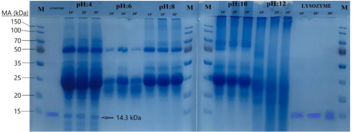
Fresh egg white 4.0, 6.0, 8.0, 10.0 and 12.0 were diluted with 5 different pH of pure water and the solutions were prepared and each solution was divided into 3 groups, each was kept at 100 °C for 10, 20 and 30 minutes. The samples were then electrophoresed in 12% SDS-PAGE. In electrophoretic findings, it was observed that only egg whites lysozymes treated at pH 4.0 yielded bands of 14.3 kDa in line with the commercial egg white lysozyme band loaded on the gel as a control (Figure 1). The density of the bands revealed no difference between the 10, 20 and 30 min treated samples. The same band was
208
not observed in egg white samples treated at other pHs. On the other hand, samples obtained by ethanol precipitation were loaded onto the gel but no lysozyme bands were observed as a result of staining of the gel.
Figure 1: Electrophoresis result of egg white lysozyme on SDS-PAGE (M: Marker) The Effect of Lysozyme Enzymes Isolated from Egg White on Bacterial Growth
Samples of egg whites containing lysozyme enzyme treated with distilled water were periodically instilled into LB-agar plates on which the B. subtilis bacteria were seeded by glass-smear method with commercial lysozyme used as a control and the optimum growth temperature of the bacteria was 37 °C until the next day were left to incubate. On the next day, although the growth of bacteria was observed on the whole plaque surface, it was observed that there was no bacterial growth in the regions where both the commercial lysozyme sample (control) and the egg white solution obtained during the study were dropped, and the bacteria could not grow lysed (Figure 2).
Figure 2: Image of LB-agar plates with B. subtilis bacteria in which lysozyme samples were dropped and bacterial growth was not observed (1: Commercial lysozyme enzyme instilled regions, 2: Egg
white solution instilled regions) DNA Isolation from Bacteria Using Egg White Solution
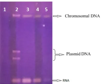
Using the egg white solution, it was investigated whether the DNA was isolated from B. subtilis bacteria. Commercial lysozyme was used as control, and no lysozyme was used in the other control. Electrophoresis results showed no DNA residue as expected in the sample where was used no lysozyme. In the sample using commercial lysozyme, genomic DNA and RNA bands were observed. Genomic
209
DNA, plasmid DNA and RNA bands were observed in electrophoresis in samples using egg white solution prepared within the scope of this study (Figure 3).
Figure 3: DNA isolation gel image using egg white solution (1: Control with no lysozyme added, 2: Plasmid DNA isolation using egg white solution, 3: Genomic DNA isolation using commercial lysozyme, 4-5: Egg white solution (Genomic DNA isolation using 100 and 50 µl
respectively)
DISCUSSION AND CONCLUSION
In the study, lysozyme enzyme was purified from egg whites by using pure water and it was investigated usability in laboratory studies. Thus, it is aimed to make a contribution to the national economy, albeit partially, by reducing the dependence on commercial lysozyme. Accordingly, white parts from the eggs obtained by commercial purchase were separated from the yolks as precisely as possible, and lysozyme enzymes were purified from these obtained egg whites using purified water. The method of dissolving egg whites in distilled water Wang et al. (1990). In this method, lysozyme enzyme could be obtained from egg whites treated under different pH conditions only from the sample treated at pH 4.0. Lysozyme enzyme was purified from egg white using different methods and it was determined enzymatic character (Alderton et al. 1945; Chang et al., 2000; Gemili et al., 2007; Krilova and Vitins, 2012; Luding et al., 2011; Rathnasamy et al., 2014; Rizwana et al, 2006; Safaric et al. Strang et al., 1984; Yan et al., 2011). Wang et al. (2009), after the purification at different pHs, they perform isolation in all pH values they work, although they reported that they perform lysozyme at pH 6.0. Wang et al. (2009) 's findings and the findings obtained in this study may be due to differences in breeding and/or differences in the feeding regimens of the chickens from which the eggs were obtained. This can only be demonstrated by using egg materials from chickens of different breeds using different feeding regimens. Moreover, the effect of the addition of lysozyme as a feed additive on egg lysozyme level can be investigated in an additional study using eggs obtained from chickens whose lysozyme enzyme has been added as a feed additive to their rations.
ACKNOWLEDGEMENTS
This study was supported by the Department of Scientific Research Projects of Çukurova University with the project number FBA-2016-5020. We would like to thank Çukurova University Rectorate and BAP Coordination Unit for their support.
210
Akınalp, AS., Asan, M., Ozcan, N. (2007). Expression of T4 Lysozyme Gene (gene e) in Streptococcus salivarius subsp. Thermophilus, African Journal of Biotechnology Vol. 6 (8): 963-966.
Alderton,G., Ward,W.H. ve Fevold,H.L.(1945). Isolation of Lysozyme from egg-white. J. Biol. Chem., 157:43-58.
Canfield, R.E. (1963) The amino acid sequence of egg White lysozyme. The Journal of Biological Chemistry, 288(8): 2698-2707.
Chang, H-M., Yang, C-C., Chang, Y-C. (2000). Rapid separation of lysozyme from chicken egg White by reductans and thermal treatment. J. Agric. Food. Chem, 48: 161-164.
Cutting, S.M., van der Horn, P.B., 1990. Molecular Biology Methods for Bacillus (Ed. By C.R. Harwood and S.M. Cutting), (pp. 27-74).
Gemili, S., Umdu, E.S., Yaprak, N., Üstok, F.I., Yener, F.Y.G., Mecitoğlu Güçbilmez, Ç., Alsoy Altınkaya, S., Yemenicioğlu, A. (2007). Partial Purification of Hen Egg White Lysozyme by Ethanol Precipitation Method and Determination of the Thermal Stability of Its Lyophilized Form. Turk. J. Agric. For., 31: 1-10.
Güzel, A. İ. (2000) Bakteriyofaj T4 lizozim geninin(GenE) plazmid vektörlerle klonlanması, Yüksek lisans tezi, Ç.Ü Sağlık Bilimleri Enstitüsü, Tıbbi Biyoloji A.B.D. danışman Prof. Dr. H. Kasap, Prof. Dr. N. Özcan), Adana, Türkiye.
Güzel, A.İ, Özcan,N., Kasap,H.(2002). bakteriyofaj T4 lizozim geninin PCR amplifikasyonu ile E. Coli'de klonlanması ve Expresyonu Turk., J. Vet. Anim. Sci.26:133-138.
Güzel,A.İ.,Özcan, N ve ark.(2008). Supression of tumerogenicty of B-16V melonoma cell via lysozyme gene. Cancer Bioterapy and Radiopharmaceuticals 23(5):603-608
Krilova, V., Vitins, V. (2012). Sorptive isolation of lysozyme from hen egg White. Material Science and Applied Chemistry, 25: 52-56
Laemmli,U.K. (1970). Cleavage of structural proteins during the assemly of the head of bacteriophage T4 Nature227:680-685.
Luding, Y., Shaochuan, S., Junxian, Y., Kejian, Y. (2011). Isolation of lysozyme from chicken egg White using polyacrylamide-based cation-exchange cryogel. Chinese Journal of Chemical Engineering, 19(5): 876-880.
Oberto, J., Davison, J. (1985). Expression of chicken egg white lysozyme by S. cerevisiae. Gene, 40: 57-65. Rathnasamy, S., Vedhahari, B.N., Kumaresan, R. (2014). Partition and purification of lysozyme from quail egg
white using aqueous two phase system. Biotechnology an Indian Journal, 9(4): 166-170.
Rizwana, I, Kite,J. et al. (2006) Affinity purification of hen egg lysozyme using sephadex G75. African Journal of Biology 5(20):1903-1908.
Safaric,I et al.(2007). Magnetik kation exchange ısolation of lysozyme from native hen Egg white. Biotechnol. 45(4):355-359.
Strang,R. H.(1984). purification of egg-white lysozyme by ion-exchange cromotography. Biochemical education,12(2):57-59.
Stryer,L.(1988). Biochemistry (3rd Ed.) ,W.H. Freeman and Company,,201-203.New York, USA.
Wang, W., Yue, H., Yuan, Q. (2009). A primary study on partial purification of lysozyme from chicken egg White using foam separation method. Biotechnology and Biotechnological Equipment, 23(2): 1237-1241. Yan, L. et al.(2011) Isolation of lysozyme from chicken egg white using polyacrilamide- based cation exchange