Contents lists available atScienceDirect
Clinical Neurology and Neurosurgery
journal homepage:www.elsevier.com/locate/clineuroCervico-medullary compression ratio: A novel radiological parameter
correlating with clinical severity in Chiari type 1 malformation
Ebru Doruk
a,c, Rafet Ozay
a,⁎, Zeki Sekerci
a,d, Hasan Ali Durmaz
b, Serra Ozbal Gunes
b,
Sahin Hanalioglu
a, Mehmet Sorar
aaMinistry of Health, Health Sciences University, Diskapi Yildirim Beyazit Training and Research Hospital, Department of Neurosurgery, Ankara, Turkey bMinistry of Health, Health Sciences University, Diskapi Yildirim Beyazit Training and Research Hospital, Department of Radiology, Ankara, Turkey cMinistry of Health, Health Sciences University, Bagcilar Training and Research Hospital, Istanbul, Turkey
dMedipol University Hospital, Department of Neurosurgery, Istanbul, Turkey
A R T I C L E I N F O
Keywords: Chiari malformation Herniated cerebellar tonsil Foramen magnum
Cervico-medullary compression ratio Syringomyelia
Hydrocephalus
A B S T R A C T
Objectives: Chiari malformation type 1 (CM-1) is associated with cough headache, intracranial hypertension, cerebellar and spinal cord symptoms/signs. Herniated cerebellar tonsil length (HCTL) is widely used radiological parameter to determine the severity of CM-1, but with limited utility due to its weak correlation with some clinico-radiologicalfindings. In this study, we aimed to evaluate a novel, practical parameter (cervico-medullary compression ratio;“CMCR”) for its relationship with clinico-radiological findings in CM-1.
Patients and methods: Thirty-five adult patients (17 F, 18 M) with CM-1 were included in this retrospective study. Head CT and craniospinal MR images were assessed. CMCR was calculated as the ratio of herniated cerebellar tonsil surface area to foramen magnum surface area, and HCTL was measured. These two parameters were correlated with clinical and radiologicalfindings.
Results: The mean CMCR was 0.60 ± 0.15 and mean HCTL was 8.91 ± 3.4 mm with no significant difference between gender and age groups for both parameters. For cough headache (0.64 ± 0.14 vs 0.52 ± 0.15, p = 0.043) and syringomyelia (0.67 ± 0.11 vs 0.56 ± 0.16, p = 0.039), only CMCR; for intracranial hy-pertension (CMCR: 0.64 ± 0.14 vs 0.55 ± 0.16, p = 0.049; HCTL: 9.66 ± 3.59 mm vs 7.79 ± 3.03 mm; p = 0.045) and cerebellar symptoms (CMCR: 0.65 ± 0.14 vs 0.54 ± 0.16, p = 0.048; HCTL: 10.4 ± 3.5 mm vs 7.4 ± 2.8 mm, p = 0.041), both CMCR and HTCL were significantly different between patients with and without respectivefindings. However, neither CMCR nor HTCL was different between patients with and without spinal cord symptoms and hydrocephalus.
Conclusion: CMCR is a superior numerical parameter than HCTL for the assessment of clinical severity in CM-1 cases and needs further validation with larger studies.
1. Introduction
Chiari malformation type 1 (CM-1) has been described as the her-niation of the cerebellar tonsils from the foramen magnum (FM) to the cervical spinal canal and its prevalence in adult population is as high as 1% [1,2]. It might be associated with congenital pathologies, such as basilar invagination, platybasia, craniosynocytosis, posterior fossa hy-poplasia, atresis of foramina of Magendie and Luschka, larger cerebellar structures and clival anomalies [3–5]. Congenital biomechanical de fi-ciencies as well as secondary or iatrogenic factors such as trauma, hy-drocephalus, tethered cord and intraspinal hypotension are blamed in the etiology [5–11]. Most cases are usually asymptomatic in the early
period. Symptoms begin to emerge between the ages of 25–40, and they could progress from mild headaches and dysesthesias to respiratory failure and tetraplegia [1–4].
Herniated cerebellar tonsil length (HCTL) should be at least 5 mm below the FM for the CM-1 diagnosis [2,7]. However, HCTL for symptomatic CM-1 could be as little as 2–3 mm [12,13]. On the other hand, several studies suggested a correlation between HCTL and the severity of clinical symptoms [12–15]. CM-1 severity has been classified according to HCTL: mild: 5–9 mm, moderate: 9–14 mm, and severe: > 14 mm [2,7,14]. However, this view is still controversial and HCTL is thought to be inadequate to determine neither response to nor timing of the surgical treatment for CM-1 [15–18]. Interestingly, nearly 20% of
https://doi.org/10.1016/j.clineuro.2018.09.016
Received 3 April 2018; Received in revised form 6 September 2018; Accepted 9 September 2018
⁎Corresponding author at: Ministry of Health, University of Health Sciences, Diskapi Yildirim Beyazit Training and Research Hospital, Department of Neurosurgery, Diskapi, 06110, Altindag/Ankara, Turkey.
E-mail address:[email protected](R. Ozay).
Available online 10 September 2018
0303-8467/ © 2018 Published by Elsevier B.V.
cases with HCTL greater than 5 mm, even with HCTL up to 27 mm, might be asymptomatic [15,16]. Inverse relationship between age and tonsil length has also been reported such that the size of the herniation diminishes but the clinicalfindings progress as the age increases [17]. As a result, there was no consensus about the relationship between the clinical symptoms of CM-1 cases and numerical value of HCTL and hence conquest for a better clinical and/or radiological marker still continues [18].
Classical headache of CM-1 is the most common symptom and de-scribed as cough headache because it increases during a Valsalva maneuver. This situation is due to the pressure gradient between the cranial and spinal compartments induced by the maneuver [18–20]. Later, progressive pathophysiological process ensues and results in bulbar, cerebellar and spinal cord symptoms and signs including par-esthesia, dyspar-esthesia, motor disturbances, trigeminal and glossophar-yngeal neuralgia, hemifacial spasm, phonation disorders, sleep apnea, swallowing difficulty, ataxia, dysmetria and dysdiadokokinesia, sensory discrimination, sphincter insufficiency and impotence [6,8,11,21–28]. Thesefindings are thought to be related to ischemia and syringomyelia/ syringobulbia, especially at the level of foramen magnum (FM), re-sulting from tonsillar herniation [8,21]. Progressive nature of CM-1 has also been reflected in recently described Chiari Severity Index [18]. In conclusion, all theories attempting to explain clinical symptomatology in CM-1 patients suggest that the etiopathogenesis is closely related to the pressure on the cervicomedullary compartment at the cranio-vertebral junction.
In this study, we evaluated a possible relationship between clinico-radiologicalfindings of patients and a novel, simple numerical para-meter (“cervicomedullary compression ratio” at the FM level). In ad-dition, the results obtained were compared with HCTL values that had been previously validated and widely used in the literature.
2. Patients and methods 2.1. Study design and data collection
This study has been conducted at Diskapi Yildirim Beyazit Training and Research Hospital, Ankara, Turkey upon the approval by the Ethics Committee for Clinical Investigations (Date: 27/04/2015, No: 22/14). A total of 35 patients who were treated at our institution between April 2013 and April 2015 were included and retrospectively evaluated in this study (17 females, 18 males; mean age: 37, range: 18–65 years). Patients were divided into 5 groups according to their age (18–25, 26–35, 36–45, 46–55, 56–65) (Table 1). Patients with systemic or other congenital pathologies have been excluded from the study. Demo-graphic groups of patients participating in the study were homo-geneous. Magnetic resonance imaging (MRI) and computed tomo-graphy (CT) data and clinicalfindings were obtained retrospectively from the archives of the Departments Radiology and Neurosurgery. Demographic characteristics of the patients were given in theTable 1.
2.2. Measurement and calculation of CMCR
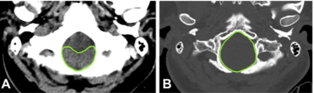
Topographic area calculations were performed on axial head CT images of the patients (Toshiba Alexion TSX-034A), at the level of FM opening in the craniovertebral junction. Section properties Pitch factor 0.688, helical pitch value 11. Average 120 kV, 110 mas, rotation time 0.75, FOV 240 were used. Images were obtained from CT images with 1 mm cross-sectional thickness and 0.8 mm cross-section intervals. Foramen magnum surface area (FMSA) and herniated cerebellar tonsil surface area (HCTSA) were measured as cm2 at the bone and par-enchymal windows (General Electric AW volume Share 5 workstation), respectively. The ratio of the HCTSA to the FMSA (HCTSA/FMSA) was calculated and described as the CMCR (Fig. 1A, B) (Table 1).
2.3. Measurement of the HCTL
Herniated cerebellar tonsil length (HCTL) below the FM was ob-tained from mid-sagittal T2W MRI images. All MRI was performed on 1.5 T scanner (Philips Intera, Philips Medical Systems, Best, Netherlands) with a multi-channel cervical-thoracic-lumbar spine coil and included the following sequence: sagittal T2-weighted image (TR/ TE = 3000–4000 ms/70–80 ms, FOV = 24 cm, matrix 480 × 480, sec-tion thickness/gap = 4/1 mm). Measurements were made using mid-sagittal MRI sections on the PACS system (K-PACS, Kuratorium OFFIS e.V. Healthcare Information and Communication Systems, Oldenburg/ Germany). The McRae line (from basion to opisthion at the level of the FM) was considered as the reference point. HCTL was measured on a direct line from McRae line to the tip of herniated tonsil and determined as mm (Fig. 2A). According to the severity of HCTL, patients were di-vided into 4 groups: 2–5 mm (suspicious), 5–9 mm (mild), 10–14 mm (moderate), and more than 14 mm (severe) (Table 2) [8].
2.4. Clinical evaluation of symptom and signs
The presence of headache aggravated by cough was regarded as cardinal symptom of CM-1. Triad of cough headache, nausea/vomiting and papillary edema were accepted as increased intracranial pressure syndrome (IIPS). The presence of any of the findings of dysmetria, dysdiadokokinesia, and nystagmus was determined as cerebellar in-volvement (ataxia was excluded because it may also be due to syr-ingomyelia). Pain-temperature discrimination of the extremities and/or presence of motor weakness were considered as spinal cord symptoms. Clinical evaluation results were marked as either present or absent, and the patients were grouped accordingly. Clinical signs were correlated with CMCR and HCTL (Table 3).
2.5. Radiological evaluation of hydrocephalus and syringomyelia The presence of hydrocephalus and syringomyelia were evaluated on the parenchymal axial head CT or MR images (for hydrocephalus; temporal horn width≥2 mm or Evan’s ratio > 30%) and on sagittal T2 cervical-thoracic MRI sections (for syringomyelia) (Fig. 2B). The results were recorded as either present or absent, and the patients were Table 1
Distribution of radiological measurements according to demographic char-acteristics. Demographic characteristics HCTSA (cm2) FMSA (cm2) CMCR HCTL (mm) Age 18–25 yo (n = 9) 5.47 ± 1.31 8.47 ± 1.57 0.58 ± 0.14 8.42 ± 3.77 26–35 yo (n = 10) 4.35 ± 1.34 8.51 ± 1.22 0.62 ± 0.12 7.75 ± 2.37 36–45 yo (n = 5) 5.60 ± 1.26 10.30 ± 1.02 0.68 ± 0.16 7.96 ± 1.96 46–55 yo (n = 6) 5.66 ± 1.42 8.19 ± 0.89 0.59 ± 0.11 12.03 ± 4.51 56–65 yo (n = 5) 5.40 ± 1.32 7.89 ± 1.61 0.64 ± 0.15 9.78 ± 3.82 P value: 0.578 0.126 0.432 0.446 Gender Female (n = 17) 4.98 ± 1.13 7.90 ± 1.34 0.63 ± 0.13 9.18 ± 3.93 Male (n = 18) 5.40 ± 1.52 9.35 ± 1.26 0.57 ± 0.18 8.63 ± 2.98 P value: 0.378 0.002* 0.302 0.654 TOTAL (n = 35) 5.18 ± 1.35 8.60 ± 1.45 0.60 ± 0.15 8.91 ± 3.68
CMCR: cervico-medullary compression ratio calculated as the ratio of HCTSA to FMSA; FMSA: foramen magnum surface area; HCTL: herniated cerebellar tonsil length; HCTSA: herniated cerebellar tonsil surface area.
grouped accordingly. Whether the results were correlated with CMCR and HCTL was investigated (Table 3).
2.6. Statistical analysis
After all data were obtained, IBM SPSS Statistics Version 22 for Windows was used for analysis. Frequency analysis was used for de-mographic analysis. The descriptive statistics were analyzed as mean ± S.D., minimum, maximum and percentage (%). The Kolmogorov–Smirnov test was used to determine the normalization of data. One-way ANOVA and Kruskal–Wallis tests were used for com-parisons between multiple groups while Student t-test and Mann–Whitney U tests were used to compare the means of two groups with parametric and non-parametric variables, respectively. Spearman's correlation test was used to compare the correlation between con-tinuous numerical variables and grouping variables. The value of p < 0.05 was considered to be statistically significant.
3. Results
3.1. Measurements of HCTSA and FMSA and calculation of CMCR The average HCTSA was 5.18 ± 1.35 cm2. No statistically sig-nificant difference was found between genders (F: 4.98 ± 1.13 vs M: 5.40 ± 1.52, p = 0.378). In addition, there was no statistically sig-nificant difference between age groups (p = 0.578) (Table 1). The average FMSA was found to be 8.60 ± 1.45 cm2. When genders are compared, it was found that FMSA in males was significantly higher than females (F: 7.90 ± 1.34 vs M: 9.35 ± 1.26), p = 0.002). How-ever, there was no significant difference between age groups (p = 0.126) (Table 1). When the CMCR (HCTSA/FMSA ratio) was ex-amined, the mean value was 0.60 ± 0.15, there was no statistically significant difference between gender and age groups (F:0.63 ± 0,13vs M:0.57 ± 0.18, p = 0.302) (For age groups; p = 0,432) (Table 1).
3.2. Measurement of HCTL
The average HCTL was 8.91 ± 3.4 mm. There was no statistical difference between male and female patients in terms of HCTL (F: 9.18 ± 3.93 vs M:8.63 ± 2.98, p = 0.654). When age groups were compared for the HCTL, it was found that there was no statistically significant difference (p = 0.446). As the severity of HCTL increased, the CMCR also increased gradually, but the difference between groups were not significant (p = 0.435) (Table 2).
3.3. Clinical evaluation of symptoms and signs
Mean CMCR of patients with cough headache (n = 24) was sig-nificantly higher than patients without headache (n = 11) (0.64 ± 0.14 vs 0.52 ± 0.15, p = 0.043). However, when the HCTL was examined, there was no statistically significant difference between the patients with and without cough headache (mean values for HCTL; 8.99 ± 3.66 mm vs 8.74 ± 3.06 mm, respectively; p = 0.835). There were significant differences in terms of both CMCRs and HCTLs of 21 patients who had IIPS, and 14 patients without IIPS (mean values for CMCR: 0.64 ± 0.14 vs 0.55 ± 0.16, p = 0.049, respectively; mean values for HCTL were 9.66 ± 3.59 mm vs 7.79 ± 3.03 mm, Fig. 1. Radiological measurements at the level of foramen magnum on axial CT images. A: Herniated cerebellar tonsil surface area measured on parenchymal window of head CT; B: Foramen magnum surface area measured on bone window of head CT.
Fig. 2. Radiological measurements and syr-ingomyelia on mid-sagittal T2W MR images. A: McRae line from basion to opisthion (dashed line) and herniated cerebellar tonsil length measured between McRae line and the tip of herniated tonsil (uninterrupted line); B: Arrow shows syringomyelia in cervical mid-sagittal T2W MR image.
Table 2
Cervico-medullary compression ratio in different cerebellar tonsil herniation severity groups based on HCTL. Although there is a trend of increase in CMCR with the extent of her-niation (severity), difference among severity groups is not statistically significant (p = 0.435).
Severity (HCTL) CMCR
Suspicious (2–5 mm) 0.57 ± 0.13 Mild (5–9 mm) 0.59 ± 0.14 Moderate (10–14 mm) 0.63 ± 0.15 Severe (≥14 mm) 0.63 ± 0.14
CMCR: cervico-medullary compression ratio; HCTL: herniated cerebellar tonsil length.
respectively; p = 0.045). Similarly, both parameters were found to have statistically higher values in patients with cerebellar symptoms (n = 19) compared to patients without cerebellar symptoms (n = 16) (mean values for CMCR: 0.65 ± 0.14 vs 0.54 ± 0.16, p = 0.048, and mean values for HCTL: 10.4 ± 3.5 mm vs 7.4 ± 2.8 mm, p = 0.041, respectively). Although CMCR was more sensitive compared to HCTL measurement in patients with (n = 17) and without (n = 18) spinal cord symptoms, this sensitivity was not found to be statistically sig-nificant (mean CMCR: 0.65 ± 0.13 vs 0.56 ± 0.16, p = 0.126; mean HCTL: 9.07 ± 3.2 mm vs 8.78 ± 3.7 mm, p = 0.83, respectively) (Table 3).
3.4. Radiological evaluation of hydrocephalus and syringomyelia The mean CMCR of patients with hydrocephalus (n = 9) was 0.66 ± 0.15, while the mean value of those without hydrocephalus (n = 26) was 0.58 ± 0.15, with no statistically significant difference between the two groups (p = 0.17). Similarly, there was no statistically significant difference in HCTL (mean HCTL of patients with and without hydrocephalus was 10.47 ± 3.34 mm vs 8.37 ± 3.4 mm, p = 0.128). The mean CMCR of patients with syringomyelia (n = 14) was 0.67 ± 0.11, while those without syringomyelia (n = 21) were found to be 0.56 ± 0.16, p = 0.039). However, there was no statistically significant difference between the two groups in terms of HCTL (mean HCTL of patients with and without syringomyelia was 9.45 ± 3.4 mm vs 8.55 ± 3.5 mm, p = 0.454) (Table 3).
4. Discussion
In this study, we calculated the ratio of herniated cerebellar tonsil surface area to foramen magnum surface area (cervico-medullary compression ratio, CMCR) on computed tomography images of Chiari malformation type 1 (CM-1) and evaluated its correlation with clinical symptoms and signs of CM-1 patients. We also critically analyzed clinical and radiological results with CMCR in comparison to widely used, albeit controversial, herniated cerebellar tonsil length on mid-sagittal T2 weighted magnetic resonance images.
Foramen magnum (FM), which connects cranial cavity and ver-tebral canal, is composed of the posterior part of the skull, basilar, lateral and squamous parts of the occipital bone [29,30]. FM is the largest of foramina at the skull base (mean diameter; from anterior to posterior: 30–40 mm, transverse: 30–35 mm), which is bordered by basion in the front, opisthion in the posterior and occipital condyles on both sides [29–32]. Medulla oblongata (MO), vertebral arteries, ante-rior and posteante-rior spinal arteries, and spinal roots of nervus accessorius pass through the FM [29,31]. Even if it has different shapes and sizes, it has a standard average effective diameter for critical structures within
itself such as MO. In a classical study, it was reported that the effective diameter of FM should be at least 19 mm [33]. In the presence of pathologies that reduce the effective diameter such as basilar in-vagination, tumour, infection, tonsillar herniation, foraminal stenosis and craniodiaficial dysplasia, neurological deficits occur due to com-pression caused by these pathologies to MO [33–41]. For these pathologies, surgical strategies have been developed such as transoral odontoidectomy (for anteriorly compressing pathologies) or FM pos-terior wall resection (for pospos-teriorly compressing pathologies) aimed at alleviating the stress on the brain stem to counteract the etiopatho-genesis [35–39].
When the etiopathogenesis of pathologic findings in Chiari mal-formation is examined, one encounters a chain of problems particularly caused by spatial compression at the level of FM rather than vertical elongation of cerebellar tonsils per se. However, in most studies con-ducted until now, findings in relation to anterior or posterior com-pression have only been evaluated with single dimension (i.e. diameter) but not two dimensions (i.e. area) [33]. For this reason, in our study regarding CM-1 cases, we aimed to investigate a numerical value (i.e. CMCR) which better explains the severity of the compression to brainstem. We anticipated that these values should be correlated with the clinical and radiologicalfindings in CM-1 cases. Furthermore, we evaluated whether CMCR is correlated with HTCL, a commonly used measure in CM-1 studies.
In the past, FMSA was calculated in people from different ethnic backgrounds in different countries using radiographic images such as MR and CT or data obtained from cadaver and skull, and similar values were obtained in the case of cross-sectional differences. In males and females, mean FMSA was found to be 829–909 mm2and 781–819 mm2, respectively [2,41–45]. In our study, the FMSA results were 935 ± 120 mm2 for males and 760 ± 130 mm2 for females (total average: 860 ± 145 mm2), similar to literature findings. In order to eliminate discrepancies between studies, HCTSA/FMSA ratio, termed as "cervico-medullary compression ratio (CMCR)", which is independent of these variables, was used in our study. In addition, it was examined whether this radiological value was independent of developmental, sexual and genetic differences for the reliability of the study and it was found that there were no statistically significant differences between the age and gender groups (Table 1). To the best of our knowledge, there are no other studies in the English literature where the HCTSA measurement is performed and the HCTSA/FMSA ratio is calculated.
Patients with CM-1 suffer from a number of symptoms that result from the pressure of the MO, cervical spinal cord and cranial nerves [11,18]. In fact, the most common accompanying symptom in both congenital and acquired CM-1 is cough headache which is exacerbated by Valsalva maneuvers or physical exertions and felt in the suboccipital area [11,19,20]. Various authors suggested that headaches occurred Table 3
Association of clinical and radiologicalfindings with CMCR and HCTL.
CMCR P value HCTL (mm) P value
Clinical symptom and signs present absent present absent
Cough Headache 0.64 ± 0.12 0.52 ± 0.12 0.043* 8.99 ± 2.95 8.74 ± 3.12 0.835
IIPS 0.64 ± 0.14 0.55 ± 0.16 0.049* 9.66 ± 3.59 7.79 ± 3.03 0.045*
Cerebellar symptom and signs 0.65 ± 0.14 0.54 ± 0.16 0.040* 10.40 ± 3.50 7.46 ± 2.80 0.040*
Spinal cord symptom and signs 0.65 ± 0.13 0.56 ± 0.16 0.080 9.07 ± 3.20 8.78 ± 3.70 0.830
CMCR P value HCTL (mm) P value
Radiologicalfindings present absent present absent
Hydrocephalus 0.66 ± 0.15 0.58 ± 0.15 0.170 10.47 ± 3.34 8.37 ± 3.40 0.128 Syringomyelia 0.67 ± 0.11 0.56 ± 0.16 0.039* 9.45 ± 3.34 8.55 ± 3.57 0.454 CMCR: cervico-medullary compression ratio; HCTL: herniated cerebellar tonsil length; IIPS: increased intracranial pressure syndrome.
due to sudden increase in CSF pressure and the tension of the veins or meninges which include pain-sensitive nerve dendrites induced by displacement of the tonsils during a cough or Valsalva action [11,20,45,46]. In our study, the CMCR of cases which have cough headache was statistically significantly higher than the cases without cough headache (0.64 ± 0.14 vs 0.52 ± 0.14, respectively; p = 0.043). On the other hand, HCTL failed to distinguish the patients with and without cough headache (8.99 ± 3.66 mm vs 8.74 ± 3.06 mm, respectively; p = 0.835). Our results suggest that, contrary to what Pascual et al. [19] reported, the cough headache is not directly correlated with HCTL. A herniated cerebellar tonsil may indeed increase intracranial pressure that leads to cough headache before se-vere ischemia and resultant atrophy develop. One can assume that as the CMCR, rather than HCTL, increases, the CSF flow obstruction should become more severe.
In our study, headache, nausea-vomiting attacks and papillary edema were accepted as IIPS. Even if hydrocephalus was reported oc-casionally (10–20%) in the CM-1 cases, it is argued to be one of most important etiopathogenetic factors of headache, whose prevalence is much higher [22,27,33]. However, it is known that CM-1 could produce idiopathic intracranial hypertension due to diffuse intracerebral edema without radiologically diagnosed hydrocephalus [46,47]. The theory was supported in CM-1 cases by acetazolamide treatment which leads to decrease in the degree of tonsillar herniation [47]. In our study, it was found that the CMCR of the patients who were diagnosed with IIPS was significantly higher than the results of patients with no IIPS (0.64 ± 0.14 vs 0.55 ± 0.16, respectively; p = 0.049). However, there was no statistically significant difference for hydrocephalus (0.66 ± 0.15 vs 0.58 ± 0.15, respectively; p = 0.173). Same results were obtained for the HCTL values (HCTL for IIPS: 9.66 ± 3.59 mm vs 7.79 ± 3.03 mm; p = 0.045, HCTL for hydrocephalus: 10.47 ± 3.34 mm vs 8.37 ± 3.4 mm; p = 0.128). According to our results, the CMCR might be used as an efficient numerical factor for the decision of surgical treatment in CM-1 cases regardless of the presence of hydrocephalus.
In the literature, different morphometric measurements have been performed on radiological imaging of CM-1 cases for determining prognosis and survival of cases [48–53]. Dufton et al. reported that the clivus length was shorter and the FM was wider in CM-1 cases than normal healthy subjects [48]. In similar studies, increased tentorial angle, expanded basal opening, decreased depth of posterior fossa and smaller diameter of posterior fossa [49–52]. It has been thought that while cerebellar tissues naturally develop, the bony structures of pos-terior fossa could not comply with this development [50,50,51,52]. Interestingly, a recent study found no clinically useful 2D or 3D radi-ological measurements to reliably distinguish symptomatic and asymptomatic CM-1 cases in children [53]. However, none of these studies have focused on cerebellar impairment which is one of the main sources of clinical symptoms resulting from compressive and ischemic damage following tonsillar herniation. In our study, both CMCR and HCTL of cases were significantly higher in patients with impaired cer-ebellar tests than those without (For CMCR: 0.65 ± 0.14 vs 0.54 ± 0.16, respectively, p = 0.048; for HCTL: 10.4 ± 3.5 mm vs 7.4 ± 2.8 mm, respectively, p = 0.041). It is possible to say according to our results that the disruption of cerebellar functions in CM-1 cases is not only due to cerebellar compression caused by small posterior fossa, but may also be due to compression on spinocerebellar or cere-bellospinal tracts at the craniovertebral junction.
It is stated that syringomyelia causes scoliosis, pain, sensation dis-order (especially pain-temperature discrimination), weakness and ataxia in CM-1 cases [8,21,54]. Recent studies have demonstrated with cine-mode MR images that compliance at both spinal and intracranial CSF sites decreases in the presence of syringomyelia in CM-1 cases [55–57]. As a result, increased CSF pressure will cause posterior spinal venous pressure to increase, which in turn disturbs the absorption of CSF to the intravenous compartment. The inhibition of CSFflow results
in the rupture of the central canal and formation of syrinx. Various authors believe that this condition is triggered by restriction of the subarachnoid space by herniated cerebellar tonsils at the cranio-vertebral junction [56–58]. However, to the best of our knowledge, there are no quantitative studies in the literature investigating severity of restricted subarachnoid space at the level of FM. In our study, it was observed that the CMCR was more sensitive than HCTL measurement in CM-1 cases with spinal cord symptoms, but there was no statistically significant difference in terms of both parameters. However, when CMCR of patients with and without syringomyelia were examined, it was found that there was a significant difference between the two groups; the higher the ratio is, the more commonly syringomyelia is seen (Table 3). In addition, there was no statistically significant dif-ference between the groups despite the fact that patients with syr-ingomyelia had higher HCTLs than those without (Table 3). While syringomyelia is diagnosed in a case whose HTCL values is 3.16 mm, it was not diagnosed in two other patients whose HTCL values were 16.3 and 13.8 mm [12–15]. Our study demonstrated on the contrary to what Milhorat et al. [14], Stovner et al. [45] and Godzik et al. [54] claimed that there is not a certain relation between syringomyelia and HTCL values, and CMCR ratio is a more sensitive value.
Due to retrospective design of the study as well as limited study population with only surgically treated cases, this study needs further confirmation. Future studies with larger cohorts and repeated CMCR measurements during longer follow-up periods are needed. By super-imposing CT and MRIfindings, CMCR measurements can be refined and thus radiologicalfindings can be better correlated with disease specific symptoms and CSFflow perturbation in future studies.
5. Conclusions
Our study results show that HCTSA/FMSA ratio or CMCR is a su-perior numerical value than HCTL for clinical severity in CM-1 cases. CMCR successfully distinguished patients with cough headache, in-creased intracranial pressure syndrome, cerebellar signs and syr-ingomyelia. CMCR is a promising radiological marker that can possibly be used for determining prognosis and selecting patients for surgical treatment.
Funding
This research did not receive any specificgrant from funding agen-cies in the public, commercial, or not-for-profit sectors.
References
[1] E. Schijman, History, anatomic forms, and pathogenesis of Chiari I malformations, Childs Nerv. Syst. 20 (5) (2004) 323–328, https://doi.org/10.1007/s00381-003-0878-y.
[2] M.W. Vernooij, M.A. Ikram, H.L. Tanghe, A.J. Vincent, A. Hofman, G.P. Krestin, W.J. Niessen, M.M. Breteler, A. van der Lugt, Incidentalfindings on brain MRI in the general population, N. Engl. J. Med. 357 (18) (2007) 1821–1828,https://doi.org/ 10.1056/NEJMoa070972.
[3] T.H. Milhorat, M. Nishikawa, R.W. Kula, Y.D. Dlugacz, Mechanisms of cerebellar tonsil herniation in patients with Chiari malformations as guide to clinical man-agement, Acta. Neurochir. (Wien) 152 (7) (2010) 1117–1127,https://doi.org/10. 1007/s00701-010-0636-3.
[4] G.N. Dyste, A.H. Menezes, J.C. Van Gilder, Symptomatic chiari malformations. An analysis of presentation, management, and long term outcome, J. Neurosurg. 71 (2) (1989) 159–168,https://doi.org/10.3171/jns.1989.71.2.0159.
[5] J.L.D. Atkinson, E. Kokmen, G.M. Miller, Evidence of posterior fossa hypoplasia in the familial variant of adult Chiari I malformation: case report, Neurosurgery 42 (2) (1998) 401–404,https://doi.org/10.1097/00006123-199802000-00129. [6] K.S. Paul, R.H. Lye, F.A. Strang, J. Dutton, Arnold Chiari malformation, J.
Neurosurg. 58 (2) (1983) 183–187,https://doi.org/10.3171/jns.1983.58.2.0183. [7] A.D. Elster, M.Y. Chen, Chiari 1 malformations: clinical and radiologic reappraisal,
Radiology 83 (2) (1992) 347–353,https://doi.org/10.1148/radiology.183.2. 1561334.
[8] W.J. Oakes, Chiari malformations, hydromyelia, syringomyelia, Neurosurgery 3 (1996) 3411–3418.
Case report, Neurosurgery 32 (2) (1993) 306–309.
[10] T.J. Buell, J.D. Heiss, E.H. Oldfield, Pathogenesis and cerebrospinal fluid hydro-dynamics of the Chiari I malformation, Neurosurg. Clin. N. Am. 26 (4) (2015) 495–499,https://doi.org/10.1016/j.nec.2015.06.003.
[11] P.P. Huang, S. Constantin,“Acquired” Chiari I malformation. Case report, J. Neurosurg. 80 (6) (1994) 1099–1102,https://doi.org/10.3171/jns.1994.80.6. 1099.
[12] P.K. Pillay, L.A. Awad, J.R. Little, J.F. Hahn, Symptomatic Chiari malformation in adults: a new classification based on magnetic resonance imaging with clinical and prognostic significance, Neurosurgery 28 (5) (1991) 639–645,https://doi.org/10. 1227/00006123-199105000-00001.
[13] J. Sahuquillo, E. Rubio, M.A. Poca, A. Rovira, A. Rodriguezbaeza, C. Cervera, posterior fossa reconstruction: a surgical technique for the treatment of Chiari I malformation and Chiari I syringomyelia complex. Preliminary results and mag-netic resonance imaging quantitative assessment of hindbrain migration, Neurosurgery 35 (5) (1994) 874–884, https://doi.org/10.1227/00006123-199411000-00011.
[14] T.H. Milhorat, M.W. Chou, E.M. Trinidad, R.W. Kula, M. Mandell, C. Wolpert, et al., Chiari 1 malformation redefined: clinical and radiographic findings for 364 symp-tomatic patients, Neurosurgery 44 (5) (1999) 1005–1017,https://doi.org/10.1097/ 00006123-199905000-00042.
[15] H. Sakamoto, M. Nishikawa, A. Hakuba, T. Yasui, S. Kitano, N. Nakanishi, et al., Expansive suboccipital cranioplasty for the treatment of syringomyelia associated with Chiari malformation, Acta Neurochir. (Wien) 141 (9) (1999) 949–961,https:// doi.org/10.1007/s007010050401.
[16] J. Meadows, M. Kraut, M. Guarnieri, R.I. Haroun, B.S. Carson, Asymptomatic Chiari type I malformations identified on magnetic resonance imaging, J. Neurosurg. 92 (6) (2000) 920–926,https://doi.org/10.3171/jns.2000.92.6.0920.
[17] H. Sabuncuoğlu, I.S. Keskil, An overlooked reason in adult upper cervical spinal cord compression: type 1 Chiari malformation [Turkish], J. Turk. Spinal Surg. 17 (2006) 17–23.
[18] J.K. Greenberg, C.K. Yarbrough, A. Radmanesh, J. Godzik, M. Yu, D.B. Jeffe, et al., The Chiari Severity Index: a preoperative grading system for Chiari malformation type 1, Neurosurgery 76 (3) (2015) 279–285,https://doi.org/10.1227/NEU. 0000000000000608.
[19] J. Pascual, A. Oterino, J. Berciano, Headache in type 1 Chiari malformation, Neurology 42 (8) (1992) 1519–1521.
[20] C.A. Sansur, J.D. Heiss, H.L. DeVroom, E. Eskioğlu, R. Ennis, E.H. Oldfield, Pathophysiology of headache associated with cough in patients with Chiari I mal-formation, J. Neurosurg. 98 (3) (2003) 453–458,https://doi.org/10.3171/jns. 2003.98.3.0453.
[21] T.H. Milhorat, R.M. Kotzen, H.T.M. Mu, A.L. Capocelli, R.H. Milhorat, Dysesthetic pain in patients with syringomyelia, Neurosurgery 38 (5) (1996) 940–946,https:// doi.org/10.1097/00006123-199605000-00017.
[22] P. Rosetti, R.O. Ben Taib, J. Brotchi, O. De Witte, Arnold Chiari type 1 malformation presenting as a trigeminal neuralgia: case report, Neurosurgery 44 (5) (1999) 1122–1124,https://doi.org/10.1097/00006123-199905000-00105.
[23] M.E. Colpan, Z. Sekerci, Chiari type 1 malformation presenting as hemifacial spasm: case report, Neurosurgery 57 (2) (2005) E371,https://doi.org/10.1227/01.NEU. 0000166688.69081.8B.
[24] K. Hida, Y. Iwasaki, I. Koyanagi, Y. Sawamura, H. Abe, Surgical indications and results of foramen magnum decompression versus syringosubarachnoid shunting for syringomyelia associated with Chiari I malformation, Neurosurgery 37 (4) (1995) 673–678,https://doi.org/10.1227/00006123-199510000-00010. [25] C. Eggers, J. Hamer, Hydrosyringomyelia in childhood. Clinical aspects,
patho-genesis and therapy, Neuropadiatrie 10 (1) (1979) 87–99,https://doi.org/10.1055/ s-0028-1085317.
[26] Y. Kanpolat, A. Unlu, A. Savas, F. Tan, Chiari type 1 malformation presenting as a glossopharyngeal neuralgia: case report, Neurosurgery 48 (1) (2001) 226–228,
https://doi.org/10.1007/s101430100178.
[27] I. Koyanagi, K. Houkin, Pathogenesis of syringomyelia associated with Chiari type 1 malformation: review of evidences and proposal of a new hypothesis, Neurosurg. Rev. 33 (3) (2010) 271–284,https://doi.org/10.1007/s10143-010-0266-5. [28] E.H. Oldfield, K. Muraszko, T.H. Shawker, N.J. Patronas, Pathophysiology of
syr-ingomyelia associated with Chiari I malformation of the cerebellar tonsils. Implications for diagnosis and treatment, J. Neurosurg. 80 (1) (1994) 3–15,https:// doi.org/10.3171/jns.1994.80.1.0003.
[29] S. Standring (Ed.), Gray’s Anatomy: The Anatomical Basis of Clinical Practice, 41st ed., Elsevier, 2016, pp. 711–714.
[30] J. Lang, O. Schafhauser, S. Hoffmann, Postnatal development of transbasal skull openings: carotid canal, jugular foramen, hypoglossal canal, condylar canal and foramen magnum [German], Anat. Anz. 153 (4) (1983) 315–357.
[31] E. Avci, A. Dagtekin, A.H. Ozturk, E. Kara, N.C. Ozturk, K. Uluc, et al., Anatomical variations of the foramen magnum, occipital condyle and jugular tubercle, Turk. Neurosurg. 21 (2) (2011) 181–190, https://doi.org/10.5137/1019-5149.JTN.3838-10.1.
[32] S.H. Zaidi, S.S. Dayal, Variations in the shape of foramen magnum in Indian skulls, Anat. Anz. 167 (4) (1988) 338–340.
[33] R. Fischbein, J.R. Saling, P. Marty, D. Kropp, J. Meeker, J. Amerine, et al., Patient-reported Chiari malformation type I symptoms and diagnostic experiences: a report from the National Conquer Chiari Patient Registry database, Neurol. Sci. 36 (9) (2015) 1617–1624,https://doi.org/10.1007/s10072-015-2219-9.
[34] D.P. Noske, B.J. van Royen, J.L. Bron, W.P. Vandertop, Basilar impression in
osteogenesisimperfecta: can it be treated with halo traction and posterior fusion? Acta Neurochir. (Wien) 148 (12) (2006) 1301–1305,https://doi.org/10.1007/ s00701-006-0870-x.
[35] D. Li, Z. Wu, C. Ren, S.Y. Hao, L. Wang, X.R. Xiao, J. Tang, et al., Foramen magnum meningiomas: surgical results and risks predicting poor outcomes based on a modified classification, J. Neurosurg. 126 (3) (2017) 661–676,https://doi.org/10. 3171/2016.2.JNS152873.
[36] T.C. Burns, S.A. Mindea, A.V. Pendharkar, N.B. Lapustea, I. Irime, J.V. Nayak, Endoscopic transnasal approach for urgent decompression of the craniocervical junction in acute skull base osteomyelitis, J. Neurol. Surg. Rep. 76 (1) (2015) 7–42,
https://doi.org/10.1055/s-0034-1395492.
[37] R. Özay, E.D. Doruk, M.S. Balkan, M.F. Ergüngör, Tratment of craniodiaphyseal dysplasia presenting with Chiari type-1, Arq. Bras. Neurocir. 35 (3) (2016) 228–233,https://doi.org/10.1055/s-0036-1586155.
[38] J.L. Zhao, M.H. Li, C.L. Wang, W. Meng, A systematic review of Chiari I mal-formation: techniques and outcomes, World Neurosurg. 88 (2016) 7–14,https:// doi.org/10.1016/j.wneu.2015.11.087.
[39] O. Öz, V.S. Bek, K. Hamamcıoğlu, Ö. Arslan, Z. Gökçil, Z. Odabaşı, Chiari type 1 malformation presenting with hemifacial spasm, J. Neurol. Sci. Turk. 25 (2008) 72–74.
[40] J.G. Piper, A.H. Menezes, Chiari malformation in the adult, in: A.H. Menezes, V.K.H. Sonntag (Eds.), Principles of Spinal Surgery, vol. 1, McGraw-Hill, New York, 1996, pp. 379–394.
[41] Y. Günay, M. Altinkök, The value of the size of foramen magnum in sex determi-nation, J. Clin. Forensic Med. 7 (3) (2000) 147–149,https://doi.org/10.1054/jcfm. 2000.0430.
[42] S. Uysal, D. Gokharman, M. Kacar, I. Tuncbilek, U. Kosa, Estimation of sex by 3D CT measurements of the foramen magnum, J. Forensic Sci. 50 (6) (2005) 1310–1314,
https://doi.org/10.1520/JFS2005058.
[43] F. Burdan, J. Szumilo, J. Walocha, L. Klepaez, B. Madej, A. Dworzanska, et al., Morphology of the foramen magnum in young eastern European adults, Folia Morphol. (Praha) 71 (4) (2012) 205–216.
[44] F. Govsa, M.A. Ozer, S. Celik, N.M. Ozmutaf, Three-dimensional anatomic land-marks of the foramen magnum for the craniovertebral junction, J. Craniofac. Surg. 22 (3) (2011) 1073–1076,https://doi.org/10.1097/SCS.0b013e3182107610. [45] L.J. Stovner, Headache associated with the Chiari type I malformation, Headache
33 (4) (1993) 175–181,https://doi.org/10.1111/j.1526-4610.1993. hed33040175.x.
[46] S. Istek, Chiari type 1 malformation in a pseudotumour cerebri patient: is it an acquired or congenital Chiari malformation? BMJ Case Rep. (2014),https://doi. org/10.1136/bcr-2013-201845Jun 4;2014. pii: bcr2013201845.
[47] M.S. Vaphiades, R. Braswell, Resolution of Chiari I malformation following acet-azolamide therapy, Semin. Ophthalmol. 22 (1) (2007) 9–11,https://doi.org/10. 1080/08820530601162966.
[48] J.A. Dufton, S.Y. Habeeb, K.S. Heran Manraj, D.J. Mikulis, O. Islam, Posterior fossa measurements in patients with and without Chiari 1 malformation, Can. J. Neurol. Sci. 38 (3) (2011) 452–455,https://doi.org/10.1017/S0317167100011860. [49] R.F. Sekula, P.J. Jannetta, K.F. Casey, E.M. Marchan, L.K. Sekula, C.S. McCrady,
Dimensions of the posterior fossa in patients symptomatic for Chiari I malformation but without cerebellar tonsillar descent, Cerebrospinal Fluid Res. 18 (2005) 1–7,
https://doi.org/10.1186/1743-8454-2-11.
[50] F. Karagöz, N. Izgi, S. Kapıcıoglu, Morphometric measurements of the cranium in patients with Chiari type 1 malformation and comparison with the normal popu-lation, Acta Neurochir. (Wien) 144 (2) (2002) 165–171,https://doi.org/10.1007/ s007010200020.
[51] W. Schady, R.A. Metcalfe, P. Butler, The incidence of craniocervical bony anomalies in the adult Chiari malformation, J. Neurol. Sci. 82 (1) (1987) 193–203,https://doi. org/10.1016/0022-510X(87)90018-9.
[52] S. Aydin, H. Hanimoglu, T. Tanriverdi, E. Yentur, M.Y. Kaynar, Chiari type I mal-formations in adults: a morphometric analysis of the posterior cranial fossa, Surg. Neurol. 64 (3) (2005) 237–241,https://doi.org/10.1016/j.surneu.2005.02.021. [53] S.S.S. Khalsa, N. Geh, B.A. Martin, P.A. Allen, J. Strahle, F. Loth, et al.,
Morphometric and volumetric comparison of 102 children with symptomatic and asymptomatic Chiari malformation type I, J. Neurosurg. Pediatr. 21 (1) (2018) 65–71,https://doi.org/10.3171/2017.8.PEDS17345.
[54] J. Godzik, M.P. Kelly, A. Radmanesh, D. Kim, T.F. Holekamp, M.D. Smyth, et al., Relationship of syrinx size and tonsillar descent to spinal deformity in Chiari mal-formation type I with associated syringomyelia, J. Neurosurg. Pediatr. 13 (4) (2014) 368–374,https://doi.org/10.3171/2014.1.PEDS13105.
[55] N. Alperin, A. Sivaramakrishnan, T. Lichtor, Magnetic resonance imaging-based measurements of cerebrospinalfluid and blood flow as indicators of intracranial compliance in patients with Chiari malformation, J. Neurosurg. 103 (1) (2005) 46–52,https://doi.org/10.3171/jns.2005.103.1.0046.
[56] J.D. Heiss, N. Patronas, H.L. DeVroom, T. Shawker, R. Ennis, W. Kammerer, et al., Elucidating the pathophysiology of syringomyelia, J. Neurosurg. 91 (4) (1999) 553–562,https://doi.org/10.3171/jns.1999.91.4.0553.
[57] Y. Akiyama, I. Koyanagi, K. Yoshifuji, T. Murakami, T. Baba, Y. Minamida Minamida, et al., Interstitial spinal-cord oedema in syringomyelia associated with Chiari type 1 malformations, J. Neurol. Neurosurg. Psychiatry 79 (10) (2008) 1153–1158,https://doi.org/10.1136/jnnp.2007.133959.
[58] I. Koyanagi, K. Houkin, Pathogenesis of syringomyelia associated with Chiari type 1 malformation: review of evidences and proposal of a new hypothesis, Neurosurg. Rev. 33 (3) (2010) 271–285,https://doi.org/10.1007/s10143-010-0266-5.