O R I G I N A L A RT I C L E
Received: 21 November 2014 / Accepted: 4 March 2015 / Published online: 27 March 2015 © Springer-Verlag Berlin Heidelberg 2015
Auditory Pathway Features Determined by DTI in Subjects with
Unilateral Acoustic Neuroma
S. Kurtcan · A. Alkan · R. Kilicarslan · A.A. Bakan · H. Toprak · A. Aralasmak · F. Aksoy · A. Kocer
Results The subjects’ ADC values measured from the
con-tralateral side were significantly higher at the lateral lem-niscus, inferior colliculus, and corpus geniculatum mediale compared with those of the controls. Also, decreased FA values were noted at the inferior colliculus for both the con-tralateral and ipsilateral sides. The highest ADC values were detected in the inferior colliculus of the auditory pathway.
Conclusions In the auditory pathway of subjects with AN,
the contralateral side is more affected than the ipsilateral side, the most affected region being the inferior colliculus. DTI is an advanced neuroimaging technique that can be used to determine the presence of microstructural damage to the auditory pathway in subjects with AN, whereas con-ventional MRI is not sensitive enough to detect damage.
Keywords Acoustic neuroma · DTI · Auditory pathway Introduction
Acoustic neuromas (AN), benign neoplasms arising from the eighth cranial nerve, mostly occur in the cerebellopon-tine angle [1]. The annual diagnosed incidence of AN is esti-mated to be 1 per 100,000, but in recent years, as a result of the increased use of magnetic resonance imaging (MRI), the ratio is two times greater [2, 3].
Patients with AN typically have nonspecific symp-toms when their tumors are in the early stages. While the most common symptom with approximately 90 % of these patients is slowly progressive unilateral hearing impairment, approximately 25 % of AN patients experience sudden sen-sorineural hearing loss [4]. Day et al. [5] have reported that in approximately 11 % of all AN patients, hearing could become completely normal.
Abstract
Purpose In the studies concerning the pathology of the
auditory pathway in the vestibulocochlear system, few use advanced neuroimaging applications of magnetic resonance imaging (MRI) such as diffusion tensor imaging (DTI). Those who did use reported DTI changes only at the lateral lemniscus and inferior colliculus level. The aim of our study was to determine diffusion changes in the bilateral auditory pathways of subjects with unilateral acoustic neuroma (AN) and compare them with healthy controls.
Material and Methods A total of 15 subjects with unilateral
AN along with 11 controls underwent routine MRI and DTI. Apparent diffusion coefficient (ADC) and fractional anisot-ropy (FA) values obtained from the lateral lemniscus, infe-rior colliculus, corpus geniculatum mediale, and Heschl’s gyrus of the auditory pathway were then compared.
S. Kurtcan () · A. Alkan · A.A. Bakan · H. Toprak · A. Aralasmak Department of Radiology, Faculty of Medicine,
Bezmialem Vakif University, 34093 Istanbul, Turkey e-mail: [email protected] R. Kilicarslan
Department of Radiology, Faculty of Medicine, Medipol University,
Istanbul, Turkey F. Aksoy
Department of Otorhinolaryngology,
Bezmialem Vakif University School of Medicine, Istanbul, Turkey
A. Kocer
Department of Neurology, Faculty of Medicine, Medeniyet University,
Neural hearing loss may be caused by lesions in the cer-ebellopontine angle or in the intra-axial auditory pathway. Unilateral sensorineural hearing loss may be due to dam-age solely of the cochlear nerve or of the cochlear nuclei. Lesions in the proximal intra-axial auditory pathway of the midbrain, thalamus, and temporal lobe cause bilateral sen-sorineural hearing loss, which is greater on the contralateral side [6, 7].
The preferred method for the diagnosis and follow-up of AN is MRI [8]. Specifically, diffusion tensor imaging (DTI) is used to assess the auditory tract in the brainstem in those who have sensorineural hearing loss [9–11]. More com-prehensively, our study used DTI to evaluate the auditory pathway of the lateral lemniscus, inferior colliculus, corpus geniculatum mediale, and Heschl’s gyrus in subjects with unilateral AN. The purpose of our study was to determine any changes of diffusion in the bilateral auditory pathway in subjects with unilateral AN, to compare them with a control group, and to assess which areas in the auditory pathway are most affected.
Material and Methods
This prospective study included 15 subjects (6 male, 9 female; mean age: 49.8 ± 17 years; age range: 19–65 years) who had unilateral AN. The diagnosis of the patients was based on the findings from their MRIs performed on a 1.5-T system (Siemens, Avanto, Erlangen, Germany). The MRI protocol included T1-weighted (T1W; repeti-tion time (TR)/echo time (TE) = 460/14 ms) and T2W (TR/ TE = 2500/80 ms) sequences in the axial and coronal planes, and FLAIR images (TR/TE = 8000/90 ms) in the axial plane with 5-mm-thick sections. After that, T1W 3D Magne-tization prepared rapid gradient echo (MPRAGE) (TR/ TE/TI = 12.5/5/450 ms) sequences with and without contrast medium (Gadolinium-diethylenetriamine pentaacetic acid,
0.1 mmol/kg body weight, intravenously) were applied. The control group comprised 11 healthy volunteers with normal hearing (mean age: 48 ± 10 years; age range: 38–58 years) assessed by physical examination, hearing tests, and MRI. The procedures properly followed the guidelines of the Helsinki Declaration on human experimentation. Written informed consent was obtained from all participants, and the study was approved by the institutional ethical committee.
The DTI protocol consisted of a single-shot, spin-echo, echo-planar sequence with the fat suppression technique: TR/ TE = 2700/89 ms; matrix, 128 × 128; field of view, 230 mm; and slice thickness, 5 mm. Thirty diffusion-encoding direc-tions were used at b = 0 s/mm2 and b = 1000 s/mm2. The
Leon-ardo console (software version 2.0; Siemens) was used for the postprocessing of DTI data sets, after which the apparent dif-fusion coefficient (ADC) and color-coded fractional anisot-ropy (FA) maps were reconstructed. T1W 3D-MPRAGE and T2W images were used as anatomic references for the placement and tracing of the regions of interest (ROIs) along the auditory pathway. These images were coupled with cor-responding region of ADC and FA maps at the same section level. The adaptation of the sizes and placement of all ROIs were realized through simultaneous assessment by two expe-rienced radiologists (Serpil Kurtcan and Alpay Alkan), and the ROIs were drawn manually with utmost care.
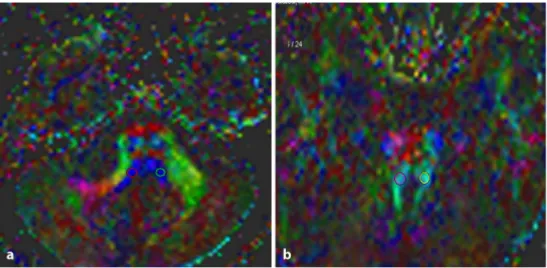
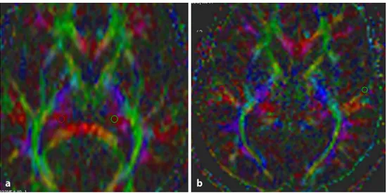
All ROI sizes were set to 4 pixels. ROIs in the auditory pathway of the lateral lemniscus, inferior colliculus, corpus geniculatum mediale, and Heschl’s gyrus were drawn on color-encoded maps of the ipsilateral and contralateral sides of the subjects with AN and also on those of the controls (Figs. 1 and 2). The following ADC and FA values were obtained automatically. In the control group, the averaged values of the left and right sides were simply recorded. ADC and FA measurements of the patients’ auditory pathways on the ipsilateral side were compared with those on the contra-lateral side. These values were then compared with those of the control group.
Fig. 1 The Axial color-coded
maps from a subject with acoustic neuroma showing placement of region of interest on the lateral lemniscus (a) and inferior
the lateral lemniscus, inferior colliculus, and corpus genicu-latum mediale (p = 0.001, 0.010, and 0.005, respectively). Conversely, the FA values were significantly lower at the inferior colliculus for both the contralateral and ipsilateral sides of the AN subjects when compared with the control group (p = 0.052 and 0.05).
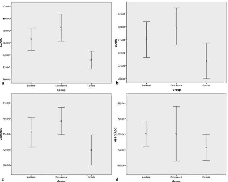
With regard to ADC and FA values obtained in the audi-tory pathway, the difference was not statistically signifi-cant between the ipsilateral and the contralateral side. On the other hand, the ADC values were significantly higher at the inferior colliculus for both the contralateral and the ipsilateral sides of the AN subjects according to the lateral lemniscus and corpus geniculatum mediale (p = 0.022 and 0.04; Fig. 3).
Discussion
In approximately 95 % of cases, AN can appear sporadically and are mostly unilateral. The hereditary form associated with type II neurofibromatosis occurs in 5 % of AN cases and is usually bilateral [12]. Additionally, AN has been detected incidentally in approximately 0.02 % of asymp-tomatic patients who were examined radiologically for other purposes [13]. Moreover, each ear provides impulses
Statistical Analysis
Statistical analysis was performed using SPSS 11.0 statisti-cal software (SPSS Inc., Chicago, IL). The Kolmogorov– Smirnov test was used to determine whether the continuous variables were normally distributed. Parametric tests were applied with all parameters showing normal distribution, and variables were given as mean ± standard deviation. The mean ADC and FA values with a 95 % confidence interval among three groups were compared using one-way analysis of variance. To compare ADC and FA values obtained from the auditory pathways in the ipsilateral and contralateral sides of the subjects and controls, Dunnett t-test was used, in which one group is treated as a control, and all other groups were compared against it. A p value < 0.05 was accepted as statistically significant.
Results
The ADC and FA values obtained from the ipsilateral and contralateral sides of the subjects with AN and of the trol group are presented in Table 1. Compared with the con-trol group, the ADC values for the contralateral side of the subjects were, to a statistically significant degree, higher at
Table 1 The ADC and FA values obtained from the ipsilateral and contralateral sides of the subjects with acoustic neuroma and of the control
group
Locations Lateral lemniscus Inferior colliculus Corpus geniculatum mediale Heschl’s gyrus
ADC FA ADC FA ADC FA ADC FA
Ipsilateral 765.7 ± 33.9 593.8 ± 84.6 775.5 ± 62.8b 725.9 ± 79b 753.7 ± 50.7 387.5 ± 67.3 751.3 ± 44 406.5 ± 106
Contralateral 785.3 ± 40.5a 544.3 ± 91.4 800.5 ± 64.8a 699.9 ± 76.7a 775.4 ± 47.4a 360.5 ± 109 750.9 ± 95.2 356.6 ± 63.4
Control 731.4 ± 21.9 657.6 ± 103 734.4 ± 51 751.3 ± 36.4 719.7 ± 43.1 401.2 ± 68.3 724.4 ± 37 301.4 ± 81
ADC apparent diffusion coefficient, FA fractional anisotropy
aStatistically significant differences between contralateral side and control group bStatistically significant differences between ipsilateral side and control group
Fig. 2 The Axial color-coded
maps from a subject with acoustic neuroma showing placement of region of interest on the corpus geniculatum mediale (a) and
processing of the auditory stimuli [16]. In intact auditory systems, neuroimaging studies show that monaural stimula-tion induces strong contralateral activity and weak ipsilat-eral activity, while on the other hand, binaural stimulation induces balanced cortical activity in both hemispheres [17–19].
In the studies concerning the pathology of the auditory pathway in the vestibulocochlear system, few use advanced neuroimaging applications of MRI such as DTI [20, 21]. Those who did use reported DTI changes only at the lat-eral lemniscus and inferior colliculus level. To our knowl-edge, no study has used DTI to evaluate the entire auditory pathway (from the lateral lemniscus, inferior colliculus, and medial geniculate body to the Heschl’s gyrus) in subjects who have unilateral AN.
by crossing at the brainstem level to the auditory cortices in both hemispheres. It is believed that unilateral AN have no effect on hearing in the ipsilateral side of the ear, but may cause minor and transient hearing loss in the contralateral side of the ear [14].
Auditory impulses come through the cochlear branch of the eighth cranial nerve. These impulse synapses first occur inside the cochlear nucleus in the medulla oblongata, then fibers project into the ipsilateral superior olivary nucleus or cross into the contralateral superior olivary nucleus, con-tinuing into the inferior colliculus through the lateral lem-niscus in the midbrain. From the inferior colliculus, efferent fibers project to the medial geniculate body in the thalamus and end at the auditory cortex in the superior temporal Hes-chl’s gyrus [15]. Contribution from contralateral pathways is more important than from the ipsilateral pathway for the Fig. 3 The apparent diffusion coefficient (ADC) values at the lateral
lemniscus (a), inferior colliculus (b), corpus geniculatum mediale (c),
and Heschl’s gyrus (d) for the ipsilateral and contralateral sides of the
subjects with acoustic neuroma compared with those of the control
group (plots with error bars representing 95 % confidence interval).
LL lateral lemniscus, CI inferior colliculus, CGM corpus geniculatum
corpus geniculatum mediale. It may be that the inferior colliculus receives multiple impulses from the lower audi-tory pathway on both sides of the brain, such that acoustic information reaches each inferior colliculus either directly by projections from the cochlear nucleus, or indirectly via cochlear nucleus projections to the superior olivary nucleus and nuclei of the lateral lemniscus [25]. Also, single-unit recordings from the inferior colliculus show strong excit-atory responses to stimulation of the contralateral side of the ear, and excitatory or inhibitory responses to stimulation of the ipsilateral side of the ear [26]. The findings identi-fied on a DTI show microstructural changes causing axonal loss and/or demyelination, and that the inferior colliculus is where injuries most often occur.
The most important limitation of our study is the rela-tively small number of patients. This situation may explain the lack of statistically significant differences with regard to patient-control group ADC values of the auditory pathway on the ipsilateral side, or when comparing the contralateral and ipsilateral sides. Such differences may arise in further studies with large series.
Conclusion
The present study demonstrates that in the auditory pathway of patients with AN, the contralateral side is more affected than the ipsilateral side, and the most affected center in the auditory pathway is the inferior colliculus. DTI is an advanced neuroimaging technique that can be used to deter-mine the presence of microstructural damage to the audi-tory pathway in subjects with acoustic neuroma, whereas conventional MRI is not sensitive enough to detect damage. Acknowledgments The authors appreciate the contributions and
edi-torial assistance by S. Delacroix, a native speaker of English.
Conflict of Interest The authors declare that they have no conflict of
interest.
References
1. Salzman KL, Davidson HC, Harnsberger HR, Glastonbury CM, Wiggins RH, Ellul S, Shelton C. Dumbbell schwannomas of the internal auditory canal. AJNR Am J Neuroradiol. 2001;22:1368–76. 2. Arthurs BJ, Fairbanks RK, Demakas JJ, Lamoreaux WT, Giddings NA, Mackay AR, Cooke BS, Elaimy AL, Lee CM. A review of treatment modalities for vestibular schwannoma. Neurosurg Rev. 2011;34:265–77.
3. Fortnum H, O’Neill C, Taylor R, Lenthall R, Nikolopoulos T, Lightfoot G, O’Donoghue G, Mason S, Baguley D, Jones H, Mul-vaney C. The role of magnetic resonance imaging in the identifica-tion of suspected acoustic neuroma: a systematic review of clinical and cost effectiveness and natural history. Health Technol Assess. 2009;13:1–154.
DTI is one of the most popular MRI modalities because it provides a noninvasive measure of structural integrity of white matter tracts and also evaluates anatomical connectiv-ity between regions of the brain. DTI enables the assess-ment and quantification of the diffusion properties of white matter, and has been used in detecting the subtle changes in the structure of tissues associated with brain development, degeneration, and injury [22]. ADC and FA are DTI param-eters that have been found to be sensitive to white matter damage and thus have been used in medical studies [23].
Lin et al. [24] have reported reduced FA values and increased radial diffusivity bilaterally at the lateral lem-niscus and inferior colliculus of patients with sensorineu-ral hearing loss. Additionally, they also detected reduced FA values and increased radial diffusivity at the lateral lemniscus and inferior colliculus on the contralateral side of their patients who had profound unilateral hearing loss according to the ipsilateral side, which suggests the pres-ence of a dysmyelinating process. In another study, Wu et al. [21] reported reduced FA values with increased radial diffusivity on both the ipsilateral and contralateral sides of patients with cochlear nerve deficiency, which could have been attributed to axonal loss and/or demyelination of the subcortical auditory tract.
Our study determined changes in the diffusion charac-teristics of the auditory pathway for the ipsilateral and con-tralateral sides of the subjects with AN. Their ADC values were significantly higher than those of the control group at the contralateral side of the lateral lemniscus, inferior col-liculus, and corpus geniculatum mediale. Also, the average ADC values of the lateral lemniscus, inferior colliculus, corpus geniculatum mediale, and Heschl gyrus on the ipsi-lateral side of the patients were higher than in those of the control group, although the differences were not statistically significant. We thought that higher ADC values might be related to microstructural changes such as loss of myelin or demyelination. On the other hand, the average ADC val-ues of the lateral lemniscus, inferior colliculus, and corpus geniculatum mediale on the contralateral side were higher than those on the ipsilateral side, but again, the differences were not statistically significant. Previous studies reported that of the fibers of the nerve bundles to the auditory cor-tices, 20–30 % come from the ipsilateral side and 70–80 % from the contralateral side [21]. This situation may explain our higher ADC values at the contralateral side in subjects with unilateral AN.
This study determined that compared with the controls, FA values of the AN patients were significantly lower at the inferior colliculus on the contralateral and ipsilateral sides. In addition, we determined significantly increased ADC values at the inferior colliculus of the contralateral and ipsilateral sides according to the lateral lemniscus and
17. Khosla D, Ponton CW, Eggermont JJ, Kwong B, Don M, Vasama JP. Differential ear effects of profound unilateral deafness on the adult human central auditory system. J Assoc Res Otolaryngol. 2003;4:235–49.
18. Langers DR, van Dijk, Backes WH. Lateralization, connectivity and plasticity in the human central auditory system. Neuroimage. 2005;28:490–9.
19. Jäncke L, Wüstenberg T, Schulze K, Heinze HJ. Asymmetric he-modynamic responses of the human auditory cortex to monaural and binaural stimulation. Hear Res. 2002;170:166–78.
20. Lin Y, Wang J, Wu C, Wai Y, Yu J, Ng S. Diffusion tensor imaging of the auditory pathway in sensorineural hearing loss: changes in radial diffusivity and diffusion anisotropy. J Magn Reson Imaging. 2008;28:598–603.
21. Wu CM, Ng SH, Wang JJ, Liu TC. Diffusion tensor imaging of the subcortical auditory tract in subjects with congenital cochlear nerve deficiency. AJNR Am J Neuroradiol. 2009;30:1773–7. 22. Mukherjee P, Miller JH, Shimony JS, Philip JV, Nehra D, Snyder
AZ, Conturo TE, Neil JJ, McKinstry RC. Diffusion-tensor MR im-aging of gray and white matter development during normal human brain maturation. AJNR Am J Neuroradiol. 2002;23:1445–56. 23. Nakayama N, Okumura A, Shinoda J, Yasokawa YT, Miwa K,
Yoshimura SI, Iwama T. Evidence for white matter disruption in traumatic brain injury without macroscopic lesions. J Neurol Neu-rosurg Psychiatry. 2006;77:850–5.
24. Lin YC, Wang CC, Wai YY, Wan YL, Ng SH, Chen YL, Liu HL, Wang JJ. Significant temporal evolution of diffusion anisotropy for evaluating early response to radiosurgery in patients with ves-tibular schwannoma: findings from functional diffusion maps. AJNR Am J Neuroradiol. 2010;31:269–74.
25. Anderson RA, Knight PL, Merzenich MM. The thalamocortical and corticothalamic connections of AI, AII, and the anterior audi-tory field (AAF) in the cat: evidence for two largely segregated systems of connections. J Comp Neurol. 1980;194:663–701. 26. Bruckner S, Rübsamen R. Binaural response characteristics in
isofrequency sheets of the gerbil inferior colliculus. Hear Res. 1995;86:1–14.
4. Suzuki M, Hashimoto S, Kano S, Okitsu T. Prevalence of acoustic neuroma associated with each configuration of pure tone audio-gram in patients with asymmetric sensorineural hearing loss. Ann Otol Rhinol Laryngol. 2010;119:615–18.
5. Day AS, Wang CT, Chen CN, Young YH. Correlating the cochleo-vestibular deficits with tumor size of acoustic neuroma. Acta Oto-laryngol. 2008;128:756–60.
6. Mark AS, Seltzer S, Harnsberger HR. Sensorineural hearing loss: more than meets the eye? AJNR Am J Neuroradiol. 1993;14:37–45. 7. Gebarski SS, Tucci DL, Telian SA. The cochlear nuclear com-plex: MR location and abnormalities. AJNR Am J Neuroradiol. 1993;14:1311–8.
8. Welling DB, Glasscock ME, 3rd, Woods CI, Jackson CG. Acoustic neuroma: a cost-effective approach. Otolaryngol Head Neck Surg. 1990;103:364–70.
9. Chang Y, Lee SH, Lee YJ, Hwang MJ, Bae SJ, Kim MN, Lee J, Woo S, Lee H, Kang DS. Auditory neural pathway evaluation on sensorineural hearing loss using diffusion tensor imaging. Neuro-report. 2004;15:1699–703.
10. Lin Y, Wang J, Wu C, Wai Y, Yu J, Ng S. Diffusion tensor imaging of the auditory pathway in sensorineural hearing loss: changes in radial diffusivity and diffusion anisotropy. J Magn Reson Imaging. 2008;28:598–603.
11. Wu CM, Ng SH, Liu TC. Diffusion tensor imaging of the subcorti-cal auditory tract in subjects with long-term unilateral sensorineu-ral hearing loss. Audiol Neurootol. 2009;14:248–53.
12. Taiwo O, Galusha D, Tessier-Sherman B, Kirsche S, Cantley L, Slade MD, Cullen MR, Donoghue AM. Acoustic neuroma: po-tential risk factors and audiometric surveillance in the aluminium industry. Occup Environ Med. 2014;71:624–8.
13. Lin D, Hegarty JL, Fischbein NJ, Jackler RK. The prevalence of “incidental” acoustic neuroma. Arch Otolaryngol Head Neck Surg. 2005;131:241–4.
14. Yoshiura T, Higano S, Rubio A, Shrier DA, Kwok WE, Iwanaga S, Numaguchi Y. Heschl and superior temporal gyri: low signal intensity of the cortex on T2-weighted MR images of the normal brain. Radiology. 2000;214:217–21.
15. Bernal B, Altman NR. Auditory functional MR imaging. AJR Am J Roentgenol. 2001;176:1009–15.
16. de Bode S, Sininger Y, Healy EW, Mathern GW, Zaidel E. Dich-otic listening after cerebral hemispherectomy: methodological and theoretical observations. Neuropsychologia. 2007;45:2461–6.