ONLINE FIRST
This is a provisional PDF only. Copyedited and fully formatted version will be made available soon.
ISSN: 0015-5659 e-ISSN: 1644-3284
Evaluation of olfactory bulbus volume and olfactory sulcus
depth development with 3 Tesla MRI in childhood
Authors: B. Güney, N. Çullu, M. Y. Özdemir
DOI: 10.5603/FM.a2021.0022 Article type: Original article Submitted: 2020-12-15 Accepted: 2021-02-01
Published online: 2021-03-01
This article has been peer reviewed and published immediately upon acceptance.
It is an open access article, which means that it can be downloaded, printed, and distributed freely, provided the work is properly cited.
Articles in "Folia Morphologica" are listed in PubMed.
Evaluation of olfactory bulbus volume and olfactory sulcus depth development with 3 Tesla MRI in childhood
B. Güney et al., Pediatric olfactor development between 1 month and 17 years
B. Güney, N. Çullu, M.Y. Özdemir
Department of Radiology, Muğla Sıtkı Koçman University Medical Faculty, Muğla, Turkey
Address for correspondence: Dr Bünyamin Güney, Assistant Professor, Muğla Sıtkı Koçman University Medical Faculty, Department of Radiology, tel: 90 533 4911152, fax: 90 252 2111345, e-mail: [email protected]
ABSTRACT
Background: In this study, it is aimed to reveal the change in OBV and OSD in healthy
Turkish pediatric individuals between 1 month and 18 years of age with 3 Tesla MRI taking into account different age groups and gender factors.
Materials and methods: In this retrospective study, 190 pediatric individuals who underwent
cranial MRI were evaluated. Healthy pediatric cases were divided into four groups as infantile period (first 24 months when cerebral myelinization was completed), early childhood (2-6 years), childhood (6-12 years) and adolescence (12-18 years). OBV and OSD measurements were performed on coronal T2-weighted brain MR images by 3T MR scan. The mean, right and left olfactory bulb volume and sulcus depths were used for evaluation.
Results: The mean age was 9.9 ± 7.5 months for the infantile period, 4.5 ± 1.3 years for early
childhood, 9.3 ± 1.7 years for childhood and 15.2 ± 1.7 years for adolescence. Mean, right and left OBV was found to be slightly larger in male children than female children (P= 0.015, P= 0.033 and P= 0.010, respectively). There was no statistical difference between the genders for mean, right and left OSD (P= 0.559, P= 0.536 and P= 0.598, respectively). Among the age groups, the values of the 3rd and 4th groups in terms of mean, right and left OBV were higher than the other two groups (P<0.001). In terms of OSD, mean, right and left values were higher in group 2, 3 and 4 than group one (P<0.001).
Conclusions: These data differ by pediatric age group and gender for the development of
OBV and OSD. Normal values for the pediatric age group and gender should be calculated to detect olfactory dysfunction.
INTRODUCTION
The sense of smell is one of the five senses that have an important function in human life, and the olfactory bulb (OB) is considered to be the most important transmission station in processing the sense of smell. Anatomical structures of the sense of smell begin to develop early in the human fetus. There are studies and anatomical atlases on the development and anatomical change of the primordial olfactory bulbus, which was first observed anatomically in a 41-day (4.5 week) embryo in the fetal development period, from gestation to birth [2, 13, 15,17]. However, the number of studies on the anatomical development and change of the olfactory bulbus in the pediatric period is limited. In a study conducted by Schneider et al about the maturation of the olfactory bulbus in the postnatal period, they found that olfactory bulbus showed a similar maturation parallel to the maturation of the cerebral white matter until the end of the second year, when the myelinization and maturation of the cerebral white matter was completed. In this study, it was found that the olfactory bulbs took the adult shape at the end of the postnatal 24 months [18]. The first study on the anatomical development and change of olfactory bulbus volume in the pediatric age group was conducted by Hummel et al [10]. A positive relationship was found between increased OBV and increased olfactory function in this study performed in children aged 1 to 17 years. It was found that both OBV and olfactory function increased with age.
Another parameter used in evaluating olfactory functions is the depth of the olfactory sulcus. Some diseases such as major depression, anxiety disorder, Behçet's disease,
Parkinson's and Alzheimer's disease have been shown to reduce the depth of the olfactory sulcus and cause a decrease in the sense of smell [1, 5, 11]. There are limited studies on the change of olfactory bulbus volume and olfactory sulcus depth with age in pediatric cases [16]. OBV and OSD are known to vary with age (4, 10). The age-related change and normal values of both OB and OSD in healthy pediatric individuals are not clearly known.
In this study, it is aimed to reveal the change of OBV and OSD over time in healthy Turkish pediatric individuals between 1 month and 18 years of age with 3 Tesla MRI taking into account different age groups and gender factors.
MATERIALS AND METHODS
After being approved by the ethics committee of our university, the research was started retrospectively in the Radiology department. Only children with normal brain MRI
were included in the study. Pediatric individuals with any brain abnormality (developmental anomaly, myelinization disorder, maturation disorder, etc.) or disease were excluded from the study. While evaluating the patients, the patient files registered in the hospital information system were taken as basis. Those with a genetic disease in their family, those with suspected congenital neurometabolic disease, chronic diseases such as diabetes mellitus and
hypertension, and asthma patients were excluded from the study. Patients who had MRI examination for control purposes and patients with nonspecific symptoms and no pathology were included in the study. The cases had nonspecific complaints such as headache and dizziness in order of frequency. No disease was detected in these pediatric individuals and no disease developed during clinical follow-up in our hospital.
In this study, 190 pediatric individuals who underwent cranial MRI between 2017-2019 were evaluated retrospectively. Cases with good image resolution and no motion artifact were included in the study, while cases with poor image resolution and artifacts were
excluded. 95 of our cases were women and 95 were men. Healthy pediatric cases were divided into four groups according to their age and gender: group 1: infantile period (1-24 months), group 2: early childhood (2-6 years), group 3: childhood (7-12 years) and group 4: adolescence (13-18 years); group A consisting of 95 women; group B consisting of 95 men. The study was conducted on the basis of and in accordance with the declaration of Helsinki. OBV and OSD measurements were made on T2-weighted brain MR images in the coronal plane (figure 1) obtained on a 3 Tesla MR (Magnetom Skyra, Siemens, Germany) device. Our imaging parameters were 256x256 matrix and 22 cm field of view (FOV), repetition time = 3500 ms (TR 3500 ms), echo time = 75 ms (TE 75 ms), excitation number = 2 (NEX 2), and a slice thickness of 4 mm. OBV and OSD measurements were made by two experienced
radiologists who had no knowledge of the cases.
Volumetric measurement of OBV was made using 3D Slicer software (3D Slicer software ver. 4.2.2-1, http://www.slicer.org). The Slicer volumetric measurement program is a free open source software package developed by Harvard University and approved for
medical research. After dividing the olfactory bulbus into sections with appropriate threshold values in the coronal image, separate MR numbers were assigned to each image with the Slicer software. ROI (region of interest) was adjusted to not exceed the anatomical contours of the bulb. After each slice containing the relevant OB sections was revealed, a three-dimensional graphical model of the OB was created and volume calculation was made. Intra-observer variability was set at less than 5%.
Ethics Committee Approval
Muğla Sıtkı Koçman University Human Research Ethics Committee. Ethics Committee Number: 200236
Consent to participate
Approval was obtained from the parent or legal guardian of each case participating in the study.
Statistical analysis
IBM SPSS version 20.0 software (IBM Corp, Armonk, NY, USA) was used for statistical evaluation and normal distribution was checked using Kolmogorov-Smirnov test. Data are presented as mean ± standard deviation. Statistical comparison of the right and left OBV values and the depth of the right and left olfactory sulcus was made using the paired t test. Independent-sample t test was used to evaluate the statistical differences between groups formed by considering gender, while one-way ANOVA test was used to evaluate the
statistical differences between groups formed according to age. Multiple comparisons were made with the Tukey test and a P value of 0.05 was considered statistically significant.
RESULTS
A total of 190 patients (95 males, 95 females) were included in the study. The mean age was 9.9 ± 7.5 months for the infantile period, 4.5 ± 1.3 years for early childhood, 9.3 ± 1.7 years for childhood and 15.2 ± 1.7 years for adolescence. The OBV was 42.03 ± 5.96 (range 29,2-57,6) mm3 on the right and 42.33 ± 6.06 (range 28,8-62,4) mm3 on the left side.
There was no statistical difference between right and left side OBV (P>0.167). Mean, right and left OBV was found to be slightly larger in male children than female children (P= 0.015, P= 0.033 and P= 0.010, respectively) (Table 1). OSD values were 8.34±0.92 on the right and 8.32 ± 0.89 mm on the left. There was no statistical difference between right and left in terms of OSD (P>0.481). There was no statistical difference between the genders for mean, right and left OSD (P= 0.559, P= 0.536 and P= 0.598, respectively) (Table I).
The distribution of mean OBV and OSD according to age groups are given in Table 2. Among the age groups, the values of the 3rd and 4th groups in terms of mean, right and left OBV were higher than the other two groups (Table II) (P<0.001). In terms of OSD, mean, right and left values were higher in group 2, 3 and 4 than group one (Table II) (P<0.001).
DISCUSSION
There are several important results of our study. First, in the pediatric age group, the olfactory bulbus volume increases as the age increases, but the most significant volume increase is in the 7-12 age group. Second, OSD is lower in the infantile patient group (1-24 months) compared to other pediatric age groups. After 3 years of age, there is no statistically significant change in the depth of the olfactory sulcus until the age of 18. Third, there was no statistically significant difference in OSD measurements based on gender in the normal healthy pediatric population, but OBV was slightly larger in male children than in female children.
MR imaging method has been used successfully in adults to analyze the normal anatomy of OBs [4, 6, 12, 20, 23]. In the study conducted by Schneider et al, Cranial MRI examinations of 121 pediatric cases aged between 1 and 19.6 years were retrospectively re-evaluated in order to detect the maturation of the olfactory bulb. Three anatomical patterns have been described defining different anatomical shapes for the olfactory bulbus. Whatever the anatomical shape of the olfactory bulbs, they found that the gradual rearrangement of the peripheral neuronal layers and central structures of the bulbus resulted in an adult-like appearance in all children at the end of the second year at the latest, in parallel with the maturation changes of the cerebral white matter [18]. Therefore, in our study, we grouped pediatric individuals of the first 2 years of age separately. However, we found that OB volume increased minimally in pediatric individuals aged 3-6 years compared to the first 2-year-old case group. We found the olfactory sulcus depth to be lower in the first 2-year-old pediatric group compared to the other three pediatric groups. This finding supports this study by Schneider et al on olfactory maturation.
In a study conducted by Croy et al on 27 depressed female individuals, 15 of whom were maltreated during childhood, the OB volume measured in individuals who were maltreated during childhood was found to be lower than those who were not exposed to childhood maltreatment [3]. In the first hypothesis they put forward for this result; It suggests that major stress exposure in childhood affects neurogenesis in human OB, as previously demonstrated in animal studies [7]. For these reasons, we think that, as in our study, it is important to know the normal OB volume and OSD measurement intervals in pediatric individuals to determine the connection between the abnormalities that may occur in adulthood and the pediatric period.
In a study conducted by Hummel et al on 87 pediatric individuals aged between 1 and 17 years without olfactory dysfunction (mean = 8 ± 5.5 years, 46 boys and 41 girls), it was
found that OB volume and olfactory function increased with age [9]. In this study, OB volumes of male children (left: 71 mm3, right: 68 mm3) were found to be larger than female
children (left: 65, right: 66 mm3). It was also found that for both boys and girls, the right and
left OB volumes gradually increased from the age of 1 year. According to the study, the average OB volume for 1 year old boys is; left: 65, right: 64 mm3, mean OB volume for girls: left: 61, right: 62 mm3. When the mean OB volume was evaluated in 17 years old pediatric individuals, in boys: left: 79 mm3, right: 77 mm3; in girls: left: 73 mm3, right: 71 mm3. As a result of our study, OBV was found to be slightly higher in boys compared to girls, similar to the study of Hummel et al. However, although OBV increased gradually with age, it was found that the statistically more significant increase in OB volume was between the ages of 7-12, regardless of gender.
In studies conducted in patients with congenital anosmia and psychiatric disorders such as schizophrenia or psychosis, OSD was found to be lower than normal [9, 14, 21, 22]. Although standard values for the depth of the olfactory sulcus have been published mostly for adults, few studies have published standard values for the pediatric age group. In the study conducted by Smitka et al on 40 normosmic children aged between 6 and 18 years, it was found that OSD was 8 mm and above in all children over 9 years old [19]. It was stated that the recommended cut-off value of 8 mm, which indicates anosmia, can be used safely for children aged 9 years and above, and different limit values should be considered for children aged 8 years and under. In the study conducted by Huart et al in 106 individuals (36 anosmic individuals and 70 healthy individuals) aged between 7 and 79 years, sulcus depth below 8 mm was found to be an important indicator for the development of anosmia [8].
The first study in the literature examining the age-related change of both OSD and OBV in healthy pediatric individuals was conducted by Şahin et al. A total of 90 pediatric patients aged between 3 and 17 years were included in this study and a 1.5 tesla MRI device was used [16]. Our study is fundamentally different from 3 aspects according to the study of Şahin et al. The first is that healthy pediatric individuals in the 1 month-2 age group were included in the study and the study was conducted with 190 individuals, the second is the use of 3 Tesla MRI device and the third is that the OBV measurement method is different. Thus, the change in OBV and OSD in all pediatric age groups between 1 month and 17 years was examined. Studies including OSD and OBV measurements of healthy pediatric individuals in the literature are summarized comparatively in Table III.
According to our research, our study is the first study to separately show the
the age of 18 in healthy pediatric individuals. It was found that OSD gradually increased with age in both healthy male and female individuals in the pediatric age group. The infantile group(0-24 months) had the lowest OSD and the mean OSD in this group was found to be less than 8 mm. For 3 years and above, mean OSD was over 8 mm for each age group. Another difference of our study from other studies is that the images were obtained with a 3T MR scanner. We think that we may have obtained more accurate results since 3 T MR images provide clearer and thinner-slice images with higher resolution.
Our study had some limitations. The first of these is that our study was retrospective and there was no evaluation of olfactory function. In addition, inter-observer variability was not taken into account in our study. Another limitation is that some of the individuals
participating in the study were very young in age and the patient backgrounds were obtained from the hospital information system.
CONCLUSIONS
In conclusion, there are still very few studies showing the development of OBV and OSD with age and time changes considering gender for normal healthy pediatric individuals. We think that more studies should be done on this subject, since these data show differences in both the pediatric age group and adults according to age and that OBV and OSD can be an indicator of the development of olfactory dysfunction. Therefore, within the pediatric age group, normal values should be calculated considering age and gender.
REFERENCES
1. Asal N, Bayar Muluk N, Inal M, et al. Olfactory bulbus volume and olfactory sulcus depth in psychotic patients and patients with anxiety disorder/depression. Eur Arch Otorhinolaryngol. 2018;275(12):3017-24.Doi: 10.1007/s00405-018-5187-x
2. Azoulay R, Fallet-Bianco C, Garel C, et al. MRI of the olfactory bulbs and sulci in human fetuses. Pediatr Radiol. 2006;36(2):97-107.Doi: 10.1007/s00247-005-0030-0
3. Croy I, Negoias S, Symmank A, et al. Reduced olfactory bulb volume in adults with a history of childhood maltreatment. Chem Senses. 2013;38(8):679-84.Doi: 10.1093/chemse/bjt037
4. Çullu N, Yeniçeri İ, Güney B, et al. Evaluation of olfactory bulbus volume and olfactory sulcus depth by 3 T MR. Surg Radiol Anat. 2020;42(9):1113-8.Doi: 10.1007/s00276-020-02484-w
5. Doğan A, Bayar Muluk N, Asal N, et al. Olfactory bulb volume and olfactory sulcus depth in patients with Behçet's disease. J Laryngol Otol. 2018:1-5.Doi: 10.1017/s0022215118002141
6. Held P, Seitz J, Fründ R, et al. MRI detection of olfactory bulb and tract. J Neuroradiol. 2000;27(2):112-8.Doi:
7. Hitoshi S, Maruta N, Higashi M, et al. Antidepressant drugs reverse the loss of adult neural stem cells following chronic stress. J Neurosci Res. 2007;85(16):3574-85.Doi: 10.1002/jnr.21455
8. Huart C, Meusel T, Gerber J, et al. The depth of the olfactory sulcus is an indicator of congenital anosmia. American journal of neuroradiology. 2011;32(10):1911-4.Doi:
9. Hummel T, Damm M, Vent J, et al. Depth of olfactory sulcus and olfactory function. Brain research. 2003;975(1-2):85-9.Doi: 10.1016/S0006-8993(03)02589-7
10. Hummel T, Smitka M, Puschmann S, et al. Correlation between olfactory bulb volume and olfactory function in children and adolescents. Exp Brain Res. 2011;214(2):285-91.Doi: 10.1007/s00221-011-2832-7 11. Hummel T, Urbig A, Huart C, et al. Volume of olfactory bulb and depth of olfactory sulcus in 378 consecutive patients with olfactory loss. J Neurol. 2015;262(4):1046-51.Doi: 10.1007/s00415-015-7691-x 12. Iida Y, Naito M, Asahina N, et al. Magnetic resonance imaging of the olfactory apparatus. Arch Otolaryngol Head Neck Surg. 1994;120(8):869-72.Doi: 10.1001/archotol.1994.01880320069015 13. Müller F, O'Rahilly R. Olfactory structures in staged human embryos. Cells Tissues Organs. 2004;178(2):93-116.Doi: 10.1159/000081720
14. Nishikawa Y, Takahashi T, Takayanagi Y, et al. Orbitofrontal sulcogyral pattern and olfactory sulcus depth in the schizophrenia spectrum. European archives of psychiatry and clinical neuroscience. 2016;266(1):15-23.Doi: DOI 10.1007/s00406-015-0587-z
15. O'Rahilly R, Muller F, Bossy J. [Atlas of the stages of development of the nervous system in the intact human embryo]. Arch Anat Histol Embryol. 1982;65:57-76.
16. Sahin S, Baykan A, Altunisik E, et al. Quantitative analysis of healthy olfactory sulcus depth, olfactory tract length and olfactory bulb volume in the pediatric population: a magnetic resonance study. Folia
Morphologica. 2020.
17. Sarnat HB, Flores-Sarnat L. Olfactory Development, Part 2: Neuroanatomic Maturation and Dysgeneses. J Child Neurol. 2017;32(6):579-93.Doi: 10.1177/0883073816685192
18. Schneider JF, Floemer F. Maturation of the olfactory bulbs: MR imaging findings. AJNR Am J Neuroradiol. 2009;30(6):1149-52.Doi: 10.3174/ajnr.A1501
19. Smitka M, Hummel T. The Depth of the Olfactory Sulcus in Normosmic Children and Adolescents. Neuropediatrics. 2015;46(S 01):WS07-1.Doi: 10.1055/s-0035-1550753
20. Suzuki M, Takashima T, Kadoya M, et al. MR imaging of olfactory bulbs and tracts. AJNR Am J Neuroradiol. 1989;10(5):955-7.
21. Takahashi T, Nakamura Y, Nakamura K, et al. Altered depth of the olfactory sulcus in first-episode schizophrenia. Progress in Neuro-Psychopharmacology and Biological Psychiatry. 2013;40:167-72.Doi: 10.1016/j.pnpbp.2012.10.001
22. Turetsky BI, Moberg PJ, Quarmley M, et al. Structural anomalies of the peripheral olfactory system in psychosis high-risk subjects. Schizophrenia research. 2018;195:197-205.Doi: 10.1016/j.schres.2017.09.015 23. Yousem DM, Geckle RJ, Bilker WB, et al. Olfactory bulb and tract and temporal lobe volumes.
Normative data across decades. Ann N Y Acad Sci. 1998;855:546-55.Doi: 10.1111/j.1749-6632.1998.tb10624.x.
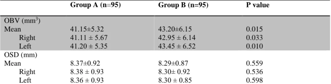
Table I. The distribution of age, mean and both sides olfactory bulb volüme (OBV) and
olfactory sulcus dept in according to sexes.
Group A (n=95) Group B (n=95) P value
OBV (mm3) Mean Right Left 41.15±5.32 41.11 ± 5.67 41.20 ± 5.35 43.20±6.15 42.95 ± 6.14 43.45 ± 6.52 0.015 0.033 0.010 OSD (mm) Mean Right Left 8.37±0.92 8.38 ± 0.93 8.36 ± 0.93 8.29±0.87 8.30± 0.92 8.30 ± 0.85 0.559 0.536 0.598
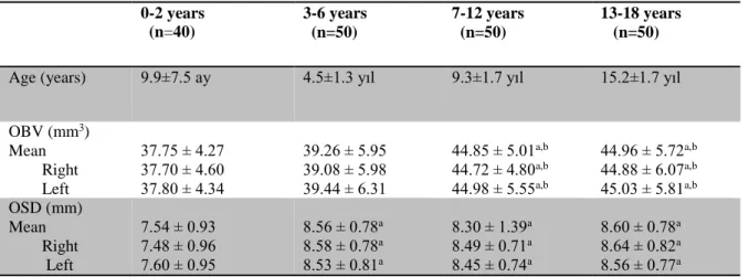
Table II. The distribution of age, mean and both sides olfactory bulb volume (OBV) and
olfactory sulcus dept in according to age groups. Data are n of participants, mean ± SD.
aP<.001 compared with 0-2 years group (Oneway ANOVA-Tukey test). bP<.001 compared with 3-6 years group(Oneway ANOVA-Tukey test).
Table III. Comparison of OBV and OSD Studies in Healthy Pediatric Cases
Figure 1. The coronal T2-weighted image shows the olfactory bulb volume measurement and
the olfactory sulcus depth measurement. A measurement example of olfactory sulcus depth (small hollow arrow) and a measurement example of olfactory bulb surface area (large hollow arrow). 0-2 years (n=40) 3-6 years (n=50) 7-12 years (n=50) 13-18 years (n=50)
Age (years) 9.9±7.5 ay 4.5±1.3 yıl 9.3±1.7 yıl 15.2±1.7 yıl
OBV (mm3) Mean Right Left 37.75 ± 4.27 37.70 ± 4.60 37.80 ± 4.34 39.26 ± 5.95 39.08 ± 5.98 39.44 ± 6.31 44.85 ± 5.01a,b 44.72 ± 4.80a,b 44.98 ± 5.55a,b 44.96 ± 5.72a,b 44.88 ± 6.07a,b 45.03 ± 5.81a,b OSD (mm) Mean Right Left 7.54 ± 0.93 7.48 ± 0.96 7.60 ± 0.95 8.56 ± 0.78a 8.58 ± 0.78a 8.53 ± 0.81a 8.30 ± 1.39a 8.49 ± 0.71a 8.45 ± 0.74a 8.60 ± 0.78a 8.64 ± 0.82a 8.56 ± 0.77a
Comparison of OBV and OSD Studies in Healthy Pediatric Cases
First Author
Year Country Measurement Method N Age (Years)
Magnetic Field
(Tesla) OBV (mm3) OSD (mm)
Hummel T. 2011 France
AMIRA 3-D visualization and modeling system (Visage Imaging, Carlsbad, USA)
87 1-17 Years 1,5 Tesla M:21-98, R:68, L:71 No measurement F:21-121, R:66, L:65 Sahin S. 2020 Turkey Semi-automatically method, Philips workstation 90 3-17 Years 1,5 Tesla M:32,7-98,6 R:64, L: 65,3 R: 8,85 (min 6 max 13,6) F: 31,8-99,6 R:67,1 L:72 L: 8,8 (min 1,8 max 16)
Our Study 2020 Turkey
3D Slicer software (3D Slicer software ver. 4.2.2-1, USA) 190 1 Month- 17 Years 3 Tesla M:30,1-62,4 R:41,11 L:41,20 R: 8.34±0.92 F: 29,2-55,2 R:42,95 L:43,45 L: 8,32± 0.89