Original Article / Özgün Araştırma
© Meandros Medical and Dental Journal, Published by Galenos Publishing House.
This is article distributed under the terms of the Creative Commons Attribution NonCommercial 4.0 International Licence (CC BY-NC 4.0).
Ad dress for Cor res pon den ce/Ya zış ma Ad re si: Hande Pamukçu MD,
Başkent University Faculty of Dentistry, Department of Orthodontics, Ankara, Turkey Phone: +90 533 683 54 50
E-mail : [email protected]
Received/Geliş Ta rihi : 16.10.2016 Accepted/Ka bul Ta ri hi : 16.11.2016 Anah tar Ke li me ler
Hızlı maksiller ekspansiyon, yarı hızlı maksiller ekspansiyon, bilgisayarlı tomografi, kök rezorpsiyonu
Keywords
Rapid maxillary expansion, semi-rapid maxillary expansion, computed tomography, root resorption
Hızlı ve Yarı Hızlı Maksiller Ekspansiyon Uygulamalarının
Dentofasiyal ve Periodontal Etkilerinin Karşılaştırılması
Şefika Ruzin Gönüldaş1, Ömür Polat Özsoy2, Hande Pamukçu2, Feride Kural3
1Private Practice, Ankara, Turkey
2Başkent University Faculty of Dentistry, Department of Orthodontics, Ankara, Turkey 3Başkent University Faculty of Medicine, Department of Radiology, Ankara, Turkey
Öz Abstract
Objective: The purpose of this study was to compare the skeletal, dentoalveolar and periodontal effects that occurred after rapid maxillary expansion (RME) and semi-rapid maxillary expansion (SRME) with an acrylic splint bonded expansion appliance and also to compare the degree of root resorption in the supporting teeth during expansion.
Materials and Methods: The sample of the study included 19 patients having maxillary constriction requiring bilateral upper premolar extraction. The semi-rapid group consisted of 10 patients and the rapid group consisted of 9 patients. Lateral and posteroanterior (PA) cephalometric radiographs were taken at the beginning of the treatment, at the end of expansion and after 3 months of retention period. Eleven patients underwent low-dose computed tomography (CT) at the beginning of treatment and at the end of retention period, after giving informed consent. The evaluation of root resorption was made using a scanning electron microscope from the extracted teeth.
Results: Both lateral and PA radiographs showed similar changes between the groups. Posterior nasal cavity width, palatal maxillary width, the distance between palatinal roots and the distance between incisor roots showed significant increase in the CT measurements. Buccal bone thickness decreased and palatal bone thickness increased in all molar and premolar teeth examined. Root resorption was seen in both groups, but no significant differences were observed between the groups. Conclusion: Some differences were seen in short-time effects between SRME and RME but these differences were temporary and disappeared at the end of the retention period. The qualitative evaluation of the roots showed that the location and degree of the root resorption were similar between the groups.
Amaç: Bu çalışmanın amacı, akrilik splintli bonded ekspansiyon apareyi ile yapılan yarı hızlı maksiller ekspansiyon (SRME) ve hızlı maksiller ekspansiyon (RME) ile meydana gelen iskeletsel, dentoalveolar ve periodontal etkileri; ayrıca tedavi sırasında destek dişlerde meydana gelen kök rezorpsiyonunun şiddetini karşılaştırmaktır.
Gereç ve Yöntemler: Maksiller darlığa sahip ve üst 1. premolar çekimi planlanan 19 birey çalışma grubunu oluşturmuştur. SRME grubunu oluşturan 10 birey ve hızlı
Comparison of Dentofacial and
Periodontal Effects of Rapid and
Semi-rapid Maxillary Expansion
Introduction
Transverse maxillary deficiency manifest itself clinically as unilateral or bilateral posterior crossbite and maxillary expansion is used for its treatment (1). Several methods have been developed for maxillary expansion. The interval of the expansion screw’s activation, magnitude of the force, treatment duration and the patients’ age are important factors for the selection of the proper method and, several protocols, such as slow, semi-rapid maxillary expansion (SRME) and rapid maxillary expansion (RME) have been suggested (2).
In the literature, some undesirable effects of the maxillary expansion treatment have been reported (3-5). These were tipping of the maxillary teeth, periodontal membrane damages, bending of the alveolar bone, fenestration of the buccal cortex, necrosis of the palatal tissue, pain during the expansion, pulpal hyperemia, relapse, micro trauma of the temporomandibular joint, micro-fracture in the midpalatal suture and, especially, external resorption of the teeth (3-5).
In 1998, İşeri et al. (6) suggested that RME causes a significant amount of deformation and stress accumulation in the facial bones which may cause relapse in the long-term. They stated that a slower rate of expansion may create less resistance in the nasomaxillary complex, thus, preventing relapse. From this view, SRME protocol has been suggested for growing patients and young adults, thus, the development of the nasomaxillary complex could be stimulated and the incidence of relapse could be reduced (6).
The purpose of this study was to compare the skeletal, dentoalveolar and periodontal effects that
occurred after RME and SRME with acrylic splint bonded expansion appliance and also to compare the degree of root resorption of the supporting teeth during expansion.
Materials and Methods
Before initiation of the study, a legal approval was received from the institutional review board and the study was approved by Başkent University Local Ethics Commitee (protocol number D-KA 10/07). The treatment group included 19 patients (11 males and 8 females). Inclusion criteria for the treatment group were as follows:
1. Presence of unilateral or bilateral posterior crossbite,
2. Class 1 or class 2 dental malocclusion,
3. Indication for fixed orthodontic treatment with extraction of two upper premolars,
4. Continuing growth and development,
5. Permanent dentition and no caries or restorations on the first upper premolars,
6. History of no previous orthodontic treatment, 7. Presence of no systematic diseases,
8. Presence of no congenital, genetic or traumatic facial deformities.
Nineteen patients were randomly divided into two groups:
Group 1 (SRME): This group consisted of 10 patients (5 females and 5 males) treated with a SMRE protocol. The mean age of the patients was 14.05±1.46 years at the start of the treatment. The expansion treatment period for this group was 61.7±20.4 days.
Group 2 (RME): This group consisted of 9 patients (3 females and 6 males) treated with a classical RME protocol. The mean age of the patients was 13.96±0.71 years at the start of the treatment. The expansion treatment period for this group was 33.4±9.6 days.
maksiller genişletme grubunu oluşturan 9 bireyden tedavi başı, genişletme sonu ve 3 aylık pekiştirme dönemi sonunda lateral ve posteroanterior (PA) sefalometrik radyograflar alınmıştır. Kabul eden 11 hastadan tedavi başı ve pekiştirme dönemi sonunda düşük dozlu bilgisayarlı tomografi (BT) kayıtları alınmıştır. Ayrıca genişletme tedavisi bitiminde çekilen dişlerde taramalı elektron mikroskobu ile kök rezorpsiyonu incelemesi yapılmıştır.
Bulgular: Her iki grupta da PA ölçümlerinde nazal kavite genişliği ve total maksiller genişlikte hem aktif tedavi sonunda hem de tüm retansiyon sonunda istatistiksel olarak anlamlı artış, sağ ve sol molar dişlerde ise anlamlı tiping meydana gelmiştir. BT görüntülerinde yapılan ölçümlerde ise iki tedavide de posterior nazal kavite genişliği ve palatal maksiller genişlikte önemli artış gözlenmiş, molar ve premolar dişlerin hepsinde bukkal kemik kalınlığı azalmış, palatinal kemik kalınlığı artmıştır. Kök rezorpsiyonu açısından iki grupta da benzer derinlik ve miktarda rezorpsiyon kriterleri görülmüştür.
Sonuç: SRME ve RME protokollerinin kısa dönemde farklı etkilerinin bulunmaktadır ancak retansiyon dönemi sonunda bu farklılıklar ortadan kalkmaktadır. Her iki gruptaki kök rezorbsiyonunun lokasyonu ve derecesi yönünden kalitatif değerlendirmede bir fark bulunamamıştır.
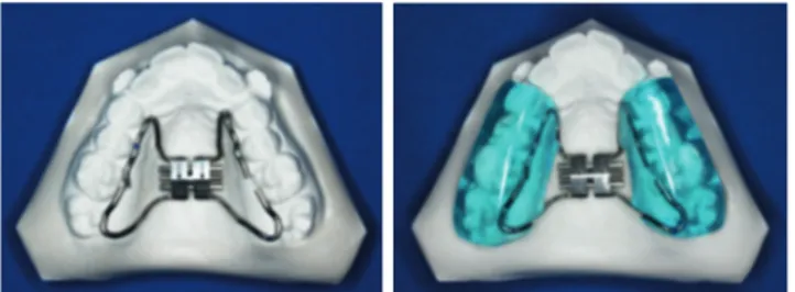
An acrylic splint bonded expansion appliance was used for both groups (Figure 1). The appliance was fixed using glass ionomer cement (Meron, Voco GmbH, Cuxhaven, Germany). For the first 7 days, the screw has been activated twice a day (2x1/4 turn=0.5 mm) in both groups. After 1 week, occlusal radiography was performed to ensure the opening of the midpalatal suture. If a gap was seen between the two halves of palatal processes of the maxillary bone, the screw activation protocol was reduced to a quarter tour (1/4 turn=0.25 mm) per day every other day in SRME group. RME group continued the activation with the same rate. The expansion ceased when the
posterior crossbite was eliminated and overcorrected. A stainless steel ligature was tied around the screw after the expansion period and the same appliance was used as a removable retainer for 3 months.
Collection of Radiographs
Lateral and posteroanterior (PA) cephalometric radiographs were taken at the beginning of the treatment (TO), end of the expansion (T1) and at the end of the 3-month retention period (T2). PA
Figure 1. Acrylic splint bonded expansion appliance
Figure 2a. Angular measurements used in the evaluation of the lateral cephalometric radiographs 1. SNA0, 2. SNB0, 3. ANB0, 4. SN-PP0, 5. MP-PP0, 6. SN-MP0, 7. U1-SN0, 8. L1-MP0. (1. SNA0 expresses the degree of protrusion and retrusion of the maxilla in relation to the cranial base, 2. SNB0 establishes the degree of protrusion and retrusion of the mandible in relation to the cranial base, 3. ANB0 measures sagittal relationship between the maxilla and mandible. 4. SN-PP0: angle between the cranial base and palatal plane , 5. MP-PP0: angle between the mandibular plane and palatal plane, 6. SN-MP0: angle between cranial base and mandibular plane, 7. U1-SN0 angle between the cranial base and upper most protrusive incisor, 8. L1-MP0 angle between the mandibular plane and lower most protrusive incisor)
Figure 2b. Linear measurements used in the evaluation of the lateral cephalometric radiographs. 1. HR ANS, 2. HR PNS, 3. VR
A, 4. VR B, 5. U1i VR, 6. L1i VR, 7. U1İ PP, 8. N-ANS, 9. ANS-Me, 10. Ls-E, 11. Li-E, 12. Overjet, 13. Overbite. (1. HR ANS: Perpendicular distance between the anterior nasal spine and horizontal reference plane, 2. HR PNS: Perpendicular distance between posterior nasal spine and horizontal reference plane, 3. VR A: Perpendicular distance between a point and vertical reference plane, 4. VR B: Perpendicular distance netween B point and vertical reference plane, 5. U1i VR: Perpendicular distance between the incisal edge of the upper most protrusive incisor and vertical reference plane, 6. L1i VR: Perpendicular distance between the incisal edge of the lower most protrusive incisor and vertical reference plane, 7. U1i PP: Perpendicular distance between the incisal edge of the upper most protrusive incisor and palatal plane, 8. N-ANS: Distance between nasion and anterior nasal spine, 9. ANS-Me: Distance between mentone and anterior nasal spine, 10. Ls-E: Distance between the most protrusive point of the upper lip and E plane, 11. Li-E: Distance between the most protrusive point of the lower lip and E plane. Overjet: Measurement between the upper incisal edge upper and the labial surface of the lower most protrusive insisors. Overbite: Distance between the incisal edges of the upper and lower most protrusive incisors)
radiographs were taken in the open-mouth position and acrylic occlusal cappings containing 0.9 mm x 10 mm wires were placed on the occlusal surface of the upper first molars to measure the amount of tipping (7). The acrylic cappings were stored in their boxes until the next radiographs were taken.
Angular and linear measurements used for the evaluation of dentofacial changes on the lateral cephalometric radiographs are given in Figure 2a, 2b. PA measurements are shown in Figure 3.
In T0 and in T2, low-dose computed tomography (CT) of the upper jaw was taken from the patients whose parents gave permission for the projection (11 patients). After the consent of the parents were obtained, 11 patients underwent CT using a 16-channel multidetector CT (Somatom Sensation 16, Siemens, Erlangen, Germany) in the radiology department at Başkent University. An 80 kV low-dose tube voltage was used and 0.75 mm collimation sections on the axial plane were taken. These sections were reconstructed in 0.5 mm thickness and bone algorithm. The radiographs were taken with the patients lying according to Camper’s plane perpendicular to the floor. The field of view was 150 and total radiation dose was 0.0736 mSv.
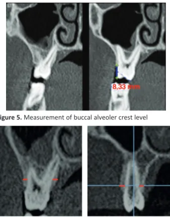
In the axial cross-sectional images, the upper posterior buccal bone thicknesses (BABT) and lingual bone thicknesses (LBT) were measured. The measurement was made from the buccolingual axis of the teeth and the midpoint of the root toward the outer contour of the bone (Figure 4). The measurements of the buccal alveolar crest level (BACL) were done on the corresponding coronal images of the upper first molars’ mesiobuccal tubercle cusps (Figure 5).
Figure 3. 1. NC-CN: Nasal cavity width, 2. Right maxillary width, 3. Left maxillary width, 4. Total maxillary width, 5. U6R0: Angle between upper first molar’s axes and horizontal reference plane, 6. U6L0: Angle between lower first molar’s axes and horizontal reference plane
Figure 4. Measurements of the upper buccal bone thickness
and lingual bone thickness Figure 6. palatal alveoler bone thickness measurementsUpper first molars’ buccal alveolar bone thickness and Figure 5. Measurement of buccal alveoler crest level
The measurements of the upper first molars buccal alveolar bone thickness (BABT) and palatal alveolar bone thickness (PABT) were made from the furcation of the teeth to outer corner of the bone in the coronal sections in which mesiobuccal and palatal roots were both seen (Figure 6). The measurements of the premolar BABT and PABT were made from the 2 mm apical of the top of the crest to outer corner of the bone in the coronal sections (Figure 6). The width between the root apex of the upper centrals (WUCA) and anterior nasal cavity width (ANCW)
were measured in the clearest coronal section which the roots of upper centrals can be seen (Figure 7). Posterior nasal cavity width (PNCW), palatal maxillary width (PMW M1) and palatal root apex width (PRAW M1) were measured in the clearest coronal section in which the palatal roots of the upper first molars can be seen (Figure 8).
Scanning Electron Microscope Analysis and Evaluation of Root Resorption
After the end of expansion (T1), the upper first premolar teeth were extracted according to the treatment plan and kept in 10% formaldehyde until they were investigated using a scanning electron microscope (SEM) for the analysis of root resorption.
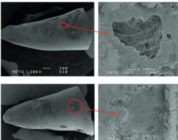
Figure 10. Scanning electron microscope images of the group rapid maxillary expansion
Figure 9. Scanning electron microscope images of the group semi-rapid maxillary expansion
Figure 8. Posterior nasal cavity width, palatal maxillary width (PMW M1) and palatal root apex width (PRAW M1)
Figure 7. The width of the upper centrals’ root apex and anterior nasal cavity width measurements
In order to prepare the teeth for SEM evaluation, the roots of the teeth were separated from the crown (if the tooth had two roots, just the buccal one was separated). Then, to remove the inorganic components from the root surface, all the specimens were submerged to 5% sodium hypochlorite for 2 hours, afterwards; they were dehydrated with increasing percentages of ethanol and were air-dried. All the root surfaces were coated with gold and the root surfaces were evaluated using a SEM microscope (SEM-EDS, JEOL, Tokyo, Japan). The images were digitalized with the SemAfore software.
The buccal root surfaces were examined in three parts: apical, medial and cervical. The presence of resorption cavity was evaluated in all the three parts. All the SEM images were taken at the same magnification; the assessments between groups and within group were made qualitatively. SRME and RME groups’ images are shown in Figure 9 and Figure 10.
Statistical Analysis
Data analysis was made using SPSS (Statistical Package for Social Science, SPSS Inc., Chicago, IL, United States) 11.5 for Windows. The reliability of the measurements made by the observer was evaluated by intraclass correlation coefficient at 95% confidence interval. The distribution of continuous variables was investigated using the Shapiro-Wilk test. Descriptive statistics for continuous variables were described by mean±standard deviation or median (the smallest - the largest). Nominal variables were described by percentages. The average differences between the groups were investigated with Student’s t-test and the significance of the difference in median values
was investigated with the Mann-Whitney U test. Nominal values were evaluated by Fisher’s exact test. Follow-up times of groups were evaluated by the dependent-sample t-test. A p value of less than 0.05 was considered statistically significant. However, a Bonferroni adjustment was made to control type 1 error in all possible multiple comparisons.
Results
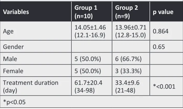
The demographic characteristics of the patients and the duration of the treatment are shown in Table 1. There was no statistically significant difference between the two groups in terms of age and gender. There was a significant difference in the duration of treatment between the groups (p<0.05)
Evaluation of Lateral Cephalometric Measurements
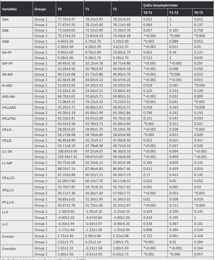
The statistical evaluation of the changes in the lateral cephalometric radiograph measurements are given in Table 2.
In the SRME group (group 1), ANB, MP-PP, SN-MP angles, ANS-Me, and VR A distances were increased significantly where U1-SN angle and overbite were decreased statistically significantly (p<0.0083) at T1-T0. During the retention period (T2-T1), significant decreases in MP-PP and SN-MP angles and significant increases in overbite were found (p<0.0083). At T2-T0, significant increases in N-ANS, HR ANS, VR A distances and significant decreases in U1-SN angle (p<0.0083) were detected.
Significant increases in ANB, MP-PP, SN-MP angles, ANS-Me, HR PNS, VR A, PP U1i measurements and overjet and significant decreases in SNB, U1-SN angles, and VR B, VR L1i measurements and overbite were found in the RME group (group 2) during the expansion period (T1-T0) (p<0.0083). During the retention period (T2-T1), significant decreases in SN-MP angle and significant increases in SNB angle and overbite value were detected (p<0.0083). During the total treatment period (T2-T0), significant decreases in SNB, U1-SN angles and VR L1i distance and significant increases in ANS-Me and PP U1i distances were found (p<0.0083).
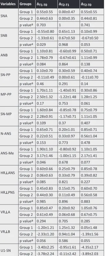
The statistical evaluations of lateral cephalometric changes between the groups in the observation period are given in Table 3. According to these evaluations, there was a significant difference only in PP U1i distance on T1-T0 and there were no significant differences in other variables.
Table 1. The demographic characteristics of patients and the duration of treatment
Variables Group 1 (n=10) Group 2 (n=9) p value Age 14.05±1.46 (12.1-16.9) 13.96±0.71 (12.8-15.0) 0.864 Gender 0.65 Male 5 (50.0%) 6 (66.7%) Female 5 (50.0%) 3 (33.3%) Treatment duration (day) 61.7±20.4 (34-98) 33.4±9.6 (21-48) *<0.001 *p<0.05
Table 2. The statistical evaluation of the changes of the lateral cephalometric radiographs measurements
Variables Groups T0 T1 T2 Çoklu karşılaştırmalar
T0-T1 T1-T2 T0-T2 SNA Group 1 77.70±3.97 78.25±3.93 78.25±4.02 0.012 1 0.012 Group 2 77.67±5.55 78.11±5.40 78.11±5.40 0.069 1 0.137 SNB Group 1 73.10±4.05 72.55±3.90 73.20±3.76 0.057 0.103 0.758 Group 2 75.17±4.23 73.83±4.19 74.50±4.28 *<0.001 *0.004 *0.004 ANB Group 1 4.60±2.26 5.70±2.70 5.10±2.40 *0.002 0.089 0.052 Group 2 2.50±2.49 4.28±2.59 3.61±2.37 *<0.001 0.011 0.01 SN-PP Group 1 9.60±2.60 9.70±2.89 10.00±2.73 0.662 0.14 0.121 Group 2 9.39±2.96 9.28±2.75 9.28±2.74 0.512 1 0.645 MP-PP Group 1 30.45±5.18 32.15±4.78 30.75±4.88 *<0.001 *<0.001 0.297 Group 2 33.28±4.56 35.78±4.92 34.56±4.18 *<0.001 0.038 0.016 SN-MP Group 1 40.15±6.08 41.75±5.80 40.90±5.70 *<0.001 *0.008 0.015 Group 2 42.56±4.38 44.83±4.13 43.67±4.26 *<0.001 *<0.001 0.013 N-ANS Group 1 53.65±3.83 54.30±3.43 54.50±3.94 0.018 0.545 *0.004 Group 2 53.33±3.18 53.56±3.12 53.89±3.43 0.225 0.332 0.149 ANS-Me Group 1 68.70±5.65 70.60±5.71 69.80±5.53 *<0.001 0.022 0.009 Group 2 71.06±4.12 74.22±4.33 73.22±4.51 *<0.001 0.031 *0.002 HR ANS Group 1 45.20±3.71 45.80±3.63 46.05±3.72 0.018 0.343 *0.008 Group 2 45.00±3.19 45.06±2.99 45.39±2.84 0.799 0.242 0.193 HR PNS Group 1 43.10±2.41 43.55±2.06 43.70±2.36 0.121 0.541 0.024 Group 2 43.33±3.58 43.78±3.73 43.89±3.66 *0.002 0.512 0.021 VR A Group 1 58.05±5.65 58.90±5.75 59.10±5.78 *<0.001 0.509 *0.002 Group 2 58.17±6.68 58.78±6.84 58.83±6.86 *0.005 0.813 0.029 VR B Group 1 46.95±8.84 45.75±9.17 47.00±8.50 0.012 0.015 0.917 Group 2 50.11±8.19 47.78±8.38 48.72±9.10 *<0.001 0.027 0.028 U1-SN Group 1 100.65±3.95 97.25±4.67 96.30±5.10 *<0.001 0.094 *<0.001 Group 2 103.44±7.32 99.67±5.65 99.56±6.69 *<0.001 0.894 *<0.001 L1-MP Group 1 93.75±6.08 92.70±6.12 92.65±5.98 0.183 0.859 0.135 Group 2 88.50±7.14 87.06±6.83 86.89±7.46 0.011 0.659 0.024 VR U1i Group 1 61.10±6.86 60.55±7.21 60.30±7.19 0.17 0.413 0.145 Group 2 61.00±7.60 60.33±7.79 60.11±8.32 0.022 0.65 0.052 VR L1i Group 1 55.70±7.87 54.75±8.35 55.75±7.42 0.041 0.065 0.92 Group 2 58.11±7.36 56.56±7.60 57.06±7.71 *<0.001 0.053 *0.002 PP U1i Group 1 30.85±3.01 31.20±2.99 31.30±3.02 0.01 0.509 0.019 Group 2 30.67±2.78 31.72±2.66 31.33±2.67 *<0.001 0.111 *0.004 Ls-E Group 1 -1.50±3.65 -1.95±4.10 -2.25±4.14 0.324 0.239 0.143 Group 2 -4.00±1.03 -3.67±0.66 -4.00±1.03 0.419 0.195 1 Li-E Group 1 -0.30±3.94 -0.50±3.97 -0.90±4.20 0.534 0.087 0.181 Group 2 -1.17±1.44 -1.22±1.09 -1.50±0.94 0.898 0.384 0.524 Overjet Group 1 5.75±2.49 5.90±2.80 5.15±2.08 0.722 0.091 0.104 Group 2 2.61±1.75 4.22±2.14 2.89±1.73 *0.003 0.01 0.384 Overbite Group 1 1.65±2.22 -0.15±1.58 1.00±1.83 *<0.001 *<0.001 0.164 Group 2 1.00±2.56 -0.61±2.93 0.33±2.72 *0.002 *0.006 0.057 *a: Bonferroni t-test; the results are significant for p<0.0083
Evaluation of Posteroanterior Cephalometric Measurements
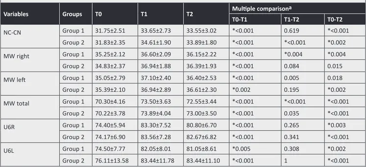
Statistical evaluations of PA cephalometric measurements are given in Table 4.
In the SRME group, significant increases in nasal cavity width (NC-CN), left and right maxillary width (MW), U6R°, and U6L° were found during T0-T1. During the retention period (T2-T1), significant decreases in right and total MW were found (p<0.0041) (p<0.0083). When the changes during the total treatment period (T2-T0) were analyzed, significant increases were detected in nasal cavity width, right MW, total MW, U6R°, and U6L°.
Significant increases in nasal cavity width (NC-CN), left, right and total MW, U6R°, U6L° measurements were found during expansion (T0-T1) in the RME
Table 3. The statistical evaluation of lateral cephalometric changes between the groups for the observation periods
Variables Groups ΔT1-T0 ΔT2-T1 ΔT2-T0 SNA Group 1 0.55±0.55Group 2 0.44±0.63 0.00±0.470.00±0.35 0.55±0.550.44±0.81
p valuea 0.703 1 0.741 SNB Group 1 -0.55±0.80 0.65±1.13Group 2 -1.33±0.61 0.67±0.50 0.10±0.99-0.67±0.50
p valuea 0.029 0.968 0.053 ANB Group 1 1.10±0.81Group 2 1.78±0.79 -0.60±0.99 0.50±0.71-0.67±0.61 1.11±0.99
p valuea 0.084 0.864 0.138 SN-PP Group 1 0.10±0.70Group 2 -0.11±0.49 0.00±0.610.30±0.59 0.40±0.74-0.11±0.70 p valuea 0.46 0.291 0.14 MP-PP Group 1 1.70±1.11Group 2 2.50±1.32 -1.40±0.91 0.30±0.86-1.22±1.48 1.28±1.25 p valuea 0.17 0.753 0.061 SN-MP Group 1 1.60±0.84Group 2 2.28±0.91 -0.85±0.78 0.75±0.79-1.17±0.71 1.11±1.05 p valuea 0.109 0.37 0.407 N-ANS Group 1 0.65±0.71Group 2 0.22±0.51 0.20±1.010.33±0.97 0.85±0.710.56±1.04
p valuea 0.153 0.773 0.478 ANS-Me Group 1 1.90±1.10Group 2 3.17±1.46 -0.80±0.92 1.10±1.05-1.00±1.15 2.17±1.41
p valuea 0.046 0.678 0.077 HR ANS Group 1 0.60±0.66Group 2 0.06±0.63 0.25±0.790.33±0.79 0.85±0.780.39±0.82
p valuea 0.085 0.821 0.227 HR PNS Group 1 0.45±0.83Group 2 0.44±0.30 0.15±0.750.11±0.49 0.60±0.700.56±0.58 p valuea 0.985 0.896 0.883 VR A Group 1 0.85±0.47Group 2 0.61±0.49 0.20±0.920.06±0.68 1.05±0.760.67±0.75 p valuea 0.294 0.705 0.285 VR B Group 1 -1.20±1.21 1.25±1.32Group 2 -2.33±1.20 0.94±1.04 0.05±1.48-1.39±1.56 p valuea 0.056 0.586 0.055 U1-SN Group 1 -3.40±2.25 -0.95±1.61 -4.35±2.17 Group 2 -3.78±2.24 -0.11±2.42 -3.89±2.03 Table 3. Continye p valuea 0.718 0.381 0.64 L1-MP Group 1 -1.05±2.30 -0.05±0.86 -1.10±2.12Group 2 -1.44±1.31 -0.17±1.09 -1.61±1.75 p valuea 0.657 0.798 0.576 VR U1i Group 1 -0.55±1.17 -0.25±0.92 -0.80±1.58 Group 2 -0.67±0.71 -0.22±1.42 -0.89±1.17 p valuea 0.798 0.96 0.892 VR L1i Group 1 -0.95±1.26 1.00±1.43 0.05±1.54 Group 2 -1.56±0.98 0.50±0.66 -1.06±0.73 p valuea 0.262 0.352 0.066 PP U1i Group 1 0.35±0.34 0.10±0.46 0.45±0.50 Group 2 1.06±0.30 -0.39±0.65 0.67±0.50 p valuea *<0.001 0.073 0.357 Ls-E Group 1 -0.45±1.36 -0.30±0.75 -0.75±1.48 Group 2 0.33±1.17 -0.33±0.71 0.00±1.41 p valuea 0.199 0.922 0.275 Li-E Group 1 -0.20±0.98 -0.40±0.66 -0.60±1.31Group 2 -0.06±1.26 -0.28±0.91 -0.33±1.50
p valuea 0.782 0.739 0.684 Overjet Group 1 0.15±1.29Group 2 1.61±1.17 -0.75±1.25 -0.60±1.05-1.33±1.20 0.28±0.91
p valuea 0.02 0.316 0.069 Overbite Group 1 -1.80±1.18 1.15±0.67Group 2 -1.61±1.02 0.94±0.77 -0.65±1.36-0.67±0.90
p valuea 0.716 0.541 0.975 *a: Bonferroni t-test; the results are significant for the p<0.017
group (group 2). During the retention period (T1-T2), a significant decrease was measured in nasal cavity width (p<0.0083). During the total treatment period (T2-T0), significant increases were detected in nasal cavity width, left MW, total MW, U6R°, and U6L° measurements.
Inter-group comparisons of PA cephalometric radiographic measurements in the observation period are given in Table 5. There were no statistically significant differences between the groups (p>0.017).
Evaluation of Computed Tomography Measurements
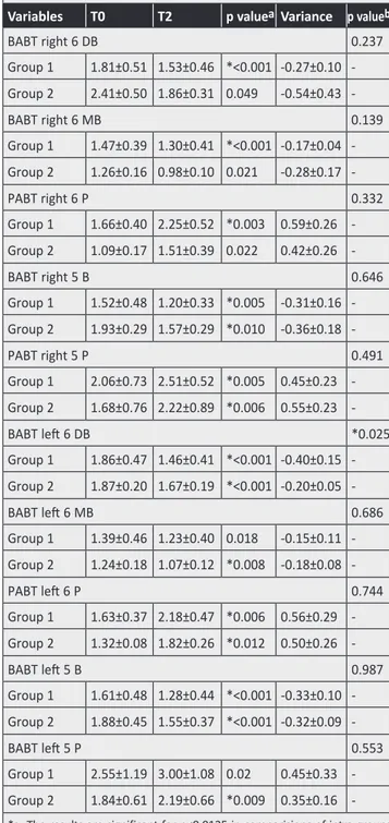
The values obtained with CT measurements are given in Table 6 and Table 7.
In SRME group, the changes during the total treatment period (T2-T0) were analyzed on the axial sections of the CT images and significant decreases in the BABT of tooth number 16 (level of the distobuccal and mesiobuccal roots), and significant increases in the palatal bone thickness of the same tooth were found (p<0.0125). Significant decreases in the BABT of tooth number 15 and significant increases in the palatal bone thickness of the same tooth (p<0.0125) were also detected. Moreover, a significant increase in palatal bone thickness of the tooth number 26 and a significant decrease in BABT of the tooth number 25 (p<0.0125) were measured.
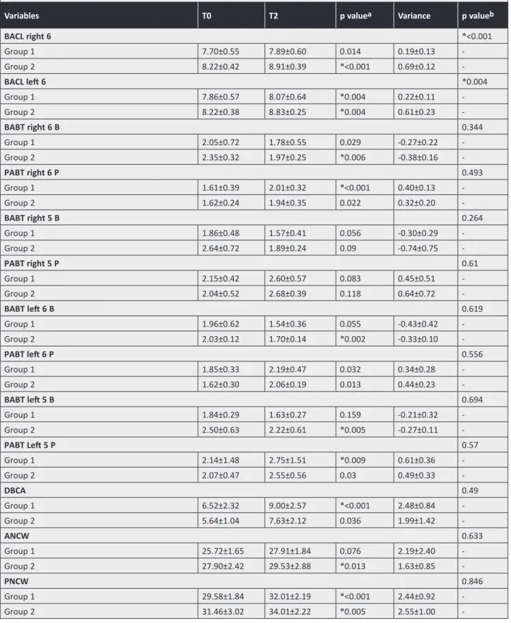
In the coronal sections of the group 1, a decrease of the BACL on the tooth number 26 on the total period was seen at T2-T0. The change in buccal and palatal bone thickness of the maxillary first molar and second premolar teeth is given in Table 7. There were statistically significant increases in the palatal bone thickness of the tooth number 16 and 25 (p<0.0125). The increase in the distance between the centrals’ apices (DBCA) during the T2-T0 period was statistically significant (p<0.025). There were significant increases in PNCW, PMW M1 and PRAW M1 (p<0.025).
In RME group, the changes during the total treatment time (T2-T0) were analyzed in the axial sections of the CT images and there were significant decreases in the BABT of the tooth number 15 and there were statistically significant increases in the palatal bone thickness of the same tooth (p<0.0125). There were significant decreases in the BABT of the tooth number 26 (level of the distobuccal and mesiobuccal roots) and significant increases in the palatal bone thickness of the same tooth (p<0.0125). Significant decreases were measured in the BABT of the tooth number 25 and significant increases in the palatal bone thickness of the same tooth (p<0.0125).
In RME group, the changes in coronal slices during the total treatment time (T2-T0) were analyzed and significant increases showing the reduction in the
Table 4. The statistical evaluation of posteroranterior cephalometric measurements for the observation period
Variables Groups T0 T1 T2 Multiple comparisona
T0-T1 T1-T2 T0-T2 NC-CN Group 1 31.75±2.51 33.65±2.73 33.55±3.02 *<0.001 0.619 *<0.001 Group 2 31.83±2.35 34.61±1.90 33.89±1.80 *<0.001 *<0.001 *0.002 MW right Group 1 35.25±2.12 36.60±2.09 36.15±2.22 *<0.001 *0.004 *0.004 Group 2 34.83±2.37 36.94±1.88 36.39±1.93 *<0.001 0.084 0.015 MW left Group 1 35.05±2.79 37.10±2.40 36.40±2.53 *<0.001 0.005 0.018 Group 2 35.39±2.10 36.94±2.89 36.61±2.30 *0.002 0.195 *0.002 MW total Group 1 70.30±4.16 73.50±3.63 72.55±3.44 *<0.001 *<0.001 *<0.001 Group 2 70.22±3.78 73.89±4.04 73.00±3.50 *<0.001 0.035 *<0.001 U6R Group 1 74.40±5.94 83.30±7.52 80.80±6.70 *<0.001 0.265 *0.003 Group 2 74.17±6.90 83.56±7.28 82.67±6.82 *<0.001 0.341 *<0.001 U6L Group 1 74.50±7.77 82.05±8.01 81.05±8.61 *0.005 0.308 *0.002 Group 2 76.11±13.58 83.44±11.78 83.44±11.10 *<0.001 1 *<0.001 MW: Maxillay width, *a: Bonferroni t-test. The results are significant for p<0.0083 (maxillary width right and maxillary width left excluding) for the maxillary width right and maxillay width left the results are significant for p<0.0041
buccal alveolar crest height of the teeth number 16 and 26 (p<0.0125) were seen. Significant decreases in the BABT of the teeth number 16, 26 and 25. (p<0.0125) were also measured. The changes during the total treatment time (T2-T0) were analyzed and significant increases in ANCW and PNCW were found, but no statistically differences were detected in PMW M1 (p>0.025).
Inter-group comparisons of the CT measurements are given in Table 6 and Table 7. The BABT of the tooth number 26 was compared between group RME and group SRME. An average decrease of 0.4 mm in group SRME and 0.2 mm in group RME were measured. Buccal crestal level of the tooth number 16 was analyzed in T2-T0 period, an average of 0.19 mm increase in group SRME and 0.69 mm increase in group RME were measured, which means a reduction in the bone level. These differences between the groups were statistically significant (p<0.025). Buccal crestal level of the tooth number 26 was analyzed
in T2-T0 period and an average of 0.22 mm increase in group SRME and 0.61 mm increase in group RME were found, which means a reduction in the bone level. This difference between the groups were found to be statistically significant (p<0.025).
Table 5. Inter-groups comparisons of posteroanterior cephalometric radiographic measurement for the observation period
Variables Groups ΔT1-T0 ΔT2-T1 ΔT2-T0 NC-CN Group 1 1.90±0.88 -0.10±0.61Group 2 2.78±1.09 -0.72±0.44 1.80±1.062.06±1.33
p valuea 0.069 0.023 0.648 MW right Group 1 1.35±0.67 -0.45±0.37Group 2 2.11±0.96 -0.56±0.85 0.90±0.741.56±1.51
p valueb 0.059 0.724 0.238 MW left Group 1 2.05±1.09 -0.70±0.59Group 2 1.56±1.01 -0.33±0.71 1.35±1.471.22±0.79
p valueb 0.322 0.234 0.82 MW total Group 1 3.20±1.21 -0.95±0.55Group 2 3.67±1.50 -0.89±1.05 2.25±1.532.78±1.58
p valuea 0.463 0.874 0.471 U6R Group 1 8.90±4.54 -2.50±6.65Group 2 9.39±3.64 -0.89±2.63 6.40±5.118.50±4.36
p valuea 0.8 0.507 0.352 U6L Group 1 7.55±6.50 -1.00±2.92Group 2 7.33±3.71 0.00±3.13 6.55±4.887.33±4.03
p valuea 0.931 0.482 0.709 *a: The results are significant for p<0.017 in Bonferroni correction, *b: The results are significant for p<0.0083 in Bonferroni correction, MW: Maxillary width
Table 6. The values for computed tomography measurements for axial sections
Variables T0 T2 p valueaVariance p valueb
BABT right 6 DB 0.237 Group 1 1.81±0.51 1.53±0.46 *<0.001 0.27±0.10 -Group 2 2.41±0.50 1.86±0.31 0.049 0.54±0.43 -BABT right 6 MB 0.139 Group 1 1.47±0.39 1.30±0.41 *<0.001 0.17±0.04 -Group 2 1.26±0.16 0.98±0.10 0.021 0.28±0.17 -PABT right 6 P 0.332 Group 1 1.66±0.40 2.25±0.52 *0.003 0.59±0.26 -Group 2 1.09±0.17 1.51±0.39 0.022 0.42±0.26 - BABT right 5 B 0.646 Group 1 1.52±0.48 1.20±0.33 *0.005 0.31±0.16 -Group 2 1.93±0.29 1.57±0.29 *0.010 0.36±0.18 -PABT right 5 P 0.491 Group 1 2.06±0.73 2.51±0.52 *0.005 0.45±0.23 -Group 2 1.68±0.76 2.22±0.89 *0.006 0.55±0.23 -BABT left 6 DB *0.025 Group 1 1.86±0.47 1.46±0.41 *<0.001 0.40±0.15 -Group 2 1.87±0.20 1.67±0.19 *<0.001 -0.20±0.05 - BABT left 6 MB 0.686 Group 1 1.39±0.46 1.23±0.40 0.018 0.15±0.11 -Group 2 1.24±0.18 1.07±0.12 *0.008 0.18±0.08 -PABT left 6 P 0.744 Group 1 1.63±0.37 2.18±0.47 *0.006 0.56±0.29 -Group 2 1.32±0.08 1.82±0.26 *0.012 0.50±0.26 -BABT left 5 B 0.987 Group 1 1.61±0.48 1.28±0.44 *<0.001 0.33±0.10 -Group 2 1.88±0.45 1.55±0.37 *<0.001 0.32±0.09 -BABT left 5 P 0.553 Group 1 2.55±1.19 3.00±1.08 0.02 0.45±0.33 -Group 2 1.84±0.61 2.19±0.66 *0.009 0.35±0.16 -*a: The results are significant for p<0.0125 in comparisions of intra-groups between T0-T2 in Bonferroni t-test. *b: The results are significant for p<0.025 in comparisions of inter-groups between T0-T2 in Bonferroni student’s t-test, BABT: Buccal alveolar bone thickness
Table 7. The values for computed tomography measurements for coronal sections
Variables T0 T2 p valuea Variance p valueb
BACL right 6 *<0.001 Group 1 7.70±0.55 7.89±0.60 0.014 0.19±0.13 -Group 2 8.22±0.42 8.91±0.39 *<0.001 0.69±0.12 -BACL left 6 *0.004 Group 1 7.86±0.57 8.07±0.64 *0.004 0.22±0.11 -Group 2 8.22±0.38 8.83±0.25 *0.004 0.61±0.23 -BABT right 6 B 0.344 Group 1 2.05±0.72 1.78±0.55 0.029 -0.27±0.22 -Group 2 2.35±0.32 1.97±0.25 *0.006 -0.38±0.16 -PABT right 6 P 0.493 Group 1 1.61±0.39 2.01±0.32 *<0.001 0.40±0.13 -Group 2 1.62±0.24 1.94±0.35 0.022 0.32±0.20 - BABT right 5 B 0.264 Group 1 1.86±0.48 1.57±0.41 0.056 -0.30±0.29 -Group 2 2.64±0.72 1.89±0.24 0.09 -0.74±0.75 - PABT right 5 P 0.61 Group 1 2.15±0.42 2.60±0.57 0.083 0.45±0.51 -Group 2 2.04±0.52 2.68±0.39 0.118 0.64±0.72 -BABT left 6 B 0.619 Group 1 1.96±0.62 1.54±0.36 0.055 -0.43±0.42 -Group 2 2.03±0.12 1.70±0.14 *0.002 -0.33±0.10 -PABT left 6 P 0.556 Group 1 1.85±0.33 2.19±0.47 0.032 0.34±0.28 -Group 2 1.62±0.30 2.06±0.19 0.013 0.44±0.23 -BABT left 5 B 0.694 Group 1 1.84±0.29 1.63±0.27 0.159 -0.21±0.32 -Group 2 2.50±0.63 2.22±0.61 *0.005 -0.27±0.11 -PABT Left 5 P 0.57 Group 1 2.14±1.48 2.75±1.51 *0.009 0.61±0.36 -Group 2 2.07±0.47 2.55±0.56 0.03 0.49±0.33 - DBCA 0.49 Group 1 6.52±2.32 9.00±2.57 *<0.001 2.48±0.84 -Group 2 5.64±1.04 7.63±2.12 0.036 1.99±1.42 -ANCW 0.633 Group 1 25.72±1.65 27.91±1.84 0.076 2.19±2.40 -Group 2 27.90±2.42 29.53±2.88 *0.013 1.63±0.85 - PNCW 0.846 Group 1 29.58±1.84 32.01±2.19 *<0.001 2.44±0.92 -Group 2 31.46±3.02 34.01±2.22 *0.005 2.55±1.00
-Evaluation of Root Resorption
The SEM images of group 1 and group 2 are given in Figure 9 and Figure 10, respectively. Resorption was observed in medial part of the root of the all specimens from group 1 but some of the specimens just showed resorption on cervical or apical parts. The resorption which included all of the three surfaces was very low in group 1, but there was no specimen with any evidence of resorption.
A great number of the specimens in group 2 had resorption in all the three parts of the root. In the apical part of some specimens (compared to the other regions), less resorption was observed. Only one tooth in this group showed no resorption. The specimens showing resorption in all of the three parts were higher in group 2 than in group 1.
The deepness of the cavities was minor in both groups. These resorption cavities included either cement or both cement and dentin in all of the groups. There were no significant differences in the depth of the resorption cavities between the groups.
Discussion
No previous studies compared the skeletal, dentoalveolar and periodontal effects that occurred after RME and SMRE with acrylic splint bonded expansion, especially using CT measurements. The present study aimed to investigate both the dentofacial and the side effects of these expansion methods thoroughly.
In the literature, some studies suggested that A point moves forward and downward with RME (3,8-13) but controversial views are also present (14-18). In the present study, some increases in SNA angle and the distance of the A point to the vertical reference plane (VR A) were measured in both groups, but these results showed no statistical significance. These
findings are in accordance with the results of Özsoy (19) (0.52°) and Ramoğlu (0.55°) (20).
Wertz and Haas have suggested that the vertical increase which was seen after RME was not permanent (3,21). In our study, a downward and backward movement of the mandible was observed in both groups, but there were no significant differences between the groups. These findings are similar with the results of Kılıç (22), who used RME and SMRE protocols with a rigid bonded RME appliance.
For the lateral cephalometric measurements, the only difference between the groups was PP U1i distance. These findings are similar with the results of previous researchers who suggested that extrusion of the upper incisors can be seen with RME (19-22). Uprighting of the upper incisors were found in both of groups; this result is not similar with the results of previous studies (19,22,23). However, there are variations of expansion appliances that have been used in different studies and the inconsistency with the previous studies may be a result of this fact.
When PA radiographs were analyzed, it was noticed that there was a higher amount of increase in the nasal cavity width in RME group which may be due to the rapid rate of activation in this group that prevented the dental movement of posterior teeth, thus, providing more bony expansion.
There have been some studies (24,25) suggesting that there was lower tipping in the posterior teeth after RME with acrylic bonded appliance and there have been some other studies (26,27) which found more tipping in RME groups than in slow expansion groups. In our study, after the active expansion period, U6R° value increased to 8.9° in SRME and 9.39° in RME groups. The more tipping of the molar in RME group can be caused by more maxillary expansion of the right segment. In SRME group, the U6L° value increased to 7.55°, and in RME, the value to increased
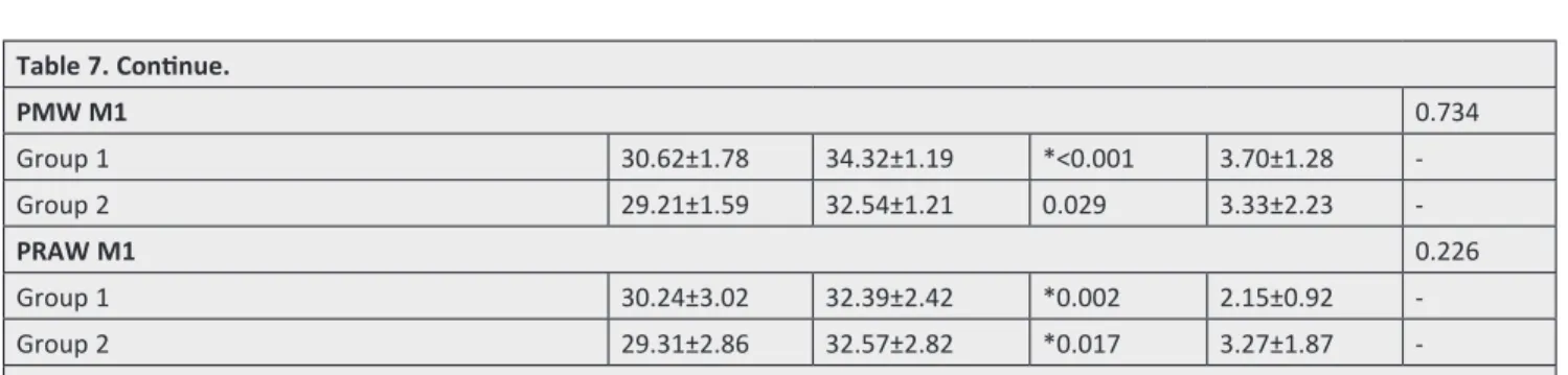
Table 7. Continue. PMW M1 0.734 Group 1 30.62±1.78 34.32±1.19 *<0.001 3.70±1.28 -Group 2 29.21±1.59 32.54±1.21 0.029 3.33±2.23 -PRAW M1 0.226 Group 1 30.24±3.02 32.39±2.42 *0.002 2.15±0.92 -Group 2 29.31±2.86 32.57±2.82 *0.017 3.27±1.87 -
*a: The results are significant for p<0.0125 in comparisions of intra-groups between T0-T2 in Bonferroni t-test. *b: The results are significant for p<0.025 in comparisions of inter-groups between T0-T2 in Bonferroni student’s t-test
7.33°. Likewise, the more tipping of the molar in SRME group can be caused by more maxillary expansion of the left segment in the same group. There was no statistically difference between the groups in the tipping of the upper molar teeth, and as a result, both of the expansion protocols can cause the same amount of the tipping.
Garib et al. (28) investigated the periodontal effects of RME using CT and concluded that there were decreases in BABT of the supporting teeth and also formation of dehiscence. There is a positive correlation between buccal movement of teeth and bone dehiscence. The animal studies made by Engelking and Zachrisson (29), Steiner et al. (30), Thilander et al. (31), and Wennstrom et al. (32) concluded that buccal tooth movement can increase the distance between BACL and cement-enamel junction. In our study, BACL increased statistically significantly in both groups that means a decrease in crestal level. There was more bone loss in group RME, which was not significant compared to the first group, but this can be due to the increased amount of tipping in this group. These findings are similar with those of Garib et al. (28), and Rungcharassaeng et al. (33).
Ballanti et al. (34) investigated the changes in the distance between the central teeth apices after RME and concluded that during the expansion period, the distance increased to 3.49 mm and decreased to 1.98 mm after 6 months. In our study, between T0 and T2, a significant increase of 2.48 mm was measured in SRME group but no significant difference was measured in RME group. This may have occurred due to the increased relapse potential after RME.
Garrett et al. (35) investigated skeletal effects of RME using cone beam computed tomography and concluded that there was a 2.67 mm increase in the PMW M1. In our study, this value was greater in both groups, but this difference can be attributed to the differences in the appliances and the need for the amount of expansion in our study group.
Several studies have investigated the incidence of root resorption after RME (9,13,36,37), but we could not find any study about the correlation between the speed of expansion and the amount of root resorption. There were varieties between resorption cavities in both groups. Some cavities were wide and superficial, some others were narrow and profound, but there were no differences between the groups. The varieties of the resorption cavities between teeth can be explained by personal differences and predispositions. However, there was one distinct fact that almost all the teeth had some degree of root
resorption. The repair of the resorption can start after the force discontinued or decreased (38,39). In our study, first premolars were extracted at the end of the expansion period to eliminate repair process. To find out the connection between the root resorption and maxillary expansion speed, future studies with larger sample sizes are required.
Conclusions
1. Some differences were seen between the short-time effects of SRME and RME, but these differences were temporary and disappeared at the end of the retention period.
2. Some decreases in the BABT of all first molars and second premolars and some increases in the palatal bone thickness of the same teeth were measured in both groups. On CT images, more decreases in the buccal alveolar crest height were detected at the level of the maxillary molars in the RME group.
3. The qualitative evaluation of the roots showed that the location and the degree of the root resorption were similar between the groups.
Ethics
Ethics Committee Approval: The study was approved by the Başkent University Local Ethics Committee (protocol number: D-KA 10/07), Informed Consent: Consent form was filled out by all participants.
Authorship Contributions
Surgical and Medical Practices: Ş.R.G., Concept: Ö.P.Ö., Design: Ö.P.Ö., Ş.R.G., Data Collection or Processing: Ş.R.G., F.K., Analysis or Interpretation: Ş.R.G., F.K., H.P., Literature Search: H.P., Ş.R.G., Writing: H.P., Ö.P.Ö., Ş.R.G.
Conflict of Interest: No conflict of interest was declared by the authors.
Financial Disclosure: The authors declared that this study received no financial support.
References
1. McNamara JA. Maxillary transverse deficiency. Am J Orthod Dentofacial Orthop 2000; 117: 567-70.
2. Sandıkçıoğlu M, Hazar S. Skeletal and dental changes after maxillary expansion in the mixed dentition. Am J Orthod Dentofacial Orthop 1997; 111: 321-7.
3. Suri L, Taneja P. Surgically assisted rapid palatal expansion: a literature review. Am J Orthod Dentofacial Orthop 2008; 133: 290-302.
4. Linder-Aronson S, Lindgren J. The skeletal and dental effects of rapid maxillary expansion. Br J Orthod 1979; 6: 25-9.
5. Akkaya S, Lorenzon S, Ücem TT. Comparison of dental arch and arch perimeter changes between bonded rapid and slow maxillary expansion procedures. Eur J Orthod 1998; 20: 255-61.
6. İşeri H, Tekkaya E, Öztan Ö, Bilgiç S. Biomechanical effects of rapid maxillary expansion on the craniofacial skeleton, studied by the finite element method. Eur J Orthod 1998; 20: 347-56. 7. Orhan M, Üşümez S. A new method for the assessment of
changes in molar inclination during RME. Cited: 15.10.2001. Availiable from: URL: http://www.vjo.it/042/molar.htm 8. Haas AJ. Rapid expansion of the maxillary dental arch and nasal
cavity by opening the midpalatal suture. Angle Orthod 1961; 31: 73-89.
9. Erverdi N, Okar I, Küçükkeleş N, Arbak S. A comparison of two different rapid palatal expansion techniques from the point of root resorption. Am J Orthod Dentofacial Orthop 1994; 106: 47-51. 10. Haas AJ. Long term posttreatment evaluation of rapid palatal
expansion. Angle Orthod 1980; 50: 189-217.
11. Chung CH, Font B. Skeletal and dental changes in the sagittal, vertical, and transverse dimensions after rapid palatal expansion. Am J Orthod Dentofacial Orthop 2004; 126: 569-75.
12. Davis WM, Kronman JH. Anatomical changes induced by splitting of the midpalatal suture. Angle Orthod 1969; 39: 126-32. 13. Langford SR. Root resorption extremes resulting from clinical
RME. Am J Orthod 1982; 81: 371-7.
14. Da Silva Filho OG, Boas MC, Capelozza Filho L. Rapid maxillary expansion in the primary and mixed dentition: a cephalometric evaluation. Am J Orthod Dentofacial Orthop 1991; 100: 171-9. 15. Asanza S, Cisneros GJ, Nieberg LG. Comparison of Hyrax and
bonded expansion appliances. Angle Orthod 1997; 67: 15-22. 16. Sarver DM, Johnston MW. Skeletal changes in vertical and
anterior displacement the maxilla with bonded rapid palatal expansion appliances. Am J Orthod Dentofacial Orthop 1989; 95: 462-6.
17. Reed N, Ghosh J, Nanda RS. Comparison of treatment outcomes with banded and bonded RPE appliances. Am J Orthod Dentofacial Orthop 1999; 116: 31-40.
18. Byrum AG. Evaluation of anterior-posterior and vertical skeletal change vs. dental change in rapid palatal expansion cases as studied by lateral cephalograms. Am J Orthod 1971; 60: 419. 19. Özsoy FS. Semi-rapid üst çene genişletmesinin dentofasiyal yapılar
üzerine olan etkilerinin incelenmesi: Ankara University, 2001. 20. Ramoğlu Sİ. Karma dentisyon döneminde modifiye akrilik bonded
aparey ile yapılan hızlı ve yarı hızlı üst çene genişletmesinin dentofasiyal yapılar üzerine etkilerinin değerlendirilmesi: Selçuk University, 2006.
21. Haas AJ. Palatal expansion: Just the beginning of dentofacial orthopedics. Am J Orthod 1970; 57: 219-55.
22. Kılıç N. Yarı hızlı ve hızlı üst çene genişletmesinin dentofasiyal yapılar ve çiğneme kaslarının tonositelerinde yaptığı değişimlerinin incelenmesi: Atatürk University, 2005.
23. Baykara C. Banded ve bonded RME apareylerinin dentofasiyal sistemdeki etkilerinin karşılaştırılması: Hacettepe University. 1999.
24. Kılıç N, Kiki A, Oktay H. A comparison of dentoalveolar inclination treated by two palatal expanders. Eur J Orthod 2008; 30: 67-72. 25. Memikoğlu UT, İşeri H. Nonextraction treatment with a rigid
acrylic, bonded rapid maxillary expander. J Clin Orthod 1997; 31: 113-8.
26. Ciambotti C, Ngan P, Durkee M, Kohli K, Kim H. A comparison of dental and dentoalveolar changes between rapid palatal expansion and nickel-titanium palatal expansion appliances. Am J Orthod Dentofacial Orthop 2001; 119: 11-20.
27. Erdinç AE, Uğur T, Erbay E. A comparison of different treatment techniques for posterior crossbite in the mixed dentition. Am J Orthod Dentofacial Orthop 1999; 116: 287-300.
28. Garib DG, Henriques JFC, Janson G, De Freitas MR, Fernandes AY. Periodontal effects of rapid maxillary expansion with tooth-tissue-borne and tooth-borne expanders: a computed tomography evaluation. Am J Orthod Dentofacial Orthop 2006; 129: 749-58.
29. Engelking G, Zachrisson BU. Effects of incisor repositioning on monkey periodontium after expansion through the cortical plate. Am J Orthod 1982; 82: 23-32.
30. Steiner GG, Pearson JK, Ainamo J. Changes of the marginal periodontium as a result of labial toorh movement in monkeys. J Periodontol 1981; 52: 314-20.
31. Thilander B, Nyman S, Karring T, Magnusson I. Bone regeneration in alveolar bone dehiscences related to orthodontic tooth movements. Eur J Orthod 1983; 5: 105-14.
32. Wennstrom JL, Lindhe J, Sinclair F, Thilander B. Some periodontal tissue reactions to orthodontic tooth movement in monkeys. J Clin Periodontol 1987; 14: 121-9.
33. Rungcharassaeng K, Caruso JM, Kan JY. Factors affecting buccal bone changes of maxillary posterior teeth after rapid maxillary expansion. Am J Orthod Dentofacial Orthop 2007; 132: 428.e1-428.e8.
34. Ballanti F, Lione R, Baccetti T, Franchi L, Cozza P. Treatment and posttreatment skeletal effects of rapid maxillary expansion investigated with low-dose computed tomography in growing subjects. Am J Orthod Dentofacial Orthop 2010; 138: 311-7. 35. Garrett BJ, Caruso JM, Rungcharassaeng K, Farrage JR, Kim JS,
Taylor GD. Skeletal effects to the maxilla after rapid maxillary expansion assessed with cone-beam computed tomography. Am J Orthod Dentofacial Orthop 2008; 134: 8.e1-8.e11.
36. Odenrick L, Karlander EL, Pierce A, Kretschmar U. Surface resorption following two forms of rapid maxillary expansion. Eur J Orthod 1991; 13: 264-70.
37. Barber AF, Sims MR. Rapid maxillary expansion and external root resorption in man: a scanning electron microscope study. Am J Orthod 1981; 79: 630-52.
38. Rygh P. Orthodontic root resorptions studied by electron microscopy. Angle Orthod 1977; 47: 1-16.
39. Casa MA, Faltin RM, Faltin K, Sander FG, Arana-Chavez. Root resorptions in upper first premolars after application of continuous torque moment. Intra-individual study. J Orofac Orthop 2001; 62: 285-95.