The role of bcl-2 family of genes during kindling
7
0
0
Tam metin
(2) 218. K. C. AKCALI ET AL.. expression of bax but not bcl-2 was increased in rat brains after kindled seizures (12). However, to our knowledge, the participation of the bcl2 family of genes has not been investigated during the formation period of the kindled form of epilepsy. In addition, it is not clear whether this neuronal death is directly involved in epileptogenesis or occurs secondary to the effects of the severe and prolonged seizures. To elucidate the role of bcl-2 family of genes and apoptosis during epileptogenesis, we investigated the expression pattern of the different bcl-2 genes both at mRNA and protein level, by monitoring at different stages of the kindling model of epilepsy in rats. Our observations provide insight into the involvement of the Bcl-2 family of proteins during epileptogenesis. METHODS Animals Male, 9-week-old, 280- to 300-g Sprague–Dawley rats were used. They were housed under controlled environmental conditions (22◦ C) with a 12-h light and 12-h dark cycle in the animal holding facility of the Department of Molecular Biology and Genetics at the Bilkent University, Turkey. All the animals received care according to the criteria outlined in the “Guide for Care and Use of Laboratory Animals” prepared by the National Academy of Science, and this study protocol complied with Bilkent University’s guidelines on humane care and use of laboratory animals. The animals were permitted unlimited access to food and water at all times. Before used in the experiments, the rats were allowed to adapt the new conditions for ≥1 week. Animals were randomly divided into three groups: one group was implanted with electrodes and received electrical impulses, the other group of animals was implanted with electrodes but did not receive electrical impulses and served as a sham group, and the last group of animals was neither implanted with electrodes nor stimulated with electrical impulses, and thus served as a control group. Animals were killed 1, 3, 7, 10, and 14 days after the electrical impulse (n = 3 at each time point) with corresponding sham and control groups of animals. Kindling procedure Rats were anesthetized by using phenobarbital (PB; 50 ml/kg, i.p.) and placed in a stereotaxic frame. A bipolar, plastic-coated stainless-steel electrode was implanted into the right ventral hippocampal CA1 region at the following coordinates: tooth-bar at 0: 4.8 mm caudal to bregma, 5.2 mm lateral to midline, and 6.5 mm ventral to dura (33). A reference electrode was placed between the skull and left temporal muscle. Ten days after surgery, we started to give one kindling stimulation (1-s, 100-Hz biphasic pulses of 1-ms duration, 400 µA peak-to-peak amplitude delivered Epilepsia, Vol. 46, No. 2, 2005. by a stimulator) every day for 14 days. Animals were killed 1, 3, 7, 10, and 14 days after the initial stimulus. Behavioral seizures were scored as follows (34): grade 0, normal behavior, wet-dog shakes, arrest; 1, facial twitches; 2, head nodding, chewing; 3, forelimb clonus; 4, rearing, falling on forelimbs; 5, falling on the side or back, hindlimb clonus. Electroencephalographic activity was monitored by a Nihon-Kohden 14-channel EEG recorder. Tissue fixation, synthesis of sense and antisense mRNAs, and in situ hybridization protocols Rats were killed and their brains were quickly removed and cryopreserved by putting them into 4% cold paraformaldeyde for 18 h and then into 30% sucrose for 24 h. The full-length rat bax, bcl-2, bcl-w, mtd, and bcl-xL were cloned into pGEM4Z (Promega, Madison, WI, U.S.A.) and pBluescript II SK+ (Stragene, La Jolla, CA, U.S.A.) expression vectors. After linearization with the appropriate restriction enzymes, digoksin-labeled antisense or sense RNAs were synthesized by using the Dig RNA labeling kit (Roche Diagnostics, Mannheim, Germany), according to the manufacturer’s protocol. After ethanol precipitation, the probes were stored at –70◦ C until used. The 15-µm cryosections were placed on the Silanetreated slides and were hybridized with 10 ng digoksinlabeled bax, bcl-2, bcl-w, mtd, and bcl-xL probe at 30 µl hybridization buffer consisting 40% deionized formamide, 10% dextran sulfate, ×1 Denhardt’s solution (0.002% Ficoll, 0.02% polyvinylpyrrolidone, 10 mg/ml Rnase-free bovine serum albumin), ×4 SSC, 10 mM DDT, 1 mg/ml yeast t-RNA, and 1 mg/ml denatured and sheared salmon sperm DNA. Sense and antisense probes were placed on the upper and lower half of every slide, respectively, and separate coverslips were used for these sections. Slides were placed horizontally in a sealed, humidified container and incubated at 42◦ C for 16 h; then the coverslips were removed, and slides were washed with ×2 SSC and ×1 SSC for 15 min each and subjected to 30-min incubation at 37◦ C with NTE buffer (500 mM NaCl, 10 mM Tris, and 1 mM EDTA, pH 8.0) containing 20 µg/ml Rnase A to digest any single-stranded RNA probe. To visualize the bound probes, slides were then incubated with APlabeled anti-digoksin antibody at 1:500 dilution at 4◦ C for 16 h. Slides were then incubated with NBT/BCIP as substrate with 10 mM levamisole to block endogenous AP activity. Then the slides were examined by using a ZeissAxioskop microscope at ×4, ×10, and ×40 magnification. To confirm tissue integrity, some slides were subjected to hematoxylin/eosin staining and examined under brightfield illumination. Immunocytochemistry Cryosections (5 µm) were incubated for 30 min in 0.3% hydrogen peroxide in methanol to quench endogenous.
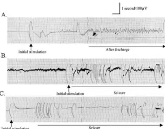
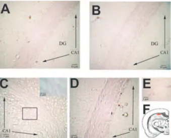
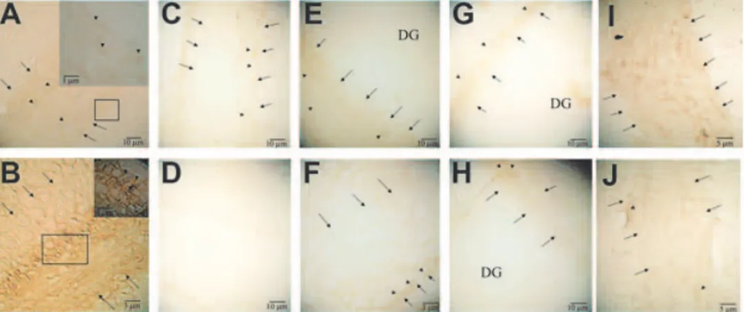
(3) bcl-2 FAMILY OF GENES DURING EPILEPTOGENESIS peroxidase activity. After washing 3 times with phosphatebuffered saline (PBS) for 10 min each, slides were incubated with preblocking serum (normal goat serum, 1.5%; bovine serum albumin, 2%; Triton-X, 0.1%) for 1 h at room temperature. The primary antibody of Bax, Bcl-xL , Bcl-2, Bcl-w, and Mtd (Santa Cruz Biotechnology, Inc., Santa Cruz, CA, U.S.A.) was applied at a concentration of 1 µg/ml in preblocking solution and kept at 4◦ C overnight. After washing 3 times with PBS, tissue sections were incubated for 1 h with 3 µg/ml of biotinylated goat antirabbit antibody (Vector Laboratories, Burlingame, CA, U.S.A.) in the preblocking solution containing 1% normal rat serum (Sigma), washed in PBS, and incubated for 1 h with an avidin-biotin complex reagent containing horseradish peroxidase (HRP) (Vector Laboratories) in PBS. After washing 10 min with PBS, slides were rinsed in 0.5% Triton-X 100/PBS for 30 s. Color development was achieved by incubation with diaminobenzidine (DAB) solution (Vector Laboratories) for 7 min. The tissues were examined by using Zeiss-Axioskop microscope at ×4, ×10, and ×40 magnification. To analyze immunoreactivity, the number of immunopositive cells in sections was semiquantitatively scored (35). The scoring was as follows: 0, not present (Fig. 4D); 1, light (Fig. 4C, F, G, H, J); 2, moderate (Fig. 4A, E, I); 3, high (Fig. 4B). In situ analysis of DNA fragmentation (terminal deoxynucleotidyl transferase–mediated dUTP nick-end labeling; TUNEL) DNA fragmentation was detected in situ with a TdT- (terminal deoxynucleotidyl transferase)–mediated fluorescein-dUTP labeling kit (Roche Diagnostics, Mannheim, Germany). Brain cryosections were fixed in 4% paraformaldehyde in PBS for 20 min at room temperature and then washed in PBS for 30 min. After incubating with a permeabilization solution (0.1% Triton X-100, 0.1% sodium citrate) for 2 min on ice, 50 µl of TUNEL reaction mixture was put into each sample and incubated for 1 h at 37◦ C in the dark in a humidified chamber. Slides were then directly analyzed with fluorescence microscopy. For evaluation by fluorescence microscopy, we used an excitation wavelength in the range of 450–500 nm and detection in the range of 515–565 nm. As negative control, we incubated the slides in the absence of TdT. For positive controls, the samples were first treated with DNase I (1,000 U/ml in 50 mM Tris-HCl, pH 7.5, 1 mg/ml BSA) for 10 min at 20◦ C to induce DNA strand breaks before labeling procedures and then incubated with 50 µl of TUNEL reaction mixture. RESULTS Induction of epilepsy in animals (kindling procedure) To investigate the participation of the bcl-2 family of genes during the formation period of kindled epilepsy, first. 219. FIG. 1. Patterns of electrographic activity during kindling. EEG traces were recorded from the electrically stimulated animals on day 1 (A); day 10 (B); and day 14 (C). After day 10, we observed stage 4 kindling seizures (rearing and falling on forelimbs).. we performed the kindling procedure to induce epilepsy. After day 10, we observed stage 4 kindling seizures (rearing and falling on forelimbs) as defined by Racine (34). As seen from EEG traces, afterdischarges were evident after applying electrical stimulus on day 1 (Fig. 1A). We observed epileptic EEG activity on day 10 (Fig. 1B) and day 14 (Fig. 1C). We did not detect any epileptic activity in the sham and control group of animals in their EEG traces (data not shown). To elucidate the role of the bcl-2 family of genes and apoptosis during epileptogenesis, we investigated the expression pattern of different bcl-2 family of genes (bax, bcl-xL bcl-2, mtd, bcl-w) at both the mRNA and protein levels, by monitoring at different stages of the kindling model of epilepsy in rats by using in situ hybridization, immunohistochemistry, and in situ analysis of DNA fragmentation (TUNEL). bax mRNA expression We observed bax mRNA expression in the hippocampal CA1 area as early as 1 day after the electrical stimulus (Fig. 2A). In the corresponding sham group of animals on day 1, bax mRNA expression also was present but to a lesser extent (Fig. 2B). Decreased expression in the sham group suggests that the increase in the expression of bax is due to electrical stimulus. Increased mRNA expression of bax was observed on day 9 (data not shown), but it was more evident at day 10 (Fig. 2C). Similar to day 10, bax expression persisted on day 14 (data not shown). Conversely, bax expression was not detectable in the sham group of animals on day 10 (Fig. 2D). By using sense bax mRNA as a control, we did not detect any specific binding (Fig. 2E). We also observed morphologic changes in the limbic lobe tissues starting on day 10 (Fig. 2C). These changes were not present in sham group animals on day 10 Epilepsia, Vol. 46, No. 2, 2005.
(4) 220. K. C. AKCALI ET AL.. FIG. 2. The expression of bax mRNA in CA1 region (F) during the formation of kindling. The expression of bax mRNA was examined by using antisense (A–D) and sense (E) bax riboprobes on day 1 (A) and day 10 (C) of electrically stimulated animals and compared to that of sham group on day 1 (B) and day 10 (D). A: bax mRNA expression was present in CA1 region of electrically stimulated rat brains on day 1. Inset, bax mRNA-positive and -negative staining area shown at high power. B: Decreased expression of bax mRNA in CA1 region of sham groups on day 1. C: bax mRNA expression increased on day 10 of electrically stimulated rat brains. Inset, bax mRNA-positive and -negative staining area shown at high power. D: No detectable bax mRNA expression in CA1 region of sham groups on day 10. E: Section probed with sense bax riboprobes revealed no specific binding. F: Cross section of brain at 35th plate, red rectangle denotes CA 1 region. Arrows, indicate the CA1 region in the brain tissues. DG, dentate gyrus.. (Fig. 2D), which rules out the local irritation of implanted electrodes. bcl-xL mRNA expression We found similar expression levels of bcl-xL mRNA at the CA1 region both in the electrically stimulated (Fig. 3A) and the sham group of animals (Fig. 3B) on day 1. This suggested that a basal level of expression existed, and this expression was not affected by electrical stimulus. Interestingly, when we examined the later stages, bcl-xL expression almost diminished on day 8 (data not shown) and day 10 of electrically stimulated animals (Fig. 3C). In contrast, on day 10, a similar level of bcl-xL expression was present in both sham (Fig. 3D) and control groups (data not shown) compared with day 1. By using sense bcl-xL mRNA, we did not detect any specific binding (Fig. 3E). The expression of bcl-xL was absent on day 14 in the electrically stimulated animals (data not shown). bcl-2, mtd, and bcl-w gene expression We did not detect any modulation in the expression of these genes in our in situ hybridization experiments (data not shown). Epilepsia, Vol. 46, No. 2, 2005. FIG. 3. The expression of bcl-x L mRNA in CA1 region (F) during the formation of kindling. The expression of bcl-x L mRNA was examined by using antisense (A–D) and sense (E) bcl-x L riboprobes on day 1 (A) and day 10 (C) of electrically stimulated animals and compared to that of sham group on day 1 (B) and day 10 (D). A: bcl-x L mRNA expression was detected in CA1 region of electrically stimulated rat brains on day 1. B: Similar levels of the expression of bcl-x L mRNA in CA1 region of sham groups on day 1. C: bcl-x L mRNA expression was not detectable on day 10 of electrically stimulated rat brains. Inset, bcl-x L mRNA-negative staining area shown at high power. D: The expression of bcl-x L mRNA was present in CA1 region of sham groups on day 10. E: Section probed with sense bcl-x L riboprobes revealed no specific binding. F: Cross section of brain at 35th plate, red rectangle denotes CA 1 region. Arrows indicate the CA1 region in the brain tissues. DG, dentate gyrus.. Bax protein expression We observed increased Bax protein expression on day 10 (Fig. 4B) compared with day 1 (Fig. 4A) with respect to electrically stimulated animals. Bax expression persisted on day 14 (data not shown). However, in the sham animals, a very light expression was noted on day 1 (Fig. 4C), and no expression was seen on day 10 (Fig. 4D). In electrically stimulated animals, the expression level of Bax on day 7 (Fig. 4I) was less than to that of day 10 but more than that of day 1. No differences were seen in the expression of control animals between days 1 and 10 (data not shown). No staining was seen in negative controls (data not shown). Bcl-xL protein expression Similar to its mRNA expression, we observed decreased Bcl-xL protein expression in the stimulated groups of animals on day 10 (Fig. 4F) compared with day 1 (Fig. 4E). Conversely, in the sham groups of animals, the expression levels were similar on days 1 (Fig. 4G) and 10 (Fig. 4H). Compared with Bax expression, an opposite pattern of expression in Bcl-xL was observed on day 7 of group of electrically stimulated animals. The level of Bcl-xL expression on day 7 (Fig. 4J) was less than that of day 1 but more than that of day 10. No expression of Bcl-xL was observed on day 14 in the electrically stimulated animals.
(5) bcl-2 FAMILY OF GENES DURING EPILEPTOGENESIS. 221. FIG. 4. The expression of Bax (A–D, I) and Bcl-xL (E–H, J) protein in CA1 region during the formation of kindling. The expression of Bax on day 1 (A) and day 10 (B) of electrically stimulated animals, compared to that of sham group on day 1 (C) and day 10 (D). Bax protein expression increased on day 10 in electrically stimulated animals. The expression of Bcl-xL protein on day 1 (E) and day 10 (F) of electrically stimulated animals compared to that of sham group on day 1 (G) and day 10 (H). The levels of Bcl-xL protein was decreased in the stimulated group on day 10, whereas in the sham groups, there was no difference between the expression on day 1 and day 10. To illustrate the effect of electrical stimulation on the expression of these proteins during kindling, the level of Bax and Bcl-xL proteins was also assessed on day 7 (I and J, respectively). Arrows indicate the CA1 region in the brain tissues. Arrowheads indicate immune reactive cells. DG, dentate gyrus.. (data not shown). No differences were seen in the expression of control animals between days 1 and 10 (data not shown). To assess the specific binding, no expression was observed when the primer antibody was omitted (data not shown). Bcl-2, Mtd, and Bcl-w protein expressions We did not find any changes in the expression levels of Bcl-2, Mtd, and Bcl-w protein in our groups (data not shown). In situ analysis of DNA fragmentation (TUNEL) Increased Bax and decreased Bcl-xL expression both in mRNA and protein levels during kindling suggested the presence of apoptosis, because the ratio between these genes has been altered to favor apoptotic death. Therefore we investigated the onset of apoptosis by examining the DNA fragmentation. However, we did not detect any TUNEL-positive cell in any of the day 1 and day 14 groups (Fig. 5A and B). Conversely, when we treated the tissue with DNase I to induce DNA strand breaks, as a positive control, we found many TUNEL-positive cells (Fig. 5C).. FIG. 5. Assessment of the presence of DNA fragmentation by TUNEL during the formation of kindling. No positive cells was seen on day 1 (A) and day 10 (B). On the other hand, there were many TUNEL-positive cells when the sections were treated with DNase I to induce DNA breaks (C).. DISCUSSION This study characterized the expression pattern of the bcl-2 family of genes at both mRNA and protein levels during epileptogenesis in a highly successful experimental murine model of kindling. By using this animal model, previously Zhang et al. (12) observed an increased ratio of bax/bcl-2 mRNA expression and apoptosis in the hippocampus of adult rats after the formation of seizures. In this study, we went one step beyond and investigated the involvement of the bcl-2 family of genes at both the mRNA and protein levels during the formation process of epileptic seizures by using in situ hybridization, immunohistochemistry, and in situ analysis of DNA fragmentation (TUNEL) experiments. Abnormal excitability has been identified in neurons or synapses in multiple sites in the kindled brain by electrophysiologic analyses of in vitro brain slices; these sites include the dentate granule cells, CA3 and CA1 pyramidal cells of the hippocampus, pyramidal neurons of the pyriform cortex, and neurons in the basolateral nucleus of the amygdala (36–39). The amygdala, which requires relatively little stimulation to induce kindling, is a particularly convenient structure because of its large size, simplifying the stereotaxic placement of a stimulating/recording electrode (39). In our study, we preferred to stimulate particularly the CA1 region to have a prolonged period of kindling to observe early apoptotic events more easily during epileptogenesis. Extensive studies performed over the last 10 years have revealed considerable information on the molecular basis of apoptosis. The mitochondria play an essential role in the apoptotic death of mammalian cells by releasing various apoptogenic proteins, including cytochrome c, into the cytoplasm (16,17). The Bcl-2 family of proteins Epilepsia, Vol. 46, No. 2, 2005.
(6) 222. K. C. AKCALI ET AL.. regulates these mitochondrial changes during apoptosis. One of the major function of the Bcl-2 protein family is to control membrane permeability directly, although the precise mechanisms by which Bcl-2 family members do so are still to be determined. It has been proposed that the ratio of the expression within the members of this family is critical in the fate of a cell; whether it should live or undergo programmed cell death (18,19). Therefore we chose to detect the expression levels of bax, bcl-2, bcl-xL , bcl-w, and mtd among the bcl-2 family of genes. Alterations within some of the Bcl-2 family of proteins have been shown both in human temporal lobe epilepsy (20,21) and in different experimental murine models of epilepsy (12,23–32). Our results provide evidence that although subthreshold electrical stimuli generate an epileptic focus during kindling, they also cause differential bax and bcl-xL gene expression in CA1 region, resulting in disruption of the delicate balance within the expression of the bcl-2 family of genes. During the formation of the kindling procedure, the expression of bax increases, whereas bclxL expression decreases at the hippocampal CA1 region, at both mRNA and protein levels. Particularly interesting is that the balance between the expression of Bax and Bcl-xL is shifted in favor of Bax on day 10 of the kindling procedure. Detection of Bcl-xL but not Bax expression in the corresponding sham and control group of animals further supports this contention. This finding implies that neuronal morphologic or functional changes or both due to epileptogenesis could be responsible for triggering apoptotic events. However, new studies are warranted to show the physical interaction of Bax-Bcl-xL , such as co-immunoprecipitation. We also observed that electrically stimulated animals developed a distinct histologic profile 10 days after the first electrical stimulus. The intensity of the current that we used during kindling formation was very low and, as defined by Engel (40), this low intensity of electricity while inducing kindling does not result in histologic damage. In addition, electrical impulses in the earlier days (before day 10) did not cause this histologic appearance. Not observing this appearance in the sham group of animals rules out local irritation due to implants. The epileptic discharges that caused stage 4 kindling seizures in these animals may be responsible for this appearance by changing the metabolic activity of the epileptic tissue. It is known that when epileptic discharges continue for a long time, morphologic changes in the tissue can be induced. Moreover, functional loss due to the epileptic discharge (Todd paresis) after a focal epileptic seizure is an indicator of the presence of histopathologic changes in the tissue (40). Tissue edema, which is considered one such change, may be associated with the appearance that we observed after epileptic seizure. Despite the lack of studies regarding to histopathologic changes after kindling seizures, this view Epilepsia, Vol. 46, No. 2, 2005. also could be related to increased hippocampal volume (41,42). Internucleosomal DNA fragmentation is considered to be a biochemical hallmark of apoptosis. Several studies have used this simple method in a wide variety of CNS disorders to identify apoptotic cells (43). However, we did not detect any DNA fragmentation with our TUNEL assay within our groups. One explanation for the absence of DNA fragmentation in our experimental system might be that we observed an early period of apoptosis. Because DNA fragmentation is a late event in the apoptotic processes, TUNEL negativity does not necessarily exclude a stage of the initial apoptotic process. Observing the TUNEL positivity in our positive control group also rules out the possibility of a methodologic flaw. Alternatively, other members of the Bcl-2 family of proteins may interact with Bax or Bcl-xL or both to prevent the release of cytochrome c from mitochondria, which ultimately blocks the activity caspases, and thus the onset of apoptosis. Nevertheless, the functional importance of cytochrome c in kindled rats is not known. In addition, TUNEL negativity may be related to the intracellular localization of Bax protein. It is known that Bax is localized in the cytoplasm, and on receiving a death signal, it is translocated to the mitochondria and starts a cascade of events resulting in the release of cytochrome c (44), leading to apoptosis. The translocation of Bax protein also has been shown to participate in neuronal cell death (45). It may be possible that Bax is not translocated into the mitochondria because of its interaction with a molecular chaperone protein 14-3-3, which negatively regulates the activity of Bax (46). Furthermore, another member of the Bcl-2 family of protein, Bad, has been shown to displace Bax from Bcl-xL , resulting in the translocation of Bax to the mitochondria (25). Therefore in our experimental system, Bad may be one of the factors responsible for the lack of DNA fragmentation. Studies to assess protein– protein interaction dynamics and dimerization responses of Bcl-2 family proteins are required to assess the role of these proteins properly during the kindling process. In summary, we investigated the expression levels of the Bcl-2 family of proteins during the formation of kindling epileptogenesis and found that differential expression of Bax and Bcl-xL at CA1 region accompanied the formation of the epileptic focus. Because these modulations occurred before the onset of apoptosis, it can be concluded that epileptic changes in neurons have the potential to induce apoptosis through regulation of Bax and Bcl-xL . Better understanding of this regulation may result in new genetic treatments to prevent the formation of epilepsy. Acknowledgment: This study was supported in part by Bilkent University Research Grant, Bilkent University Faculty Development Grant, Ege University, and Tubitak (SBAG-2239). We also thank Dr. Sakire Pogun and Dr. Gonul Peker for their invaluable help in the kindling procedure..
(7) bcl-2 FAMILY OF GENES DURING EPILEPTOGENESIS REFERENCES 1. Babb TL, Brown WJ, Pretorius J, et al. Temporal lobe volumetric cell densities in temporal lobe epilepsy. Epilepsia 1984;25:729– 40. 2. Kim JH, Guimaraes PO, Shen MY, et al. Hippocampal neuronal density in temporal lobe epilepsy with and without gliomas. Acta Neuropathol 1990;80:41–5. 3. Sagar HJ, Oxbury JM. Hippocampal neuron loss in temporal lobe epilepsy: correlations with early childhood convulsions. Ann Neurol 1987;22:334–40. 4. Ebert U, Brandt C, Loscher W. Delayed sclerosis, neuro-protection, and limbic epileptogenesis after status epilepticus in the rat. Epilepsia 2002;43:86–95. 5. Fujikawa DG, Shinmei SS, Cai B. Kainic acid-induced seizures produce necrotic, not apoptotic, neurons with internucleosomal DNA cleavage: implications for programmed cell death mechanism. Neuroscience 2000;98:41–53. 6. Kubova H, Druga R, Lukasiuk K, et al. Status epilepticus causes necrotic damage in the mediodorsal nucleus of the thalamus in immature rats. J Neurosci 2001;21:3593–9. 7. Puig B, Ferrer I. Caspase-3-associated apoptotic cell death in excitotoxic necrosis of the entorhinal cortex following intraperitoneal injection of kainic acid in the rat. Neurosci Lett 2002;321:182–6. 8. Ekdahl CT, Mohapel P, Elmer E, et al. Caspase inhibitors increase short-term survival of progenitor-cell progeny in the adult rat dentate gyrus following status epilepticus. Eur J Neurosci 2001;14:937– 45. 9. Sankar R, Shin DH, Liu H, et al. Patterns of status epilepticusinduced neuronal injury during development and long-term consequences. J Neurosci 1998;18:8382–93. 10. Bengzon J, Kokaia Z, Elmar E, et al. Apoptosis and proliferation of dentate gyrus neurons after single and intermittent limbic seizures. Proc Natl Acad Sci U S A 1997;94:10432–7. 11. Sloviter RS, Dean E, Sollas AL, et al. Apoptosis and necrosis induced in different hippocampal neuron populations by repetitive perforant path stimulation in the rat. J Comp Neurol 1996;366:516– 33. 12. Zhang LX, Smith MA, Li XL, et al. Apoptosis of hippocampal neurons after amygdala kindled seizures. Brain Res Mol Brain Res 1998;55:198–208. 13. Goddard GV, McIntyre DC, Leech CK. A permanent change in brain function resulting from daily electrical stimulation. Exp Neurol 1969;25:295–330 14. McNamara JO. The neurobiological basis of epilepsy. Trends Neurosci 1992;15:357–9 15. Dam AM. Epilepsy and neuron loss in the hippocampus. Epilepsia 1980;21:617–29. 16. Martinou JC, Green DR. Breaking the mitochondrial barrier. Nat Rev Mol Cell Biol 2001;2:63–7. 17. Zamzami N, Kroemer G. The mitochondrion in apoptosis: how Pandora’s box opens. Nat Rev Mol Cell Biol 2001;2:67–71. 18. Festjens N, van Gurp M, van Loo G, et al. Bcl-2 family members as sentinels of cellular integrity and role of mitochondrial intermembrane space proteins in apoptotic cell death. Acta Haematol 2004;111:7–27. 19. Sedlak TW, Oltvai ZN, Yang EM, et al. Multiple Bcl-2 family members demonstrate selective dimerizations with Bax. Proc Natl Acad Sci U S A 1995;92:7834–8. 20. Henshall DC, Clark RS, Adelson PD, et al. Alterations in bcl-2 and caspase gene family protein expression in human temporal lobe epilepsy. Neurology 2000;55:250–7. 21. Uysal H, Cevik IU, Soylemezoglu F, et al. Is the cell death in mesial temporal sclerosis apoptotic? Epilepsia 2003;44:778–84. 22. Nagy Z, Esiri MM. Neuronal cyclin expression in the hippocampus in temporal lobe epilepsy. Exp Neurol 1998;150:240–7. 23. Shinoda S, Schindler CK, Quan-Lan J, et al. Interaction of 14-33 with Bid during seizure-induced neuronal death. J Neurochem 2003;86:460–9.. 223. 24. Fannjiang Y, Kim CH, Huganir RL, et al. BAK alters neuronal excitability and can switch from anti- to pro-death function during postnatal development. Dev Cell 2003;4:575–85. 25. Henshall DC, Araki T, Schindler CK, et al. Activation of Bcl2-associated death protein and counter-response of Akt within cell populations during seizure-induced neuronal death. J Neurosci 2002;22:8458–65. 26. Bengzon J, Mohapel P, Ekdahl CT, et al. Neuronal apoptosis after brief and prolonged seizures. Prog Brain Res 2002;135:111–9. 27. Kondratyev A, Sahibzada N, Gale K. Electroconvulsive shock exposure prevents neuronal apoptosis after kainic acid-evoked status epilepticus. Brain Res Mol Brain Res 2001;91:1–13. 28. Henshall DC, Skradski SL, Lan J, et al. Increased Bcl-W expression following focally evoked limbic seizures in the rat. Neurosci Lett 2001;305:153–6. 29. Ananth C, Thameem Dheen S, Gopalakrishnakone P, et al. Domoic acid-induced neuronal damage in the rat hippocampus: changes in apoptosis related genes (bcl-2, bax, caspase-3) and microglial response. J Neurosci Res 2001;66:177–90. 30. Lopez E, Pozas E, Rivera R, et al. Bcl-2, Bax and Bcl-x expression following kainic acid administration at convulsant doses in the rat. Neuroscience 1999;9:1461–70. 31. Tan Z, Sankar R, Tu W, et al. Immunohistochemical study of p53associated proteins in rat brain following lithium-pilocarpine status epilepticus. Brain Res 2002;929:129–38. 32. Tuunanen J, Lukasiuk K, Halonen T, et al. Status epilepticusinduced neuronal damage in the rat amygdaloid complex: distribution, time-course and mechanisms. Neuroscience 1999;94:473–95. 33. Paxinos G, Watson C. The rat brain in stereotaxic coordinates. San Diego: Academic Press, 1986. 34. Racine RJ. Modification of seizure activity by electrical stimulation, II: motor seizure. Electroencephalogr Clin Neurophysiol 1972;32:281–94. 35. Narkilahti S, Pirttila TJ, Lukasiuk K, et al. Expression and activation of caspases 3 following status epilepticus in the rat. Eur J Neurosci 2003;18:1486–96. 36. Bragdon AC, Taylor DM, McNamara JO, et al. Abnormal hyperexcitability of hippocampal slices from kindled rats is transient. Brain Res 1988;453:267–4. 37. Gean PW, Shinnick Gallagher P, et al. Spontaneous epileptiform activity and alteration of GABA- and of NMDA-mediated neurons kindled in vivo. Brain Res 1989;494:177–81. 38. King GL, Dingledine R, Giacchino JL, et al. Abnormal neuronal excitability in hippocampal slices from kindled rats. J Neurophysiol 1985;54:1295–304. 39. McNamara JO, Wada JA. Kindling model. In: Engel J Jr, Pedley TA, eds. Epilepsy; a comprehensive textbook. Philadelphia: LippincottRaven, 1997:419–25. 40. Engel J Jr. Basic mechanisms of epilepsy. In: Engel J Jr, ed. Seizures and Epilepsy. Philadelphia: FA Davis Company, 1989:102. 41. Adams RA, Maurice V, Allan HR. Epilepsy and other seizure disorders. In: Victor M, Ropper AH, eds. Principles of Neurology. New York: McGraw-Hill, 1997:326. 42. Sutula TP. Experimental models of temporal lobe epilepsy: new insights from the study of kindling and synaptic reorganization. Epilepsia 1990;31(suppl 3):S45–54. 43. Liou AKF, Clark RS, Henshall DC, et al. To die or not to die for neurons in ischemia, traumatic brain injury and epilepsy: a review on the stress-activated signaling pathways and apoptotic pathways. Prog Neurobiol 2003;69:103–42. 44. Wolter KG, Hsu Y, Smith CL, et al. Movement of Bax from the cytosol to mitochondria during apoptosis. J Cell Biol 1997;139:1281– 92. 45. Kirkland RA, Windelborn JA, Kasprzak JM, et al. A Bax-induced pro-oxidant state is critical for cytochrome c release during programmed neuronal death. J Neurosci 2002;22:6480–90. 46. Nomura M, Shimizu S, Sugiyama T, et al. 14-3-3 Interacts directly with and negatively regulates pro-apoptotic Bax. J Biol Chem 2003;278:2058–65.. Epilepsia, Vol. 46, No. 2, 2005.
(8)
Şekil
Benzer Belgeler
Therefore we next analyzed the mRNA levels of Bcl-2, Bax, Bcl-x L and Puma in MDA-MB-231 cells treated with cisplatin, paclitaxel, HA14-1, HA14-1 plus cisplatin and HA14-1
Calpain-mediated conversion of Bax into a truncated form (arises from cleavage of N-terminal 33 amino acids, p18 Bax) enhances its pro-apoptotic properties of the protein
Hey.» li şiirleri yayılm ağa başlam ıştır.. Hiç b ir şiiri millî
Ofis-95 çeşidinde ortalama klorofil a, klorofil b ve klorofil (a+b) miktarı artan selenyum dozuna bağlı olarak kontrole göre azalma meydana getirirken, 2 ppm
[r]
Sıçan hippocampus piramidal hücreleri için Al’un nörotoksik, melatoninin ise nöron koruyucu etkilere sahip olduğunu tespit ettik. Bu çalışma iki yönlü olarak birbirini
In the 4-month-old offspring, however, the Bcl-2 protein levels in the liver and cerebellum of both male and female pups were higher in the TCDD group as compared with the
Şekil 9.30 : y noktasında düşey yer değiştirmenin zamana bağlı olarak değişimi Kesitte oluşan maksimum iç kuvvetler ise yine dikdörtgen kesitli tünelin sonuçları ile
