A. O. Vet. Fak. Derg. 35 (2-3) : 396-411, 1988
THE USE OF POLYESTER FIBRE IMPLANTS IN THE TENDON INJURIES OF MARE AND DONKEYS
Erdoğan Samsar! Attila Tanyolaç? Necdet GüzeP
i.
Ayhan Özkul4 Kadircan Özkan5 Perran Gökçe6 Eser Özgencil6 At le Eşeklerio Teodo Yaralarında Polyester Fibre Implantının Kullanılma~ıÖzet: Bu çalişl11ada bir klS/'ak ve iki eşek kullanıldi. Böylece. bu hayvanlann 12 bacağmdaki 111.flex. dig . .lup}: ve m . .llex. dig. prof
ten-dolarma operasyon yapıldı. Deneyselolarak kesilen tendolara, Polyes-ter Fibre (Mersilene Mes/ı "EtMeon") implante edildi.
En iyi sorluç, nı. flex. dig. supf veya m. flex. dig. prof: tendolarm-dan yalnızca birisine gref uygulandığmda elde edildi. M. flex. dig . .lup): ve MJlex. dig. prof tendolarmm birlikte operasyona almdığı2 olguda ise, iyi sonuç almamadı. Bu olgularda hiperextensioıı görüldü ve tendolar kalmlaşmış olarak kaldl. Yine bu olgularda, implant, minimal bir düzeyde yabancı cisim etkisi yaptı. Uygulanan handaj, 4-9 hafta arası tutuldu.
Sabit bandaj almdıktan sonra, sıkı bir sargı ile hacak desteklendi. Bu denemelerin sonucunda, hayvanlarm yürümeleri
%
70 oranmda iyiydi.Polyester fihre'm implantasyonundan sonraki değişik sürelerde ali-nan tendo materyali, ışık-ve elektron mikroskopunda aynca değerlen-dirildi.
Klinik ve laboratuvar denemelerinin ışığmda polyester fihre implan-tmm bir tendo rupturunda haşanyla kullallllabileceği sonucuna vanldı. Suınmary: In this studyone mare and tıvO donkeys ıvere used. AIso mJlex. dig. supf or mJlex dig. pro.f: tendons of
ı
2limbs of these animals were operated. Polyester Fihre (Mersilene Mesh "Ethican") implants were used in experimentally injured tendans.i Prof. Dr., AD Veteriner Fakültesi, Cerrahi Anabilim Dalı, Ankara. 2 Prof. Dr., AD Veteriner Fakültesi Histoloji-Embriyoloji Bilim Dalı, Ankara. 3 Doç. Dr., AD Veteriner Fakültesi, Cerrahi Anabilim Dalı, Ankara. 4 Doç. Dr., AD Veteriner Fakültesi Patoloji Anabilim Dalı, Ankara. 5 DOÇ. Dr., sD Veteriner Fakültesi, Cerrahi Anabilim Dalı, Konya. 6 Arş. Gör., AD Veteriner Fakültesi, Cerrahi Anabilim Dalı, Ankara.
AT VE EŞEKLERİN YARALARıNDA POLYESTER KULLANILMASI 391
The best result were obtained when only the m. flex. dig. supf or m. flex. dig. prof tendon was operated. In two cases, the prognosis was lVorse, becaıtse the nı. flex. dig. sllpf and nı. flex. dig. prof tendons were operated together. In these eases hyperexteıısion were seen and the tendons have remained thiekened. Tlze implant provoked minimal foreign body respons.
Generally, the east was maintained for 4 to 9 weeks and then repla-eed with a slIpporting bane/age. At the end of these experiments walking of the animals C1inically70
%
were good.The tendon material ıvas eolleeted at different times af ter the implan-tation of polyester fibre aııd examined under light and eleetron mieros-eope.
The c1inieall)' and laboratory results prol'ed that the polyester fibre eoııld succesfully be med in cC/seof a single tendoıı.
Introduction
In horses, eomplete rupture of the flexor digital superficial and profound tendans most frequently involves forelimbs (9). In cattle, the digital flexor tendans of the hindlimbs are most common1y affected, and the gastrocnemius tendon is the 2nd most common site of trau-matic rupturc (10).
The tendon rupturc l11ay involve one or both flexor digital ten-dons, and in severe cases the suspensory ligament is also involved. The superficial digital f1exoI' tendon usually ruptures just below the carpus or just above the fetloek. The profound digita! flexor tendans usuaııy rupture at the level of the sesamoid bone.
The treatment consists of supporting the limb, relieving pain and inflammatiOJl and enforcing rest (9).
Experimental studies on donkeys were reported that implants of homografts provide satisfactory substitues for tendans (ıo).
Auto-grafts have been used to repair the m. gastrocnemius and m. flex. sııpf. or profound tendons in dogs (Samsar, E. 1974) (l I).
In 1976 we learnt of aresearch project using carbon fibres which had bcen carried out by D.H.R. Jenkins (2, 5, 7, 8). Part of this study involved the removal of the gastrocnemius tendon from sheep and rep-lacing it with strands of carbon fibI'e, plaited to from a strong cord which was attached to the tuber calcis and the gastrocnemius muscle.
398 E. Samsar. A. Tıınyolaç - N. Güzel. İ.A. Özkul - K. Özkan - P. Gökçe - E. Özgeneil
Jenkins and his eolleaques reparted that new callagen was deposited along the earbon and a strong and functional "new tendon" was thus forıned (7).
The materia1 is reported to provoke minimal foreign body res-ponse and to induee the formatian of a new tendon like tissue (3).
Further inves,igation eanfinned these findings and showed that this material induccd the formatian of a tissue closcly resembling the original stnıeture (4). These observatioIls aroused İnterest in the use of carbon fibre for the repair of tendon and ligament injurics in human and aniınal patients. In horses İts value has been tested for the treat-ment of strains of digitaI fIexoI' tendans and for tenrlan sevel'ence. Edwards and Vaughan (3), have :ılso ıısed e::uban fibre implants to repair the lacerated collater2-! lig?ments of the fetloek jaint in horses (3, 12). Authors reportcd on the c\perimcntal rep1:lccment of a sec-tion of digital with earbon fibrc ,"nd round that a fibroBs structure resembling normal tendon developed around the implant as earyl as 30 days. In their study, a period of 30 days support in a east was thought adequate; but two horses did show temporary hyperextension of the fetlock. The authors found that when bath tendons \Yere severed, sup-port for 90 days was preferable; this is strongly recommended if con-sistently satisfactory results are to be achieved.
It usuaııy ll1eans the replaeement of the east after about 40 days and this also allows inspectian of the wound. Af ter removal of the sup-porting cast a further period of 30 days eonfinement in a loose box is advisable. Uneontrolled aetivity at a very earyl stage results in pre-mature overloading of the repair and causes further thiekening of the tendon (12).
Filamentous carbon is an expensive materia1 in our country. for this reason we preferably used Polyester Fibre in this study.
Materİal and Method
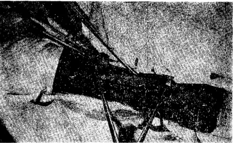
In this studyone mare and two donkeys were used. Alsa m. fIex. dig. supf. and prof. tendons of 12limbs of the se animals were used. POLYESTER FIBRE (Mersilene Mesh "Ethieon") implants (Fig. i) were used in experimentally injured tendans.
Mersilene Polyester Fibre has been used for many years as a su-ture materia1. it has also be en used in abdominal waıı and diaphrag-matie defeets.
AT VE EŞEKLERİN YARALARINDA POLYESTER KULLANILMASI 399
Fig. i. Photograph of Polyester Fibre sheet.
Mersilene Polyester Fibre Mesh can be cut with scissors to any desired shape depending upon the surgical indications and desires of on experienced surgeon and should be sterilised prior to use by au to c-laving at 121 oC for 20 minutes (1).
Polyester Fibre implants were made by folding a piece of Mersilene Mesh sheet like a tendon in shape. They were sterilised in steriliser at
120 oC for 20 minutes.
Mare and donkeys were prel11edicated with Chlorpromazine (CombeIen "Bayer") and Chloralhydrate (8 g /100 kg
ı.
V.). Volar or plantur nerves were anaesthetized with a local anaesthetic solution (Lignocain 2%).
The region from carpus to hoof (forelimb), from tarsus to ho of (hindlimb) were elipped and thoroughly prepared for an aseptic sur-gery.
A skin incision \Vas made on the posterior surface of the metacar-pus or metatarsus. The m. flex. dig. supf. and m. fIex. dig. prof. ten-dons were exposed. Tenonectomy (5 cm long) were performed on both tendons and 5 cm implants were bridged betwcen the ends of the ten-dons and sutured to the tendon ends with interrupted sutures of silk (no: O) (Fig. 2, 3). The skin wound was closed with interrupted sutures with silk (no: 1). A ca st of PVC was applied from mid-connon to the hoofs, with foot held in semiflexion to avoid the strain on sutures.
400 E.Samsar - A. Tanyolaç. N. Güzcl - t.A. Özkul - K. Özkan. P. Gökçc - E. Özgcnçi1
Fig. 2. Diagram showing the method of attaching Polyester Fibre implant between ten-don ends.
AT VE EŞEKLER iN YARALARıNDA POLYESTER KULLANıLMASı 401
The east was removed at least af ter 4-8 weeks and then several week s of rest in a loose-box was prescribed.
Tissue pieces were taken anaesthetized animals 15 days, I, 2, 3, 4, 5, 6, 8 and 9 months later operation in order to determine the histo-pathologieal tissue formation at the implanted sites. The freeze and paraffin sections were prepared fol1owing iO
%
formol fixation and dyed with H. E. and triehrom and examined histopathological1y under light microscope.For thin stmetures the taken pieces were fixed with Kamovsky's method (6) and osmie acid were blocked in Araldit M.
The sections were cut by LKB ultratom III and examined under electron microscope (Cari Zeiss Model EM 9 S-2).
Results
On reeovery from anaesthesia the mare and donkeys had no dif-fieulty in walking back to their loose-boxes. The following day, ho-wever the lameness was usually worse and some were reluctant to move or to be fed, without medication this discomfort subsided within four to five days. Af ter one week, they took full weight on the operatedleg. In four eases swelling and local heat developed at each incision and also a. light diseharge of tissue f1uid; but this subsided after 7-8 days. In others, antibiotic and Trasylol "Bayer, 25000 KIU" injeetion was applied loeally after the operation. Af ter this applieation in 8 eases swelling and diseharge of tissuc fluid was not observed.
In ease (Nr. 1) a purulent diseharge arouse from the incision. In this ease loeal antibiotie injeetion was applied for five days. This was sueeesfuI. Prompt wound healing then occured, and :n this case re-eovered good function.
In ease (Nr. 2), after 5 weeks when the cast of the right hindliınb of the mare was reınoved the wound been open (5 cm long) and implant whieh is surrounded by granulation tissue was observed on the ineision line. The gap between the eut ends of the tendon had been filled in eompletel1y and there was a little aeute infiammation at the operation site. Af ter debrideınent and irrigation with an antibiotie solution, the wound was closed with interrupted sutures and a rigid suppor tiri"g east was applied. After 10 days rigid supporting east was reınoved, the wound had healed and there was a little swelling at
402 E. Samsar - A. Tanyolaç • ~. Güzel - tA. Özknl - K. Özkan - P. Gökçc - E. Özgencil
the operation site. There was not any hyperextension of the digit when the horse had to bear weight.
In ease (Nr: 3), after 5 weeks the east of the left forelimb of the mare was removed. The wound had healcd and there was a big swelIing at the operation site; and upward tilting of the toe of the hoof when the mare had to bear weight. A neweast was applicd for one month. Mter this time to east was removed. The implants do not appear to have eaused any harm. The mare was generalIy able to walk; but stilI there was hyperextension of digit when mare had to bear weight. After 9 week s the affeeted tendons were still thiekened and loeal heat was absent. The tendons have remained thiekened. The thiekness of the affeeted tendons were twiee the normal tendon.
In eases (3, 5), the material provoked minimal foreign body res-ponse and it protruded throught the wound. The implant had to be removed beeause it had protruded throught the wound and produced a little septie diseharge.
When the implants ends were throughly buricd İn the ends of tendon no sueh eomplieation oeeured.
The best results were obtained when only the m. f1ex. dig. supf. or m. flex dig. prof. tendon was operated.
In two eases, the prognosis was worse beeausc the m. f1ex. dig. supf. and prof. tendons were operated together. In these eases hyperex-tension were seen and the tendons have remained thiekened.
GeneralIy, the east was maintained for 4 to 9 weeks and then rep-laeed with a supporting bandage. When onlyone tendon was operated the east was maintained for 4 weeks; when two tendam were operated in same time the east was maintained 9 weeks and the best results were obtained.
Three months later from the operation donkeys were ridden at the walk for a similar period and then gradualIy introdııeed to anormal programme (Iife-walking). At the and of this experiments walking of the animals cliniealIy 70
%
were good.Tendans whieh are operated together hyperextension were seen and the tendans have remained thiekencd.
Overgrowth of hom most frequent eause of hyperextension for this reason before operation ho of trimming always mu st be performed.
AT VE EŞEKLERIN YARALARINDA POLYESTER KULLANıLMASı ,103
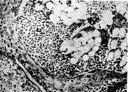
In general, an aeute inflammation and foreign body granulation tissue were observed around the 1ransplanted Mersilene Mesh in aıı the cases.
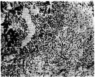
15 days after operation: N umerous leueocytes gathered around the foreign body, a few foreign body giant eeııs formcd, fibroblasts surrounded the leueoeytes and extended into the Icucoeyte masses (Fig. 4 and 5).
Fig. 4. IS dail)', ML Mersileııe Mcsh: LL Leucocytes; arrows) Fibroblasts X 2S2
ı
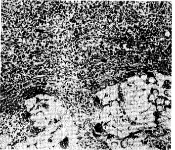
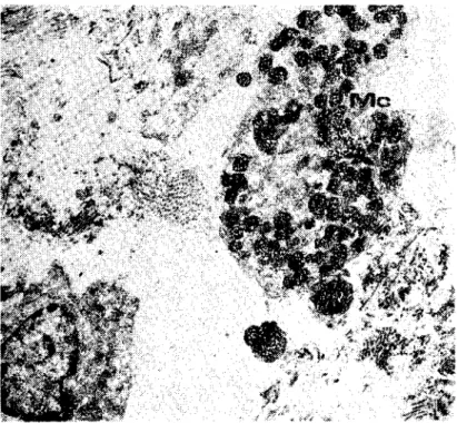
month af ter operation; A fibrous capsule, in:.:luding fibroblasts, was observed around the foreign body (Fig. 5). In the exterior of the eapsule found a vast amount of granulation tissu(' which was dominated also by leueocytcs especially Iymphoeytes. Other than mastoeytes, maerophages and, as immune eompotent eells, plasmablasts, plasmaey-tes were found (Fig. 6). These eeııs found in large numbers in this period were also observed throughout the eourse of the expcriments. The pietures also showed a large number of fibroblasts along with a spread network of eapillary vein. Foreign body giant eeııs were also found at random around the Mersilene Mesh (Fig. 6).3-4 months after operation: Fibrous eapsule became more pro-minent. A thiek layer of fibroblasts and eoııagen fibres formed and
,10. E.Samsnr. lı.Taııynlaç. N.Güzel _t.A.Özkııl -K.Özkan' P.Gökçe. E.Özgencil
Fig. 5. One monthly, asterisks) Fibrous capsule; arrows) Foreign body giant cells X ı60.
Fig. 6. One monıhly, Ma) Macrophage; Pc) Plasma cell X 2400.
Mersilene Mesh clogged by leucocytes, fibroblasts, thin collagen fibres and foreign body giant cells (Fig 9, 10,
ıı, ı
4 andı
5).5-6 months after operation: lt \Vas observed that fibroblasts found
in the fibrous capsule also formed the thick collagcn fibre bundles (Fig. 12 arrows).
AT VE EŞEKLERİN YARALARINDA POLYESTER KULLANILMASI ,HIS
lig. 7. Onc tnon!hly, Mc) Mas! cell X 7200.
406 E. Samsar. A. Tanyolaç. N. Gü7.cl . t.A. Özkul • K. Özkan. P. Gökçe. E. Ö7.genci!
Fig. 9. Three Ii,o"ıhly, L) iClICOCYlcs;asteris!,s) Fibrou5 ca~sıı:c X 160.
Fig. 10. Three monthly, L) leucocytes; n.sterisks) Fibrous capsule; arrows) Foriegn body giant ceııs X 252.
AT VE EŞEKLERiN YARALARıNDA POLYESTER KULLANıLMASı 407
Fig. II. Four montlıly, arrows) Foreign body giaııt ee!ls )( 252.
Fig. ı2. Six münihiy, arrow,) Cıllage.-, fihrcs bıı-,dles X 252.
8-9 months after operation: it was sccn th,~t the thinner fibres leaving the thick collagen fibre bundlcs wrapped the meshed tissue of the implant (Fig.
ı
3).Fig. !3. Eight r:ıo:ıthj~ .. ;ı,:cri,ks) Caliage:] fibrcs wlıich are sprcd to the pores of the foreign mateıi?! X 400.
14. ColIection of fibroblasts (Fb) "raund the Mersilcne Meslı (M) during theearly period fol!oV/ing operation X 9000.
AT VE EŞEKLERiN YAR~LARJNDA POLYESTER KULLANıLMASı 1(19
Fig. 15. Fibrous tissue elements frequently observed in the operatien area as one monthly period, Fb) Fibroblasts; Kı) Coııagen fibres: Pc) I'lasma eeııs X 36CO.
Discussİon and Conclusion
Samsar, E. (ıo) reported the successful use homograft in the re-pair of tendon, and in 1974 also Samsar, E. (ll) report ed the successful use of autograft in the repair of tendon.
In 1977-1978 Jenkins and others (2, 5, 7, 8) report that implant of filamentous carbon proyided satisfactory substitutes for ligaments and tendons in experimental sheep and rabbits stimulated its use for the reconstruction of these stmetures clinical1y İn men and animals. The material is reported to proyoke minimal foreİgn body response and to induee the formation of a new tendon like tissue (3).
In our experiments Polyester Fibre implants were used. The prog-nosis was good in ease of m. f1ex. dig. supf. or prof. The best results were aehived when onlyone tendon was operated; but when two ten-dons were operated together poor results were obtained. Also the
ten-'DO E. Samsar - A. Tanyola', - N. GLzcl • İ:A. Özkul - K. Özkau -I'. Gök"c - E O'.gcnçil
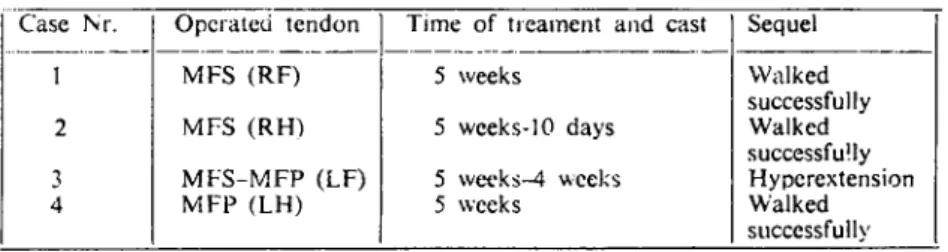
Table I. Results of Treatment in Mare -_~~~__~r~ ~.::-:.t.~~. ~~don_L~~e __~~~J~ame~_and east
1 MFS (RF) 5 weeks 2 3 4 Mf S (RH) MFS-MFP (LF) MFP (LH) 5 weeks-IO days 5 wct'ks-4 .•••.ecks 5 wccks Sequel Walked suecessfuJly Walkcd successfully Hyperextension Walked slıccessfuJly
Table 2. Resuits of Treatment in Donkeys
Case NI'. Opcraıed tendon
i
Time of treament and ca st Sequel---_. --- ---_._----" 5 MFS-MFP (RF) 5 weeks Thickened tendon and hyperextension 6 MFS-MFP (RF) 5 weeks Thickened tencton and hyperextcnsion
7 MFP (RI-I) 4 ••••eeks Walked
successfulIy ~ MFP (RH) 4 weeks Walked successfulIy 9 MFP (LH) <1 weeks Walked slıccessfuJly LO MFS (LF) 4 weeks Walked successfulIy
II MFS (LH) 2 wceks-3 days Euthanasia
12 MFP (Ll') 2 weeks-3 days Euthaııasia
MFS: M. f1cx. dig. supf. MFP: M. flex. dig. prof. RF: Right forelimb RH: Right hindlimb LF: Left fmclimb LH: Lefı hindlimb
oons have rCl11ained thiekened and the implants eaused minimal foreign body reaetions. Howevcr the Polyester Fibre implants can be uscd in singlc tendon nıptur.
Our results on formation of eapsule arouno Mersilene-Mesh by fibroblasts, reinforeement of capsule by eoııagen fibres and clogging of thinner eollagen fibres the mesh tissue of the implant resemble the findings of Goodship and Brown (5). In this work, the finding of ecııs with picnotie nuclei among leucocytes and Iymphoeytes shows the destruetion of immune reaetion or the phagocytosis period. Our results, howevcr, eonfHet with Goodship and Brown (5) in that they
AT YE EŞEKLERİN YARALARıNDA POLYESTER KULLANILMASI 411
did not observe any infiammation and found a very few foreign body giant eelIs in their work on the aehil tendon of Califomia rabbit they implanted "earbon fibre". As a result it can be said that the low price and high success in curing make Mersilene-Mesh an aecaptable implant.
Referenccs
i. Anteplioğlu, H., Sam~ar, E. (1976): Büyükbaş Ruminatlarda Gen;ş Defektli Fıtık
De-liklerinin Sentetik Creflerle Onarılması. AÜ Vet. Fak. Derg. 23, (1-2): 89-102.
2. Bredin, K. (1986): Treatment of a Strained Tendon in a Thorughbred Racehorse with
Carbon Fibre Filaments. Irİsh Yeterinary Journal 40: 139-140.
3. Edwards, G.B., Yaughan, L.C (1984): Use of Carbon Fibre Implants in the Treatment
of Fetlock loint Disloeation in Two Horses. Yeterinary Record 114, (Jan. 28): 87-88.
4. Forster, L. W., Ralis, Z.A., Mc Kibbin, B., Jenkins, D.H.R. (1978): Biologieal Reaetion
to Carbon Fibre Implants. The Formatian and Strueture of a Carbon-Indııeed "Neo ten-don" Clin. Orthop. 131: 299-307.
5. Goodship, A.E., Brown, P.N., Jenkins, D.H.R., Silver, L.A. (1980): An Assesment of
Filamentous Carbon Fibre for Treatment of Tendon Injury in the Horse. Yeterinary
Re-cord 106, (March 8): 217-221.
6. Kamovsky, M.J. (1965): A Formaldelıyde'glııtaraldelıyde Fixative of High Osmolality for Use in Eleetron Mieroseopy. J. Cell BioL, 27: 137 A-138 A.
7. Vttlc Wood, H.F. (1979): Treatment of Sprained Tendans in Horses witlı Carbon Fibre
Implants. Yeterinary Record. 105, (Sept. 8): 223-224.
8. Mc GiII, CA., Fraser, D.M. (1983): Tlıe Use ofCarbon Fibre Jmplants to Repair Severed
Digital Flexor Tendons in a Fallow Deer. Australian Yeterinary Journal 60, (10): 318.
9. Oehme, F.W., Prier, J.E. (1975): Large Animal Surgery. The Williams and Wilkins Company. Baltimore.
10. Samsar, E. (1973): At ve Merkeplerin Metaearpus ve Metatarsus Bölgelerindeki M. jlex. dig. supf ve prof Tendolarında Dikiş ve Homogref Uygulamaları Ozerinde
Deney-sel Çalışmalar. AÜ Vet. Fak. Derg. 20, (i): 14-34.
11. Samsar, E. (1974): Köpeklerde Deneysel Tendo Dikişleri ve Tendo Autogrefleri. Aü Vet. Fak. Derg. 21, (1-2): 70-79.
12. Yalde7., H., Coy, H.C, Swanson. T. (1982): Flexible Carbon Fibre for Repair of Gast.
menemius and Superficial Digital Flexor Tendons in a Hei/er and Gastroenemius Tendon in a Foal. JAYMA-181. (2): 154-157.