| The Annals of Clinical and Analytical Medicine
159
The Annals of Clinical & Analytical Medicine Case Report
Hirayama disease
Late onset hirayama disease:
Characteristic magnetic resonance imaging features hirayana disease
DOI: 10.4328/ACAM.6154 Received: 09.01.2019 Accepted: 25.01.2019 Publihed Online: 05.02.2019 Printed: 01.03.2020 Ann Clin Anal Med 2020;11(2): 159-161 Corresponding Author: Kemal Murat Haberal, Yukarı Bahçelievler Mah. Mareşal Fevzi Çakmak Cad. No:45, 06490, Çankaya, Ankara, Türkiye.
E-Mail: [email protected]
ORCID ID: https://orcid.org/0000-0002-8211-4065
Abstract
Hirayama Disease is a rare benign lower motor neuron disorder which is primarily affecting young males. It is characterized by the progressive weakness of the distal upper extremities followed by spontaneous stabilization of the symptoms. In this paper, we describe a 58 year- old female patient with a complaint of weakness in the right hand and forearm. Magnetic resonance imaging of the cervical spine established the final diagnosis of Hirayama disease. Hirayama disease when detected and intervened at an early stage of the disease process, can have a good prognosis. Clinicians and radiologists should be aware of the clinical features, as well as suspicious findings on neutral-position MR imaging and an additional neck-flexion MR imaging study should be arranged to confirm the diagnosis.
Keywords
Hirayama Disease; Motor Neuron Disorder; Myelopathy; Magnetic Resonance Imaging
Kemal Murat Haberal1, Aynur Yılmaz Avcı2, Mert Bayramoğlu1, Ahmet Muhteşem Ağıldere1 1Department of Radiology, 2Department of Neurology, Baskent University, Alanya, Turkey
| The Annals of Clinical and Analytical Medicine Hirayama disease
160
Introduction
Hirayama disease(HD) is a lower motor neuron disease charac-terized by unilateral weakness and atrophy of the distal muscles of the upper extremity in the distribution of C7-T1 myotomes. Bilateral involvement is rare. It is a rare non-progressive spi-nal muscular atrophy seen predominantly in young men in late adolescents and third decadence. The onset is insidious, the symptoms progress from three to five years, then the disease progresses spontaneously [1,2].
In this case report, a 58-year-old woman with gradually increas-ing weakness in the forearm was presented with MRI findincreas-ings.
Case Report
A 58-year-old female patient; was admitted to the neurology clinic for 25 days with complaints of the increased weakness and occasional numbness in the right forearm. Physical exami-nation of the patient was normal. Neurological examiexami-nation re-vealed numbness in the distal right arm and muscle strength was 2/5. No pathology was found in the other neurological ex-amination. Blood investigations including complete blood count, sedimentation rate, renal, liver, and thyroid function tests, cre-atine kinase, and vitamin B12 and vitamin D3 level were within normal range.
Electromyography (EMG) examination showed that the right ra-dial and ulnar nerve combined muscle action potential (CMAP) amplitude was lower than the left. Other messages were in the normal range. In the right radial and ulnar nerve innervated muscles, there was a slight increase in polyphyletic, motor unit potential (MUP) times. No denervation potential was observed. The other muscles examined were normal.
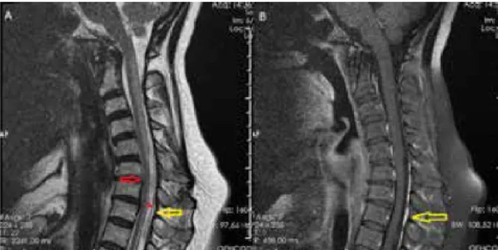
In the cervical magnetic resonance radiography (MRI), sagit-tal plane T1 and T2 weighted sequences, axial gradient echo sequence for the discs and axial and sagittal postcontrast T1 weighted sequences were applied at a neutral position. MRI re-vealed flattening in cervical lordosis, spinal cord atrophy under C5-C6 intervertebral disc in T2-weighted sagittal sections, and increased signal in the posterior spinal cord at C2-C5 levels (Figure 1). In the differential diagnosis of the patient, the cervi-cal MRI examination was refreshed with flexion in view of the Hirayama disease. Flexion MRI, in addition to neutral MRI find-ings, showed posterior dural displacement between C5-T1 lev-els, forward displacement in the spinal cord, enlargement in the posterior epidural space, and homogeneous contrast enhance-ment in the epidural distance in the postcontrast series (Figure 2). The patient was diagnosed with HD by MRI findings and was offered cervical neck and physical therapy exercises.
Hirayama disease was first described by Hirayama et al. in 1959 [1]. The exact pathogenesis of the disease is still unclear. The generally accepted hypothesis is cervical myelopathy induced by flexion. The underlying mechanism is the disproportion with the duramater and the growth of the vertebral column. While the length of the spinal canal increases with flexion, the dura-mater is stretched and separated from the vertebral canal wall. Anterior spinal artery irrigations may occur in the lower cervical spinal cord with anterior spinal cord compression and anterior displacement. Recurrent flexion may lead to chronic circulatory disorder, gliosis, and localized cord atrophy [3].
HD is more common in young males, especially among 15-25 years. Our patient is one of the few cases sampled in the litera-ture in term of age and sex. For HD, self-limiting weakness and insidious onset in the hand and forearm, which are mostly unilat-eral, are typical. In our case, there were atrophy and numbness
symptoms in unilateral forearm and hand muscles and there was no sensory or pyramidal path involvement, typical for HD. In HD, cervical radiographs revealed no specific finding ex-cept for flattening in lordosis or scoliosis. Specific features in neutral and flexion MRI have been defined in the diagnosis of HD in the literature. These are mainly spinal cord atrophy, asymmetric cord flattening, intramedullary signal changes in the cervical cord, abnormal cervical axis in neutral posi-tion, and the loss of the connection between the posterior dural sac and the lamina, forward displacement of posterior dura, the expansion in the posterior epidural space and con-trast enhancement in this region with/without signal void ar-eas suggesting dilated epidural venous plexus in flexion [4]. In our case, findings consistent with the literature were defined in the cervical MRIs in neutral-flexion positions and a diagnosis of Hirayama disease was made.
The differential diagnosis of HD includes spinal muscular atro-phy, amyotrophic lateral sclerosis, post-polio syndrome, multi-focal motor neuropathy, toxic neuropathy, and structural lesions of the cervical cord such as syringomyelia. These clinical enti-ties are characterized by their characteristic clinical, radiologi-cal, and electrophysiological characteristics [1]. In degenerative diseases, disc-osteophyte complex, which may lead to second-ary cord compression, can be seen in T1A sequences. In demy-elinating diseases, T1A hypointense, T2A hyperintense, focal or diffuse cord lesion with variable diffusion characteristics can be seen in the spinal cord. In spinal cord infarction, T1A hypoin-tense, T2A hyperinhypoin-tense, diffusion-restricting spinal cord lesion is seen in arterial irrigation area [5].
Dynamic cervical MRI, which has an important role in diagnosis in HD, is used in increasing frequency in patients who are inves-tigated in terms of positional spinal cord compression, cervical degenerative disease, cervical myelopathy, rheumatoid arthri-tis, cervical trauma [6,7].
Figure 1. T2 weighted spin echo sequence (A) and postcontrast fat saturated T1 weighted spin echo sequence(B) in neutral position. A: Atrophy in the spinal cord under the C5-C6 intervertebral disc level(yellow arrows) and increased intramedullary signal at the posterior of the spinal cord upper this level(red arrow). B: Flattening in cervical lordosis(yellow arrow).
Figure 2. T2 weighted spin echo sequence (A) and postcontrast fat saturated T1 weighted spin echo sequence(B) in flexion. A: Forward displacement in spinal cord at C5-T1 levels(red arrow), anterior displacement of posterior dura(red asteriks), enlargemnet off posterior epidural space(yellow arrow). B: Contrast enhancement in posterior epidural space(yellow arrow).
| The Annals of Clinical and Analytical Medicine Hirayama disease
161
Conclusion
Limitation of neck flexion can be extremely beneficial if the my-elopathy symptoms and signs of Hirayama occur early, even if they are nonprogressive. Clinicians and radiologists should be familiar with imaging findings of the disease, and the key role of flexion cervical MRI should be kept in mind when considering the differential diagnosis.
Scientific Responsibility Statement
The authors declare that they are responsible for the article’s scientific content including study design, data collection, analysis and interpretation, writing, some of the main line, or all of the preparation and scientific review of the contents and approval of the final version of the article.
Animal and human rights statement
All procedures performed in this study were in accordance with the ethical stan-dards of the institutional and/or national research committee and with the 1964 Helsinki declaration and its later amendments or comparable ethical standards. No animal or human studies were carried out by the authors for this article. Conflict of interest
None of the authors received any type of financial support that could be consid-ered potential conflict of interest regarding the manuscript or its submission. References
1. Hirayama K, Toyokura Y, Tsubaki T. Juvenile musculer atrophy unilateral upper extremity- a new clinical entity. Psychiatr Neurol Jpn. 1959; 61: 2190-7. 2. Hirayama, K. Juvenile Muscular Atrophy of Distal Upper Extremity (Hirayama Disease). Internal medicine (Tokyo, Japan). 2000; 39(4): 28390.
3. Kikuchi S, Tashiro K, Kitagawa M, Iwasaki Y, Abe H. A mechanism of juvenile muscular atrophy localized in the hand and forearm (Hirayama’s disease)--flexion myelopathy with tight dural canal in flexion. Rinsho shinkeigaku = Clinical neurol-ogy. 1987; 27(4): 412-19.
4. Yüksel M, Kalemci O, Yüksel KZ, Ergüden C, Yücesoy K. Hirayama Hastalığı ve Tanıda Manyetik Rezonans Görüntülemenin Önemi. J Nervous Sys Surgery. 2009; 2(4): 191-5
5. Vargas, María Catalina, Mauricio Castillo. Magnetic Resonance Imaging in Hi-rayama Disease. Journal of radiology case reports. 2011; 5(3): 17–23.
6. Giuliano V, Giuliano C, Pinto F, Scaglione M. The Use of Flexion and Extension MR in the Evaluation of Cervical Spine Trauma: Initial Experience in 100 Trauma Patients Compared with 100 Normal Subjects. Emergency radiology. 2002; 9(5): 249–53.
7. Nigro L, Donnarumma P, Tarantino R, Rullo M, Santoro A, Delfini R. Static and Dynamic Cervical MRI: Two Useful Exams in Cervical Myelopathy. Journal of spine surgery (Hong Kong). 2017; 3(2): 212-16.
How to cite this article:
Haberal KM, Avcı AY, Bayramoğlu M, Ağıldere AM. Late onset hirayama disease: Characteristic magnetic resonance imaging features hirayana disease. Ann Clin Anal Med 2020;11(2): 159-161