A T H E R O S C L E R O S I S
Prevention of atherosclerosis by bioactive palmitoleate
through suppression of organelle stress and
inflammasome activation
Ismail Çimen,1,2Begüm Kocatürk,1,2Seda Koyuncu,1* Özlem Tufanlı,1,2Umut I. Onat,1,2 Asli D. Yıldırım,1,2 Onur Apaydın,1,2Şeyma Demirsoy,1†Zaliha G. Aykut,1Uyen T. Nguyen,3 Steven M. Watkins,3Gökhan S. Hotamışlıgil,4Ebru Erbay1,2‡
De novo lipogenesis (DNL), the conversion of glucose and other substrates to lipids, is often associated with ectopic lipid accumulation, metabolic stress, and insulin resistance, especially in the liver. However, organ-specific DNL can also generate distinct lipids with beneficial metabolic bioactivity, prompting a great interest in their use for the treatment of metabolic diseases. Palmitoleate (PAO), one such bioactive lipid, regulates lipid metabolism in liver and improves glucose utilization in skeletal muscle when it is generated de novo from the obese adipose tissue. We show that PAO treatment evokes an overall lipidomic remodeling of the endoplasmic reticulum (ER) membranes in macrophages and mouse tissues, which is associated with resistance of the ER to hyperlipidemic stress. By preventing ER stress, PAO blocks lipid-induced inflammasome activation in mouse and human macrophages. Chronic PAO supplementation also lowers systemic interleukin-1b (IL-1b) and IL-18 concentrations in vivo in hyperlipidemic mice. Moreover, PAO prevents macrophage ER stress and IL-1b production in atherosclerotic plaques in vivo, resulting in a marked reduction in plaque macrophages and protection against atherosclerosis in mice. These findings demonstrate that oral supplementation with a product of DNL such as PAO can promote membrane remodeling associated with metabolic resilience of intracellular organelles to lipid stress and limit the progression of atherosclerosis. These findings support therapeutic PAO supplementation as a potential preventive approach against complex metabolic and inflammatory diseases such as atherosclerosis, which warrants further studies in humans.
INTRODUCTION
Fatty acids (FAs) can be generated de novo from a surplus of carbohy-drates and other substrates in lipogenic organs. These lipids are then released as very low density lipoprotein (VLDL) particles from the liver and stored as triglycerides (TAGs) in the adipose tissue until further
need (1). Although diet is usually a sufficient source of FA for mammals,
de novo lipogenesis (DNL) is essential during development, as exempli-fied by the embryonic lethality of genetic mouse models lacking key DNL
enzymes, such as FA synthase and acetyl–coenzyme A carboxylase 1.
Furthermore, a maternal fat-rich diet does not rescue the lethality, sug-gesting that certain structural or signaling lipid molecules essential for
embryonic development can only be generated de novo (2–4). Recent
studies have shown that several bioactive lipid species are produced by DNL, including endogenous ligands for nuclear receptors
[1-palmitoyl-2-oleoyl-sn-glycerol-3-phosphocoline and
1-stearoyl-2-oleoyl-sn-glycerol-3-phosphocoline, peroxisome proliferator–activated receptor
a (PPARa) and alkyl ether lipids, and PPARg], fatty acid–hydroxy fatty acids (FAHFAs), and palmitoleate (PAO), which have specific
biological activities and exert potent metabolic effects (5–9).
One striking observation regarding the biological impact of DNL-generated lipids was made in snakes. PAO concentrations spike in the plasma of Burmese pythons that are going through an extreme metabolic adaptation to a large meal after long periods of starvation. Palmitoleic acid ethanolamide (palmitoleoyl ethanolamide), a potent regulator of
energy balance, is markedly increased in the fed pythons’ gastrointestinal
tract. During this adaptation, the pythons undergo remarkable organ growth, including a particularly striking increase in cardiac size. On the other hand, administration of a lipid mix that includes PAO can promote
healthy heart growth in a physiological setting (10). Together, these
findings suggest a surprising beneficial metabolic impact of a mono-unsaturated fatty acid (MUFA), PAO, previously not associated with any distinct biological function.
Adipose tissue DNL and PAO production is also activated in fatty
acid–binding protein (FABP)–deficient mice, which are highly resistant
to developing insulin resistance and atherosclerosis despite developing
obesity and dyslipidemia on a high-fat diet (7, 11–15). Liver is the
primary site for FA synthesis. Adipose tissue DNL is usually inactive, and the lipid composition at this site reliably reflects dietary lipid
input (16). However, adipose tissue from FABP-deficient mice becomes
refractory to dietary input and activates DNL to generate its own FA
from glucose (7, 17, 18). PAO generated from the adipose tissue can
have regulatory effects on lipid metabolism in liver and improve glucose
utilization in skeletal muscle (7, 17, 18). Hence, PAO is considered to
be a lipokine, a lipid signal serving at the adipose tissue–liver and
adi-pose tissue–muscle axes. Additionally, exogenous PAO administration
has favorable metabolic and anti-inflammatory effects in mice, sheep,
and rats (19–22).
1Department of Molecular Biology and Genetics, Bilkent University, Ankara 06800,
Turkey.2National Nanotechnology Research Center, Bilkent University, Ankara
06800, Turkey.3Metabolon, Sacramento, CA 95691, USA.4Department of Genetics
and Complex Diseases and Sabri Ülker Center, Harvard T.H. Chan School of Public Health, Boston, MA 02115, USA.
*Present address: Department of Cellular and Molecular Medicine, Katholieke Universiteit Leuven, Leuven 3000, Belgium.
†Present address: CECAD Cluster of Excellence: Cellular Stress Responses in Aging-Associated Diseases, University of Cologne, Cologne 50931, Germany.
‡Corresponding author. Email: [email protected]
by guest on February 7, 2019
http://stm.sciencemag.org/
Interpretation of association studies to translate these preclinical findings to humans has been challenging because PAO is generated in the setting of obesity and alongside hepatic DNL. Because hepatic DNL associates with ectopic lipid accumulation and insulin resistance, the rise
in PAO in serum has been associated with metabolic disorder (23–25).
The coupling of liver DNL to PAO production in obesity may be an adaptation to help offset the hazardous effects of excess intake of saturated lipids (such as on membranes and membrane-initiated
signaling) (11, 26, 27). Another complication in the analysis of these
epidemiological studies is that PAO concentrations were measured from different lipid classes. For example, PAO in the nonesterified fatty acid (NEFA) fraction shows a positive correlation with insulin sensitiv-ity, whereas esterified PAO in cholesterol ester (CE), diacylglycerol (DAG), and TAG correlates with fatty liver disease, insulin resistance,
diabetes, and acute coronary disease (23, 24, 28–30). In humans,
cir-culating esterified PAO concentration mainly reflects hepatic DNL
(31, 32). These seemingly discrepant results imply that the free form
of PAO could have distinct bioactivity in humans, similar to mice (7).
Mouse studies using an exogenous source of PAO (independent of
hepatic DNL) provide valuable insight into PAO’s biological impact,
including enhanced insulin sensitivity, reduced inflammation, and
improved lipid metabolism (7, 21, 22, 33–35). In a recent randomized,
controlled trial in humans, purified PAO treatment markedly reduced high-sensitivity C-reactive protein and improved serum lipids, suggesting that PAO supplementation may have a beneficial impact on
athero-sclerosis (36). However, these correlations do not establish causality
or accurately reflect the real biological function of PAO in cardiovascular disease, warranting a direct analysis of the effect of PAO supplementation in a model of atherosclerosis.
Atherosclerosis is a chronic inflammatory disease of the arterial wall induced by lipid imbalance and a maladaptive immune system
(37–39). The immune system plays a crucial role in the development of
plaques in the intima, progression, and predisposition to rupture (40–42).
The activation of the Nod-like receptor (NLR) family, pyrin domain containing-3 (NLRP3) inflammasome, a multiprotein complex in the myeloid linage that plays an important role in the innate immune response to pathogens, can be induced by cholesterol crystals or saturated
FA (SFA) and promote atherogenesis (43, 44). The inflammasome is
assembled from NACHT, LRR, and PYD domains–containing protein
(NALP), apoptosis-associated speck-like protein containing a 1 recruitment domain (ASC), and 1. Upon its activation,
caspase-1 processes pro–interleukin-1b (pro–IL-1b) and pro–IL-18 into their
mature forms, resulting in their secretion (45). Inflammasomes can also
be activated in response to organelle stress, a pathological mechanism
that contributes to atherogenesis (11, 44, 46–48). Endoplasmic reticulum
(ER) stress is linked to inflammasome activation through mobilization of intracellular calcium and generation of reactive oxygen species (ROS),
which provide an activation signal for the inflammasome (44, 46, 47).
Moreover, calcium mobilization during ER stress causes mitochondria
damage and generation of ROS linked to inflammasome activation (47).
Under conditions of metabolic challenge, simultaneous ER stress and mitochondrial oxidative stress can generate a vicious cycle, potentiating inflammasome activity and establishing a pattern of chronic inflamma-tion. Macrophage ER stress, similar to inflammasome activity, contributes to the development of obesity, insulin resistance, and atherosclerosis
(11, 43). Additionally, macrophage mitochondrial oxidative stress
pro-motes atherogenesis (48). In an earlier study, we have demonstrated that
lipid-induced ER stress can be antagonized by PAO in vitro (11).
Whether PAO can inhibit inflammasome activation in macrophages or alter the course of atherosclerosis remains unknown.
Despite major interest in DNL-based therapeutic development for chronic metabolic diseases, nothing is known about the
DNL-generated lipid species’ impact on atherosclerosis. Moreover, very little
is known about the mechanism of action of these lipids in vivo, particu-larly in innate immune cells like macrophages, during the development of atherosclerosis. Here, we investigated the direct impact of a DNL product, PAO, on atherosclerosis. PAO prevented lipid-induced stress response in the ER, suppressing inflammasome activation in mouse and human macrophages and IL-1b and IL-18 production in vivo. The metabolic resilience of organelles to lipotoxic stress was associated with PAO-initiated lipidomic remodeling of ER membranes in macrophages and tissues in vivo. Overall, PAO supplementation increased the incor-poration of this FA into major lipid species represented in the mouse aortas and reduced macrophage ER stress and atherosclerotic plaque
area in apolipoprotein E–deficient (ApoE−/−) mice. These findings have
potentially important translational implications for atherosclerosis treatment through nutritional management of bioactive lipid species such as PAO.
RESULTS
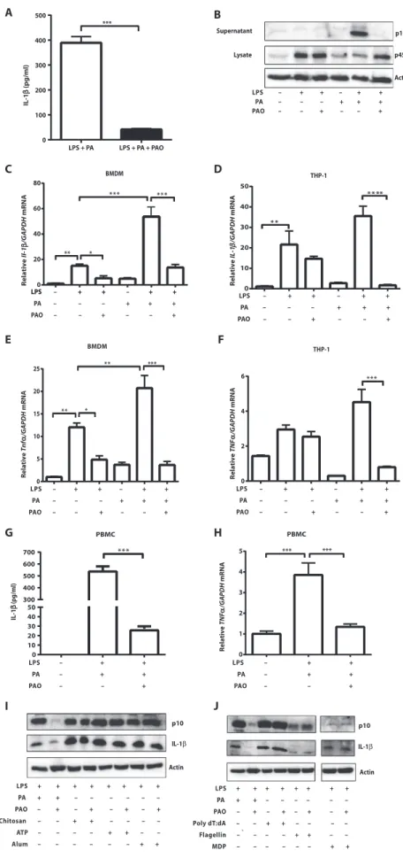
PAO blocks lipid-induced inflammasome activation and inflammation in macrophages
Here, we assessed the impact of PAO on SFA-induced inflammasome activation in macrophages. Earlier studies showed that treatment of
lipopolysaccharide (LPS)–primed mouse bone marrow–derived
macro-phages (BMDMs) with an SFA such as palmitate (PA) specifically induced the activation of NLRP3 inflammasome and the secretion of
IL-1b (44). PAO cotreatment completely blocked PA-induced IL-1b
se-cretion (P < 0.001; Fig. 1A). PA stimulation of LPS-primed BMDM
activated caspase-1, whereas cotreatment with PAO blocked this
activ-ity (Fig. 1B). Priming with LPS up-regulatespro–Il-1b mRNA, which is
expressed in low amounts in resting macrophages (49). PAO partially
blocked the LPS-inducedIl-1b mRNA increase in mouse BMDM but
not in human THP-1 macrophages (Fig. 1, C and D). PA treatment
enhancedIl-1b mRNA expression in LPS-primed BMDM and in
THP-1 macrophages, but this effect could be completely blocked with PAO cotreatment (Fig. 1, C and D). Upon release, IL-1b and IL-18 engage their respective receptors on other innate immune cells and stim-ulate a proinflammatory response characterized by the secondary release
of cytokines such as tumor necrosis factor–a (TNFa) (50). PAO
treat-ment blocked PA-stimulatedTnfa production in LPS-primed BMDM
and human THP-1 macrophages (P < 0.001; Fig. 1, E and F). We
ob-served similar inhibitory effects of PAO on PA-stimulatedIl-6 and Ccl2
production from LPS-primed BMDM and THP-1 cells (fig. S1, A to D). Finally, PAO inhibited PA-induced IL-1b secretion in LPS-primed human peripheral blood mononuclear cells (PBMCs) without reduction
inIL-1b mRNA (P < 0.001; Fig. 1G and fig. S1, E and F). Furthermore,
PAO blocked PA-inducedTNFa mRNA induction in the same cells (P <
0.001; Fig. 1H) (51). Collectively, these experiments demonstrate that
PAO is a potent inhibitor of lipid-induced inflammasome activation and proinflammatory cytokine production in mouse and human macrophages.
We next asked whether PAO could inhibit other inflammasome complexes. For this, mouse BMDMs were treated with specific activators
by guest on February 7, 2019
http://stm.sciencemag.org/
for various inflammasome complexes (52). Although PAO abolished lipid-induced NLRP3 inflammasome activation, as evidenced by the suppression of cleaved caspase-1 and secreted IL-1b, it did not inhibit NLRP3 in-flammasome induction by aluminum crystals (alum), chi-tosan, or adenosine triphosphate (ATP) (Fig. 1I). These findings showed that PAO specifically intercepts lipid-induced NLRP3 inflammasome activation. Moreover, PAO did not prevent activation of the absent in melano-ma 2 (AIM2) inflammelano-masome by poly(deoxyadenylic-deoxythymidylic) [poly(dA:dT)] acid, NLR family
caspase-1 recruitment domain (CARD)–containing
protein–4 (NLRC4) inflammasome by flagellin, or
NLRP1 inflammasome by muramyldipeptide (MDP),
supporting PAO’s specific role as an inhibitor of the
lipid-induced NLRP3 inflammasome (Fig. 1J). PAO prevents PA-induced ER stress by remodeling ER membranes
Next, we investigated the mechanism by which PAO blocks lipid-induced NLRP3 inflammasome activation. SFAs like PA can induce ER stress and activate the
inflammasome (11, 44, 53). Previous studies showed
that ER stress induces inflammasome activation through several mechanisms, including calcium mobi-lization, ROS release, and promotion of mitochondrial
damage (52). Damaged mitochondria release ROS,
which can further potentiate the inflammasome activity
(44, 47). We hypothesized that inhibition of PA-induced
Actin p45 Supernatant Lysate A B IL-1 β (pg/ml) 0 100 200 300 400 500 *** LPS + PA LPS + PA + PAO C D E F THP-1 THP-1 LPS – + + + + LPS – + + – + + PA – – – + + + PAO – – + – – + BMDM p10 BMDM LPS – + + – + + PA – – – + + + PAO – – + – – + LPS – + + – + + PA – – – + + + PAO – – + – – + LPS – + + – + + PA – – – + + + PAO – – + – – + Relative Il-1 β /GAPDH mRNA Relative TNF α /GAPDH mRNA Relative IL-1 β /GAPDH mRNA Relative Tnf α/GAPDH mRNA 0 2 4 6 ** * 0 10 20 30 40 50 * * ** * * 0 20 40 60 80 ** * ** * ** * 0 5 10 15 20 25 *** * * * * * G H *** 0 1 2 3 4 5 *** 0 10 20 30 40 50 300 400 500 600 700 IL-1 β (pg/ml) * * * PBMC PBMC I J p10 p10 Actin Actin IL-1β IL-1β LPS – + + PA – + + PAO – – + LPS – + + PA – + + PAO – – + LPS + + + + + + + + PA + + – – – – – – PAO – + – + – + – + Poly dT:dA – – + + – – – – Flagellin – – – – + + – – MDP – – – – – – + + Relative TNF α /GAPDH mRNA LPS + + + + + + + + PA + + – – – – – – PAO – + – + – + – + Chitosan – – + + – – – – ATP – – – – + + – – ` Alum – – – – – – + + LPS + + – + + PA – – – + + + PAO – – + – – +
Fig. 1. PAO prevents lipid-induced inflammasome activation and proinflammatory cytokine expression in macrophages. (A) Concentrations of secreted IL-1b in supernatants of LPS-primed BMDM stimulated with PA or PAO, alone or together (n = 8). (B) Immunoblots for
active caspase-1 (p10) from supernatant and pro
–caspase-1 (p45) and actin from cell lysate of BMDM. Blots shown are representative of three independent experiments. (C and D) Quantitative reverse transcription polymerase chain reaction (qRT-PCR) for (C) Il-1b mRNA in mouse BMDM and (D) IL-1b in human THP-1 macrophages, primed with LPS and stimulated with PA or PAO, alone or together
(n≥ 3). (E and F) qRT-PCR for (E) Tnfa in mouse BMDM and
(F) TNFa in human THP-1 macrophages, primed with LPS
and stimulated with PA or PAO, alone or together (n≥ 3).
(G and H) LPS-primed human PBMCs were analyzed (G) by enzyme-linked immunosorbent assay (ELISA) for IL-1b
se-creted into conditioned medium (n≥ 3) and (H) by
qRT-PCR for TNFa mRNA (n≥ 3). (I and J) Active caspase-1 (p10)
and mature IL-1b were detected by Western blotting in supernatant of mouse BMDMs primed with LPS and in-duced with various (I) NLRP3 agonists (ATP, chitosan, or alum) or (J) specific activators of other inflammasome complexes [poly(dA:dT), flagellin, and MDP] according
to previously published protocols (91–95). Blots shown
are representative of three independent experiments. Data are means ± SEM. *P < 0.05, **P < 0.01, ***P < 0.001. One-way analysis of variance (ANOVA) [in (C) to (H)] or two-tailed Mann-Whitney U test [in (A)] was used for statistical analysis.
by guest on February 7, 2019
http://stm.sciencemag.org/
ER stress by PAO blocks NLRP3 inflam-masome activation in macrophages. In an earlier study, we showed that PAO prevents lipid-induced ER stress but not proteotoxic
ER stress in macrophages (11). Because
inflammasome activation requires LPS pre-treatment in cultured macrophages, we assessed whether PAO can block ER stress under inflammasome-activating conditions. PAO also blocked PA-induced ER stress in LPS-pretreated BMDMs, as measured
by splicedXbp1 mRNA production (fig.
S1G). This finding shows that PAO can block lipid-induced ER stress upstream of inflammasome activation.
To understand how PAO limits the impact of lipid stress on macrophages, we next used high-resolution quantitative lipidomics to analyze PAO-induced altera-tions in macrophage lipid composition. For this purpose, we analyzed the lipid changes in mouse BMDMs treated with PA and PAO alone or in combination (PA + PAO). Analysis of the data showed increases in
total PAO concentration and MUFA/SFA ratio in cells treated with
PAO (alone or with PA;P < 0.001; Fig. 2, A and B). PAO incorporated
into all major lipid species whether applied alone or in combination with PA (Fig. 2C). PA + PAO cotreatment expanded the TAG compart-ment and its metabolic precursor, DAG, suggesting that PAO may be buffering PA-associated lipotoxicity by facilitating its effective entry into
neutral lipid stores (P < 0.001 and P < 0.01, respectively; Fig. 2D). More
PA incorporated into TAG and DAG when cotreated with PAO (P <
0.001 andP < 0.01; Fig. 2E). We also examined another membrane
con-stituent, phospholipids (PLs), because their production is mainly regu-lated by the ER with the contribution of IRE1-XBP1 signaling branch
(54). PA, but not PAO (alone or cotreated), expanded the PL
compart-ment (P < 0.01; Fig. 2D). However, PAO effectively incorporated into all
PL species, with highest concentrations seen in phosphatidylcholine
and its metabolic precursor, lysophosphatidylcholine (Fig. 2, C and F). These findings imply that desaturation of membrane lipids by PAO could
have an influence on the membrane’s biophysical properties (55).
Next, we used a mouse model of atherosclerosis,ApoE−/−mice, in
which ER stress and inflammasome activation can be induced over time
by a hyperlipidemic (Western) diet (43). Typically, PAO makes up
about 0.38% of Western diet. We decided to administer up to threefold higher PAO by oral gavage (about 1.2% of diet or 1400 mg/kg per day)
toApoE−/−mice on Western diet and analyze the effect of this chronic
exposure on lipid composition of plasma and multiple tissues (56). Four
weeks of PAO treatment increased PAO in the circulation and aortas
ofApoE−/−mice but not in liver, adipose, and muscle tissues (P < 0.05;
Fig. 3A and fig. S2A). In addition, the MUFA/SFA ratio was increased in the aortas and plasma from PAO-treated mice when compared to control
PAO concentration (mol %)
Control PA PA + PAO PAO
MUFA/SFA ratio
Control PA PA + PAO PAO
LPC LPE PC PE PG PI PS A B C D E 0 10 20 30 40 *** *** 0.0 0.2 0.4 0.6 0.8 1.0 *** *** *** Control PA PA + PAO PAO 0 10 20 30 40 Control PA PA + PAO PAO *** *** *** *** *** *** *** ** ** *** ** *** *** *** *** *** ** *** ** Control PA PA + PAO PAO
FFA TAG CE DAG PL
F 0 50 100 150 200 40,000 30,000 20,000 10,000 ** *** *** *** ** * *** ** ** 0 20 40 60 *** *** *** *** *** * *** *** *** *** *** *** ** ** ** PAO concentration (mol %)
PAO concentration in phospholipids (
mol %)
PA concentration (µ
M)
FFA TAG CE DAG PL
0 2000 40000 60,000 50,000 30,000 20,000 10,000 Control *** *** * *** ** * *** * ** ns TAG DAG PL PA PA + PAO PAO
Lipid class concentration
(µ
M)
40,000 Fig. 2. PAO regulates lipid composition in
primary mouse macrophages. Mouse BMDMs were treated with PA or PAO, alone or together, for 6 hours. (A) Mean PAO (C16:1n7) concentra-tion [mole percent (mol %); the ratio of moles of FAs to total moles of FAs]. (B) MUFA/SFA ratio. (C) PAO (C16:1n7) concentration (mol %) in various lipid classes. (D) Mean concentra-tion of each lipid class (mM). ns, not signifi-cant. (E) Mean concentration of PA (C16:0) in each lipid class (mM). (F) PAO (C16:1n7) con-centration in various PLs (mol %). Data are means ± SEM. *P < 0.05, **P < 0.01, ***P < 0.001. n = 4. Nonparametric Kruskal-Wallis test and
un-paired two-tailed Student’s t test were used
for statistical analysis. PC, phosphatidylcholine; LPC, lysophosphatidylcholine; LPE, lysopho-sphatidylethanolamine; PG, phosphatidyl-glycerol; PE, phosphatidylethanolamine; PI, phosphatidylinositol; PS, phosphatidylserine; FFA, free fatty acid.
by guest on February 7, 2019
http://stm.sciencemag.org/
0.0 0.5 1.0 1.5 MUFA/SFA ratio * Control PAO 0 5000 10000 15000 20000
Lipid class concentration (
µ
M)
Control PAO
FFA TAG CE PL DAG
FFA TAG CE DAG PL
PS PI LPC LPE PC PE PG
PAO concentration (mol %)
0 5 10 15 PAO * Control A B C D E F G 0 1000 2000 3000 4000 Control PAO Aorta-PG Aorta-PE 0 2 4 6 8 * * * Control PAO Control PAO 0 5 10 15 20 * * * *
PAO concentration in phospholipids (
mol %) Control Control Control Control PAO PAO PAO PAO PG22:4 PG20:4 PG18:4 PG20:5 PG22:1 PG22:6 PG18:2 PG18:0 PG20:1 PG22:2 PG20:2 PG18:3 PG22:5 PG14:1 PG14:0 PG15:0 PG17:0 PG20:3 PG22:0 PG16:1 PG16:0 PG18:1 −4 −2 0 2 4 6 Control Control Control Control PAO PAO PAO PAO
PE18:4 PE18:3 PE14:1 PE20:5 PE22:0 PE15:0 PE20:4 PE22:4 PE22:2 PE18:2 PE14:0 PE17:0 PE16:0 PE18:0 PE20:3 PE16:1 PE22:1 PE20:1 PE20:2 PE22:5 PE22:6 PE18:1
−2 −1 01234 Control Control Control Control PAO PAO PAO PAO
CE14:1 CE15:0 CE18:2 CE18:3 CE17 :0 CE18 :4 CE18 :0 CE20:3 CE20 :4 CE20 :0 CE22:2 CE20:5 CE12 :0 CE16 :1 CE18 :1 CE14 :0 CE22 :0 CE22 :6 CE20:2 CE22:4 CE16:0 CE22:5 CE24 :0 CE22 :1 CE24 :1 CE20 :1
−2 −1 0123 Aorta-CE H I Control Control Control Control PAO PAO PAO PAO TA G17:0 TA G14:0 TA G15:0 TA G14:1 TA G16:0 TA G20:0 TA G18:3 TA G20:4 TA G 20:5 TA G22:4 TA G 18:0 TA G 22:6 TA G22:5 TA G18:2 TA G12:0 TA G22:1 TA G 20:2 TA G 16:1 TA G18:1 TA G20:3 TA G 20:1 −4 −2 02 4 Control Control Control Control PAO PAO PAO PAO DA G14:0 D A G18:3 D A G12:0 D A G20:1 D A G14:1 D A G18:2 D A G20:2 D A G20:0 D A G16:1 D A G18:1 D A G15:0 D A G22:4 DA G18:0 D A G20:5 D A G22:5 D A G20:3 DA G22:6 D A G16:0 D A G20:4 −4 −2 0 2 4 Aorta-TAG Aorta-DAG J K Phospholipid concentration (µM)
PAO concentration (mol %
)
PC PE PS PI PG LPC LPE
Fig. 3. PAO regulates lipid composition in aortas fromApoE−/−mice.
Aortas were isolated from PAO-treated and control ApoE−/−mice on Western
diet and analyzed. (A) Mean concentration of PAO (C16:1n7) (mol %; the ratio of moles of FAs to total moles of FAs). (B) MUFA/SFA ratio. (C) Concentration of each lipid metabolite (expressed as mM). (D) Mean concentration of PAO (C16:1n7) (mol %) in various lipid metabolites. (E) Concentration (mM) of the various PL metabolites. (F) Mean concentration of C16:1n7 (mol %) in the various PL metabolites. (G to K) The heat maps display lipid class composition analysis for (G) PG, (H) PE, (I) CE, (J) TAG, and (K) DAG. In the heat maps, x axis
shows various lipid metabolites in each sample (y axis) that are up-regulated (red) or down-regulated (green), calculated by centering to the mean of the control group and scaled by the SD of all observations. In the line plots, y axis shows the t statistics from comparing two groups. The black line is the 95th percentile of the standardized statistics over 1000 permutations. The red dots are the z scores of the observed statistics that were standardized to 1000 per-mutations. Graphs show means ± SEM. *P < 0.05. n = 4. Unpaired two-tailed
Student’s t test [in (B)] or Wilcoxon test [in (A) and (C) to (F)] was used for
statistical analysis.
by guest on February 7, 2019
http://stm.sciencemag.org/
(P < 0.05, Fig. 3B; P < 0.01, fig. S2B). PAO treatment did not cause ex-pansion of any major lipid class analyzed in aortas and plasma (Fig. 3C
and fig. S2C), but PAO was increased in these samples (P < 0.05; Fig. 3D
and fig. S2D). In particular, PAO was increased in many of the PL species without expanding the PL compartment (Fig. 3, E and F, and fig. S2, E and F). PAO (C16:1n7) was one of the most elevated FAs in many of the lipid compartments (Fig. 3, G to K, and fig. S3, A to F). In conclusion, oral PAO supplementation increased plasma PAO concentration and enhanced PAO incorporation into various lipid compartments in the aortas of hyperlipidemic mice.
PAO-induced lipidomic remodeling of cellular membranes and tis-sues may underlie the resilience of ER to lipotoxic stress. Lipotoxicity, lipid-induced cellular dysfunction and death, is associated with the disruption of ER structure resulting from membrane stiffening caused
by increased SFA or cholesterol content (57, 58). Therefore, we examined
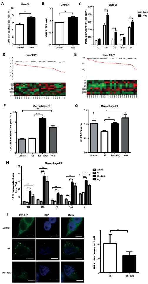
whether orally delivered PAO can enter and remodel ER membranes in mouse tissues. Lipidomic analysis of isolated liver ERs from PAO-treated mice showed a significant increase in total PAO in ER membranes
compared to control (P < 0.05; Fig. 4A and fig. S4A). PAO treatment
increased the MUFA/SFA ratio in liver ERs (Fig. 4B). PAO incorporated
into many lipid compartments, especially PL and CE, of liver ERs (P <
0.05 andP < 0.01; Fig. 4C). PAO was one of the most highly elevated
FAs in all lipid compartments of liver ER (Fig. 4, D and E, and fig. S4, B to D). Collectively, these data demonstrate that oral PAO treatment can promote systemic desaturation of ER membranes in vivo.
Lipidomic analysis of ER compartments from macrophages also reflects the increase in total PAO and MUFA/SFA ratios after PAO
treatment (P < 0.05, Fig. 4F; P < 0.01, Fig. 4G; and validation of the ER
fraction; fig. S4E). PAO is incorporated into all ER lipid species (Fig. 4H). In the macrophages, PA-induced unfolded protein response (UPR) activation (as assessed by IRE1 phosphorylation and spliced XBP1 pro-tein) was blocked by PAO (fig. S4F). Therefore, the efficient integration of PAO into PL and CE together with an increase in MUFA/SFA ratio in the ER membranes suggests that PAO treatment could promote robust changes in membrane dynamics and membrane-initiated UPR activation in macrophages. For example, the assembly of IRE1 or PERK into high-order oligomers on ER membranes is essential for transmitting
stress from within this organelle (59). Activation of these proximal UPR
kinases by saturated lipids, unlike activation by unfolded proteins, does
not require ligand binding by their luminal domains (60). Lipid
composition changes in ER membrane may alter oligomerization and activation states of these kinases. Thus, we investigated the impact of PA and PAO on IRE1 oligomerization states on ER membranes using
previously established protocols (61). PA induced green fluorescent
protein (GFP)–tagged IRE1 oligomer formation on ER and splicing
of XBP1, which was largely prevented by cotreatment with PAO (Fig. 4I and fig. S4, G to I). This result confirms that ER membrane saturation or desaturation by PA or PAO, respectively, can alter the oligomerization state of a proximal UPR sensor and affect downstream UPR signaling. PAO prevents the development of atherosclerosis in mice Atherosclerosis is a nonresolving chronic inflammatory disease of the arterial wall. It is triggered by subendothelial deposition of lipoproteins in areas of disturbed laminar flow that cause endothelial damage. These retained lipoproteins are susceptible to oxidation and other modifications that render them highly proinflammatory and toxic to the overlying en-dothelial cells. This also causes activation of the enen-dothelial cells and intramural recruitment of cells from the mononuclear lineage. In the
subendothelial space, monocytes are transformed first to phagocytes that scavenge lipid deposits and then into proinflammatory, cholesterol-laden foam cells. The foam cells eventually become engorged with cho-lesterol and lose their capacity to migrate, compromising their immune response. As a result, the plaques advance and become more complex with the contribution of other immune cells and vascular smooth muscle (VSM) cells. At this advanced stage, the plaque stability is highly influ-enced by macrophages through their secretion of proinflammatory cytokines and chemokines as well as matrix-degrading proteases. Moreover, upon their death, the release of their toxic content results
in the formation of a prothrombotic necrotic core (37–39, 62).
Atherosclerotic plaques of both mice and humans display signs of ER stress, mitochondrial oxidative stress, and inflammasome activation
in macrophages (11, 43, 63–66). Increased ER stress in lesions associates
with plaque vulnerability and acute coronary syndrome (65). Genetic,
dietary, and chemical interventions that reduce ER stress and
inflam-masome activity can prevent atherosclerosis in mice (11, 67–70).
More-over, specific and selective inhibitors for ER stress and inflammasomes that can alleviate multiple components of metabolic syndrome are
cur-rently under investigation (52, 71).
Given our findings that PAO can inhibit lipid-induced stress in the ER and subsequent activation of the inflammasome in macrophages (Fig. 1 and fig. S1), we hypothesized that chronic PAO intervention in mice could prevent atherosclerosis in vivo. For this purpose, male
ApoE−/−mice were placed on a hypercholesterolemic diet at 6 weeks of
age for a total of 16 weeks. There were no significant differences in body weights or blood glucose concentrations of mice in the experimental groups before or after PAO treatment (table S1). Starting at 12 weeks on diet, PAO was administered daily by oral gavage, whereas control mice received vehicle for a total of 4 weeks (Fig. 5A). Chronic oral
PAO treatment resulted in a significant (P < 0.01) decrease (−33%) in
the development of atherosclerotic lesions in en face aorta analysis when compared to control mice (Fig. 5B). The impact of PAO on athero-sclerosis development was also evaluated in cross-sectional lesions of the aortic root. The foam cell area in the plaques (visualized with
Oil Red O staining) was significantly (P < 0.05) decreased (−29.4%)
in the aortic sinus sections (Fig. 5C). In parallel, there was a significant
(P < 0.01) reduction in the necrotic core area in the lesions of the
PAO-treated group when compared to control mice (−31%) (Fig. 5D). These
results show that PAO treatment alleviated atherosclerosis without improving dyslipidemia and that ER stress (as evident by decrease in sXbp1 mRNA in hearts) was markedly reduced in PAO-treated mice (Fig. 5E and fig. S5, A to D). These in vivo findings demonstrate that dietary supplementation with PAO can effectively mitigate athero-sclerosis in mice.
PAO alters plaque composition by reducing macrophages in lesions
Many kinds of immune and nonimmune cell types participate in the formation and advancement of atherosclerotic plaques. For example, intramural deposition of lipoproteins is linked to endothelial damage that triggers mononuclear cell infiltration to the subendothelial space and subsequent formation of foam cells. Other immune cell types, such as dendritic cells, lymphocytes, eosinophils, and mast cells, are also
involved in plaque advancement (38, 39, 68, 72, 73). Given that PAO
reduces atherosclerosis inApoE−/−mice, we next analyzed PAO-initiated
changes in the cellular composition of atherosclerotic plaques. When
compared to control, PAO treatment reduced (−18%) the macrophage
by guest on February 7, 2019
http://stm.sciencemag.org/
content of aortic root lesions, as visualized by
MOMA-2 staining (P < 0.05; Fig. 6A and fig. S6).
This is consistent with the reduced amount of foam cells in the lesions of PAO-treated mice
(Fig. 5C, seen as Oil Red O–positive staining).
This was not, however, related to an increase in macrophage apoptosis, because PAO treatment also reduced the number of apoptotic cells
(−34.8%) in the lesion area stained with
MOMA-2, as determined by terminal deoxynucleotidyl
transferase–mediated deoxyuridine triphosphate
nick end labeling (TUNEL) assay (P < 0.05; Fig.
6B). Furthermore, we examined the T lymphocyte populations in PAO-treated and control aortic sinus lesions and found no significant differences
in CD3+cells between the two experimental
groups (Fig. 6C and fig. S6).
A
D E
G
Control PAO FFA TAG CE DAG PL
F
B C
H
Control PA PA + PAO PAO 0.0 0.5 1.0 1.5 MUFA/SFA ratio * Control PAO Liver-ER-CE Liver-ER-PC
Control PA PA + PAO PAO
TAG CE DAG PL 0 5 10 15 20 ** * * ** 0.0 0.5 1.0 1.5 * * *
Liver-ER Liver-ER Liver-ER
Macrophage-ER Macrophage-ER Control PA PA + PAO PAO 0 2 4 6 8 10 * Macrophage-ER MUFA/SFA ratio FFA PAO concentration (mol %) PAO concentration ( µ M ) PAO concentration (mol %) 0 10 20 30 40 50 ** ** ** *** *** * ** ** ** ** **
PAO concentration (mol %)
IRE1-GFP DAPI Merge
Control PA PA + PAO IRE1 foci number/cell α 0 5 10 ** PA PA + PAO Control Control Control Control Control PAO PAO PAO PAO PAO PC20:2 PC18:2 PC16:0 PC20:3 PC18:0 PC22:4 PC20:4 PC18:1 PC20:1 PC18:4 PC22:6 PC18:3 PC20:0 PC22:1 PC17:0 PC14:0 PC14:1 PC15:0 PC12:0 PC22:5 PC20:5 PC22:2 PC16:1 − 4 − 3 − 2 − 1 0123 Control Control Control Control Control PAO PAO PAO PAO PAO
CE18:1 CE22:6 CE14:0 CE12:0 CE17:0 CE18:0 CE15:0 CE18:2 CE16:0 CE20:4 CE22:1 CE20:1 CE18:4 CE22:5 CE18:3 CE20:2 CE24:1 CE20:5 CE22:2 CE22:4 CE20:0 CE20:3 CE24:0 CE22:0 CE16:1 CE14:1
−4 −2 02 I 0 1000 2000 3000 4000 5000 Control PAO ** * * *
Fig. 4. PAO regulates lipid composition in the
ERs from livers of ApoE−/−mice and from
macrophages. (A to E) Liver ER fractions were
isolated from PAO-treated and control ApoE−/−mice
on Western diet and analyzed. (A) Mean concentra-tion of PAO (C16:1n7) (mol %; the ratio of moles of FAs to total moles of FAs). (B) MUFA/SFA ratio. (C) Mean concentration (mM) of PAO (C16:1n7) in vari-ous lipid metabolites. Data are means ± SEM. *P <
0.05, **P < 0.01. n = 5. Unpaired two-tailed Student’s
t test [in (B)] and Wilcoxon test [in (A) and (C)] were used for statistical analysis. (D and E) The heat maps display lipid class composition analysis for (D) PC and (E) CE. In the heat maps, x axis shows each lipid metabolite measurement in each sample (y axis) as up-regulated (red) or down-regulated (green), as determined by centering to the mean of the control group and scaled by the SD of all observations. In the line plot, y axis shows the t statistics from compar-ing two groups. The black line is the 95th percentile of the standardized statistics over 1000 permutations. The red dots are the z scores of the observed statistics that were standardized to 1000 permutations. Data are means ± SEM. *P < 0.05, **P < 0.01. n = 5. (F to H) ER fractions were isolated from RAW 293.6 macrophages and analyzed. (F) Mean concentration (mol %) of C16:1n7. (G) MUFA/SFA ratio. (H) Mean concentration (mol %) of C16:1n7 in various lipid classes. Data are means ± SEM. *P < 0.05, **P < 0.01, ***P < 0.001. n = 4. Nonparametric
Kruskal-Wallis test and unpaired two-tailed Student’s t test
were used for statistical analysis. (I) Cells expressing GFP-IRE1 were treated with PA or PAO, alone or together, for 6 hours and analyzed for formation of discrete IRE1 oligomer foci (green dots) by imaging with a confocal microscope (blue, nuclei; green, GFP-IRE1). The graph depicts quantification of IRE1 oligomerization as reported distinct IRE1 foci (green)
per cell. Data are means ± SEM. **P < 0.01. n≥ 100
cells from four experiments. To compare PA-treated versus cotreated with PA and PAO, Mann-Whitney U test was used for statistical analysis. Scale bar,
20 mm. DAPI, 4′,6-diamidino-2-phenylindole.
by guest on February 7, 2019
http://stm.sciencemag.org/
In addition to having proatherogenic immune cell types, atherosclerotic lesions contain VSM, which can migrate from adventitia to intima and aid in sealing the fibrous cap against rupture. This migratory effect is controlled by the mediators and chemokines
re-leased from macrophages and other plaque cells (38).
Immunofluorescence staining of aortic root lesions
with a specific antibody against a–smooth muscle
actin showed no significant change in VSM+area after
PAO treatment (Fig. 6D and fig. S6). Additionally, VSM-derived collagen content showed no change in PAO-treated lesions when compared to control lesions (Fig. 6E). Overall, PAO treatment does not appear to improve the mechanical strength of the lesions. PAO prevents macrophage ER stress and inflammasome activation in lesions
Our findings demonstrated a strong impact of PAO on lesion macrophages and their viability. Our earlier studies have also indicated that PAO can block lipo-toxic, but not proteolipo-toxic, ER stress in macrophages
(11). In our current study, we observed that PAO can
block lipid-induced ER stress, subsequently leading to suppression of the inflammasome (Fig. 1 and fig. S1). Hence, we asked whether the stress and anti-inflammatory effects of PAO observed in macrophages can be recapitulated in vivo during atherogenesis. First, we examined the immunofluorescence signal intensity for two markers of activation of the ER stress re-sponse, namely, phosphorylation of eukaryotic initiation factor 2a (eIF2a) and expression of cyclic adenosine
monophosphate–dependent transcription factor 3
(ATF3) in macrophage-enriched areas of aortic root
6 weeks 18 weeks 22 weeks
Western Diet
Oral gavage once daily for 4 consecutive weeks 1% BSA in PBS : Control group PAO in 1% BSA in PBS : PAO group
H&E Oil Red O Control PAO Control PAO A B C Necrotic core D E Lesion area ( 10 3µ m 2) 0 100 200 300 * PAO Control
Lesion area (%total)
0 5 10 15 20 ** Control PAO 0 200 400 600 800 1000 ns Total cholesterol (mg/dl) Cholesterol (mg/dl) 0 200 400 600 800 ns ns ns VLDL LDL HDL Control PAO l o r et s el o h C (µ n oi t c a rf r e p g ) VLDL 100 80 60 40 20 0 LDL HDL Control PAO 1 Control PAO
Necrotic core area (%)
0 5 10 15 20 ** Control PAO 12 26 50 F Fraction number Fig. 5. PAO treatment reduces atherosclerosis in
ApoE−/−mice. (A) Experimental design for the analysis
of atherosclerosis in ApoE−/−mice on a Western diet.
BSA, bovine serum albumin; PBS, phosphate-buffered saline. (B) PAO treatment reduced atherosclerotic lesions in en face aorta (control, n = 18; PAO, n = 15; **P < 0.01). (C) Histology of plaques (marked with arrows) at the aortic sinus stained with Oil Red O and hematoxylin and eosin (H&E). Lesion area was reduced in the plaques of PAO-treated mice (control, n = 10; PAO, n = 10; *P < 0.05). Scale bar, 300 mm. (D) The necrotic core area (marked with dashed lines and arrows) was also de-creased in the plaques of PAO-treated mice as compared to control mice (control, n = 12; PAO; n = 11, **P < 0.01). Scale bar, 50 mm. (E) Serum total cholesterol and lipo-protein cholesterol [VLDL, low-density lipolipo-protein (LDL), or high-density lipoprotein (HDL)] concentrations were similar between PAO-treated and control mice. (F) Li-poprotein profiles from control (blue) and PAO-treated
(red) ApoE−/−mice show the average distribution (mg per
fraction; 1 to 13: VLDL, 14 to 31: LDL, and 32 to 50: HDL) of total cholesterol for each group. Data are means ± SEM. *P < 0.05, **P < 0.01. ns, not significant. control, n = 4; PAO, n = 6. Control versus PAO: Mann-Whitney
U test [in (B) to (D)] or unpaired two-tailed Student’s
t test [in (E)] was used for statistical analysis.
by guest on February 7, 2019
http://stm.sciencemag.org/
lesions. Quantitative analysis of ER stress by immunofluorescence staining showed a
sig-nificant (P < 0.01) reduction in p-eIF2a and
ATF3 (−42.5 and −42.6%, respectively) in
the macrophage-rich regions of lesions after chronic PAO treatment when compared to the control group (Fig. 7, A and B, and fig. S6). Moreover, PAO supplementation re-duced XBP1 splicing in the hearts from these mice (fig. S5D). These results are consistent with our earlier findings in macrophages in vitro and demonstrate the efficacy of oral PAO treatment in alleviating macrophage ER stress in atherosclerotic lesions in vivo. We next asked whether PAO could in-hibit lipid-induced inflammasome acti-vation and secretion of IL-1b and IL-18
in vivo. Previous studies showed that the hyperlipidemic diet inApoE−/−
mice causes a marked induction of the inflammasome in the lesions and systemically, as evidenced by increased secretion of IL-1b and IL-18
(43, 74). We found that the intensity of immunofluorescence staining for
IL-1b in atherosclerotic lesions was significantly (P < 0.05) reduced by
PAO (−32.8%), demonstrating effective suppression of the
inflamma-some by oral PAO treatment (Fig. 7C and fig. S6). Analysis of serum from mice supplemented for 4 weeks with PAO (threefold higher dosage than
found in Western diet) showed a significant (P < 0.05) reduction in
sys-temic IL-1b and IL-18 production (−32.2 and −26.6%, respectively) by
PAO treatment when compared to control mice (Fig. 7, D and E). Col-lectively, these findings demonstrate that chronic PAO supplementation can effectively block hyperlipidemia-induced macrophage ER stress and inflammasome activity in atherosclerotic plaques in vivo.
DISCUSSION
The contribution of DNL to obesity and its complications is complex in part because lipogenesis in various organs has different consequences MOMA-2 Control PAO α-SMA Masson’s trichrome A B C D
MOMA–2 positive area (%)
0 10 20 30 * Control PAO α-SMA–positive area (%) 0.0 0.5 1.0 1.5 2.0 2.5 ns Control PAO TUNEL 0 10 20 30 40 ns Control PAO
Collagen area (% of lesion area) CD3 CD3 cells/mm + 2 0.00 0.02 0.04 0.06 0.08 ns PAO Control TUNEL-positive cell/mm (%) 2 0 5 10 15 20 25 * Control PAO E Fig. 6. PAO reduces the macrophage area
and apoptotic cells in atherosclerotic
pla-ques. Six-week-old ApoE−/−mice were put on
Western diet (total of 16 weeks) and orally gavaged with PAO or vehicle daily (for the last 4 weeks). Proximal aorta cryosections were stained with specific antibodies for various cellular markers. The representative images are shown for (A) macrophage marker MOMA-2 (n = 10 per group). Mean percent
MOMA-2–positive area relative to total lesion
area is reported. (B) TUNEL assay. Arrowheads show apoptotic cells (control, n = 10; PAO, n = 9). Average apoptotic cells per plaque area
(mm2) are reported. (C) T cell marker, CD3
(control, n = 9; PAO, n = 10). Arrowheads show CD3-positive cells. The average numbers of
T cells per plaque area (mm2) in each section
are reported. (D) VSM cell marker a–smooth
muscle actin (a-SMA) is depicted (n = 10 per group). Dashed line outlines the lesion area. Mean percent a-SMA area relative to total lesion area is reported. Enlarged images
(four-fold; upper right) represent the a-SMA–positive
area in the intimal region. (E) Masson’s
tri-chrome stain for collagen (blue, collagen; red, cytoplasm and muscle fibers) (control, n = 13; PAO, n = 9). Mean percent collagen area rela-tive to total lesion area is reported. Data are means ± SEM. *P < 0.05. ns, not significant. Control versus PAO: Mann-Whitney U test was used for statistical analysis. Scale bars, 150 mm.
by guest on February 7, 2019
http://stm.sciencemag.org/
on systemic metabolism. For example, liver DNL is increased in obesity and frequently associates with fatty liver disease and
in-sulin resistance (75, 76). Lipogenesis in
the skeletal muscle can promote insulin resistance but is also required for optimal
muscle strength (77). Brain DNL generates
lipid signals that affect food intake and
energy metabolism (78). DNL in white
ad-ipose tissue is beneficial for systemic me-tabolism by increasing insulin sensitivity
and improving lipid metabolism (7, 79).
Therefore, developing therapeutic strate-gies based on DNL is challenging. For in-stance, a global DNL inhibitor may have beneficial metabolic effects acting on the hepatocytes, but adverse effects may be associated with blocking DNL in adipocytes,
pancreatic b cells, and macrophages (76).
It is now clear that DNL can generate nu-merous lipid signals (1-palmitoyl-2-oleoyl-sn-glycerol-3-phosphocoline, alkyl ether lipids, and PAO) that can act through dis-tinct receptors and mechanisms to exert
potent metabolic effects (75). Alternatively,
nutritional supplementation with a specific DNL product may effectively generate the beneficial metabolic effects of DNL in dis-eases afflicting target cells in metabolically
active organs (6, 7, 9–11, 21, 33, 34, 36, 80).
Despite a great interest in DNL-based ther-apeutic development for chronic metabolic
diseases, DNL-generated lipid species’
im-pact on the vasculature and atherosclerosis is not known. Circumstantial findings from several genetic or chemical models of DNL activation in mice suggest that DNL or its products could be protective against
atherosclerosis (33, 34, 36, 75, 79). Our study
provides a direct examination of PAO’s
im-pact on atherogenesis. Our findings dem-onstrate that PAO supplementation (at threefold higher dosage than found in the composition of Western diet) reduces
ath-erosclerosis inApoE−/−mice. Chronic PAO
treatment effectively mitigated macrophage ER stress in lesions and blocked inflamma-some activation in lesions and systemically, similar to our initial observation in cultured primary mouse and human macrophages. In addition, a recent PAO supplementa-tion trial in humans resulted in a marked reduction in high-sensitivity C-reactive pro-tein and improved serum lipids, suggest-ing that PAO supplementation could also
be atheroprotective in humans (36).
Col-lectively, the outcome of these studies im-plies that nutritional supplementation with
Control PAO p-eIF2α ATF3 MOMA-2/DAPI MOMA-2/DAPI MOMA-2/DAPI p-eif2α/DAPI p-eif2α/DAPI ATF3/DAPI ATF3/DAPI A B C IL-1ββ Control PAO IL-1 β (pg/ml) 0 50 100 150 200 * Control PAO Control PAO ATF3 0 10,000 20,000 30,000 40,000 * Control PAO Relative fluorescence intensity (a.u.)
IL-1β p-elF2α 0 5000 10,000 15,000 ** Control PAO Relative fluorescence intensity (a.u.)
Relative fluorescence intensity (a.u.) 0 10,000 20,000 30,000 ** Control PAO MOMA-2/DAPI IL-18 (pg/ml) 0 50 100 150 200 250 Control PAO * D E
Fig. 7. PAO mitigates ER stress in lesional macrophages and suppresses the inflammasome in lesions. Aortic root sections from PAO or vehicle-treated mice were analyzed using the following specific antibodies for immunofluorescence staining: (A) p-eIF2a (green) and MOMA-2 (red) (control, n = 8; PAO, n = 5), (B) ATF3 (green) and MOMA-2 (red) (control, n = 8; PAO, n = 4), and (C) IL-1b (control, n = 10; PAO, n = 8).
Fluorescence intensity of ER stress markers in macrophage-enriched (MOMA-2–positive) areas was
quanti-fied using ImageJ. IL-1b (control, n = 8; PAO, n = 4) (D) and IL-18 (n = 7 per group) (E) were measured in
plasma of ApoE−/−mice treated with or without PAO after 4 weeks. All data are means ± SEM. *P < 0.05, **P <
0.01. Control versus PAO: Mann-Whitney U test was used for statistical analysis. Scale bars, 150 mm. a.u., arbitrary units.
by guest on February 7, 2019
http://stm.sciencemag.org/
PAO may be a promising preventive approach in atherosclerosis through effective mitigation of ER stress and inflammasome activity. This does, however, require direct validation in future human trials.
The mechanism of action of DNL-produced lipids in vivo during
atherogenesis is not known. Here, we sought to gain insight into PAO’s
mechanism of action in vivo by studying its effects on macrophages involved in atherogenesis. Macrophages play a prominent role in the
progression of atherosclerosis to vulnerable plaques (38, 62). Previous
studies established that cholesterol crystals and SFAs specifically induce NLRP3 inflammasome and IL-1b secretion from macrophages, and genetic or pharmacological inhibition of the inflammasome alleviates
atherosclerosis (43, 74, 81). Here, we found that treatment of primary
mouse or human macrophages with PAO effectively blocked inflamma-some activation by SFAs, but not by other NLRP3 inducers like ATP and alum. Additionally, PAO did not prevent activation of any other inflammasome complexes (AIM2, NLRC4, or NLRP1) by their specific
inducers. These results underscore PAO’s specificity as an inhibitor to
lipid-induced NLRP3 inflammasome activation. Consistently, in vivo PAO treatment also lowered hyperlipidemia-induced IL-1b and IL-18 concentrations in serum and IL-1b expression in plaques. Therapies involving small-molecule inhibitors and dietary regimes aimed at blocking inflammasome activation and IL-1b secretion in a variety of diseases are
highly sought after (52, 69, 82).
The activation signal for the inflammasome can be generated by
distress in ER (44, 47, 50, 83). There are several ways in which ER can
induce inflammasome activity, including release of Ca2+or ROS and
mitochondrial damage (47, 49). Moreover, damaged mitochondria
generate ROS, which can further potentiate inflammasome activity (49).
In an earlier study, we showed that PAO is a potent inhibitor of
lipid-induced but not proteotoxic ER stress (11). Studies have also shown ER
stress being causally associated with atherogenesis, diabetes, and obesity
(84). Here, we confirmed that PAO inhibits hyperlipidemia-induced ER
stress in vivo and in the macrophages infiltrating atherosclerotic plaques, and subsequent inflammasome activation. Collectively, the outcome of
these experiments underscores PAO’s ability to strongly antagonize
lipid-induced ER stress upstream of inflammasome activation. How does PAO promote metabolic resilience to hyperlipidemia? Dietary PAO intake is quite low because PAO is not ubiquitiously found
in food sources (25). However, PAO is the fifth most abundant FA in
humans, suggesting that endogenous PAO synthesis, particularly in
obesity, is a major contributor to PAO abundance in humans (85).
One plausible explanation for the increased PAO production in obesity is that it reflects a necessary metabolic adaptation to meet the high demand for MUFA in rerouting the metabolism of SFA and cholesterol toward storage or excretion and to support membrane function by
active remodeling (86–89). Here, we showed that PAO effectively replaces
other FAs, resulting in increased MUFA/SFA ratio in macrophages, plasma, and aorta, but without expanding any of the lipid classes. The marked incorporation of PAO into multiple PL species and, in particular, cardiolipin implies dynamic membrane remodeling of organelles through desaturation by PAO. Further lipidomic analysis of isolated ER fractions from macrophages and livers confirmed that PAO effectively incorpo-rates into most lipid compartments, especially PLs and cholesterol, in this organelle and increases the MUFA/SFA ratio. Extensive ER membrane remodeling can thus be achieved through PAO-induced desaturation and may have wider implications, such as altered membrane biophysical properties and UPR signaling emanating from these membranes. Proximal UPR regulators form high-order oligomers on ER membranes
upon ER stress induction by chemicals (61). Here, we show that PA
evokes IRE1 oligomerization on ER membranes and splicing ofXBP1
mRNA, which can be blocked by PAO cotreatment. The outcome of these experiments strongly supports the idea that dynamic remodeling of membranes by desaturation with PAO underlies ER resistance to stress induced by PA. One limitation of our study is that we do not know the
extent of remodeling in other intracellular organelles’ membranes
because we only analyzed the ER via lipidomics. However, our study provides proof of principle that organelle membrane remodeling by nutritional supplementation with a bioactive lipid could be an effective strategy for the treatment of atherosclerosis.
This study and others support the conclusion that PAO supplemen-tation can mitigate more than one component of metabolic syndrome,
including insulin resistance and atherosclerosis (7, 28, 33, 36, 90). PAO
is a potent inhibitor of lipid-induced ER stress and inflammasome activation in mouse and human macrophages and atherosclerotic plaques in vivo. These beneficial changes are associated with dynamic lipidomic remodeling of ER membranes in mice in vivo. Our findings show that it is possible to modify organelle stress responses and suppress inflammasome activity through nutritional supplementation with PAO. PAO supplementation reduces atherosclerosis in mice,
and these results warrant future investigation to assess PAO’s therapeutic
potential in human disease.
SUPPLEMENTARY MATERIALS
www.sciencetranslationalmedicine.org/cgi/content/full/8/358/358ra126/DC1 Materials and Methods
Fig. S1. Lipid-induced proinflammatory cytokine expression and ER stress prevented by PAO. Fig. S2. Increased desaturation of plasma lipids in PAO-supplemented ApoE−/−mice. Fig. S3. Lipid class composition analysis of plasma from ApoE−/−mice.
Fig. S4. Analysis of liver or macrophage ER membrane lipid composition and IRE oligomerization on ER.
Fig. S5. Lipoprotein profile and ER stress status of PAO-supplemented ApoE−/−mice. Fig. S6. Representative images for negative controls in immunofluorescence staining. Table S1. Physical and biochemical characteristics of ApoE−/−mice in PAO-treated and control groups.
Table S2. Raw data and statistical analyses (provided as an Excel file). References (96–99)
REFERENCES AND NOTES
1. A. Aarsland, D. Chinkes, R. R. Wolfe, Contributions of de novo synthesis of fatty acids to total VLDL-triglyceride secretion during prolonged hyperglycemia/hyperinsulinemia in normal man. J. Clin. Invest. 98, 2008–2017 (1996).
2. E. J. Parks, M. K. Hellerstein, Carbohydrate-induced hypertriacylglycerolemia: Historical perspec-tive and review of biological mechanisms. Am. J. Clin. Nutr. 71, 412–433 (2000).
3. L. Abu-Elheiga, M. M. Matzuk, P. Kordari, W. Oh, T. Shaikenov, Z. Gu, S. J. Wakil, Mutant mice lacking acetyl-CoA carboxylase 1 are embryonically lethal. Proc. Natl. Acad. Sci. U.S.A. 102, 12011–12016 (2005).
4. S. S. Chirala, H. Chang, M. Matzuk, L. Abu-Elheiga, J. Mao, K. Mahon, M. Finegold, S. J. Wakil, Fatty acid synthesis is essential in embryonic development: Fatty acid synthase null mutants and most of the heterozygotes die in utero. Proc. Natl. Acad. Sci. U.S.A. 100, 6358–6363 (2003). 5. M. V. Chakravarthy, I. J. Lodhi, L. Yin, R. R. Malapaka, H. E. Xu, J. Turk, C. F. Semenkovich, Identification of a physiologically relevant endogenous ligand for PPARa in liver. Cell 138, 476–488 (2009).
6. S. Liu, J. D. Brown, K. J. Stanya, E. Homan, M. Leidl, K. Inouye, P. Bhargava, M. R. Gangl, L. Dai, B. Hatano, G. S. Hotamisligil, A. Saghatelian, J. Plutzky, C.-H. Lee, A diurnal serum lipid integrates hepatic lipogenesis and peripheral fatty acid use. Nature 502, 550–554 (2013).
7. H. Cao, K. Gerhold, J. R. Mayers, M. M. Wiest, S. M. Watkins, G. S. Hotamisligil, Identification of a lipokine, a lipid hormone linking adipose tissue to systemic metabolism. Cell 134, 933–944 (2008).
by guest on February 7, 2019
http://stm.sciencemag.org/