Dicle Tıp Derg / Dicle Med J Cilt / Vol 37, No 3, 282-286
Yazışma Adresi /Correspondence: Dr. Zeki Dostbil, Dicle University Medical Faculty, Department of Nuclear Medicine, Diyarbakir, Turkey. E-mail: [email protected]
Copyright © Dicle Tıp Dergisi 2010, Her hakkı saklıdır / All rights reserved
ORIGINAL ARTICLE / ÖZGÜN ARAŞTIRMA
Myocardial perfusion scintigraphy fi ndings in patients with mild coronary
atherosclerotic lesions on coronary angiography
Koroner anjiografi sinde hafi f aterosklerotik koroner lezyonu olan hastalarda
miyokard perfüzyon sintigrafi bulguları
Zeki Dostbil1, Habib Çil2, Ebru Öntürk Tekbaş2, Zuhal Arıtürk Atılgan2, Yahya İslamoğlu2 Bekir Taşdemir3, Yusuf Dağ1
Dicle University Medical Faculty, 1Department of Nuclear Medicine and 2Cardiology, Diyarbakir, Turkey 3Elazig Training and Research Hospital, Department of Nuclear Medicine, Elazig, Turkey
Geliş Tarihi / Received: 05.04.2010, Kabul Tarihi / Accepted: 12.05.2010
ÖZET
Amaç: Miyokard perfüzyon sintigrafi si (MPS) miyokard perfüzyonunun fonksiyonel değerlendirmesinde yaygın olarak kullanılmaktadır. Fakat bazı çalışma sonuçları ko-roner anjiografi ile tespit edilen koko-roner arter hastalığının şiddeti ile zıtlık gösterir. Koroner anjiografi tamamen nor-malken MPS’nde iskemiden bahsedilmesi sık karşılaşılan bir durumdur. Bu çalışmada, hafi f aterosklerotik lezyonla-rın iskemiye sebep olup olmadıklalezyonla-rını araştırmayı amaç-ladık.
Yöntem ve Gereç: Kardiyoloji Polikliniğine göğüs ağrısı şikayeti ile başvuran ve 3 ay içerisinde koroner anjiografi yapılmış olan 52 hastaya 99mTc-MIBI ile miyokard perfü-yon sintigrafi si yapıldı.
Bulgular: Hafi f derecede aterosklerotik lezyonu olan 52 hastanın 22’sinde MPS’de değişik derecelerde iske-mi bulguları gözlendi. Yapılan istatistik analizde iskeiske-mi ile cinsiyet, hipertansiyon, DM, dislipidemi, sigara içme, mitral kapak yetmezliği, sol ventrikül hipertrofi si, efor test sonucu ve aterosklerotik lezyonun bulunduğu damar ara-sında anlamlı bir ilişki tespit edilmedi.
Sonuç: Çalışma bulgularımız koroner arterlerdeki ha-fi f aterosklerotik lezyonların, çok erken dönemlerde bile olsa, miyokardda iskemiye sebep olabileceğini göster-mektedir.
Anahtar kelimeler: koroner arter hastalığı, miyokard is-kemisi, miyokard perfüzyon görüntüleme, koroner anjiog-rafi
ABSTRACT
Objectives: Myocardial perfusion scintigraphy (MPS) is widely used in functional assessment of myocardial per-fusion. But, some study results are in contradiction with severity of coronary artery disease detected by coronary angiography (CA). It is frequently encountered case that CA is completely normal whereas MPS describes isch-emia. In this study, we aimed to investigate whether mild atherosclerotic lesions cause ischemia.
Materials and methods: MPS with 99mTc-MIBI was per-formed in 52 patients who applied to cardiology clinics for history of chest pain and underwent diagnostic CA within 3 months.
Results: In 22 of 52 patients with mild atherosclerotic le-sions, ischemia in various degrees was detected on MPS. In statistical analysis, any signifi cant relationship was not found between ischemia and gender, hypertension, DM, dyslipidemia, smoking, mitral valve insuffi ciency, left ven-tricular hypertrophy, exercise testing result and affected coronary artery.
Conclusion: Our study fi ndings have shown that mild ath-erosclerotic lesions even at very early stage may cause myocardial ischemia
Key words: coronary artery disease, myocardial isch-emia, myocardial perfusion imaging, coronary angiogra-phy
INTRODUCTION
Myocardial perfusion scintigraphy (MPS) is a non-invasive modality widely used in evaluation of myo-cardial ischemia. Perfusion defects have been linked to areas supplied by signifi cantly stenosed coronary arteries.1 It has been also shown that perfusion
ab-normality might be caused by angiographically nonsignifi cantly stenosed (<50% diameter stenosis) arteries.2,3 Patients with reversible perfusion defects
on MPS suggestive of ischemia but angiographical-ly insignifi cant coronary artery disease have often been labeled as having a “false-positive” scan.1
Recent studies with intravascular ultrasonogra-phy (IVUS) and microvascular function assessment have revealed the limitations of traditional coronary angiography (CA) and underestimation of athero-sclerotic burden in areas of perfusion defects on MPS where microvascular and endothelial dysfunc-tion were shown.2,4-7
In this study, we aimed to investigate myo-cardial ischemia fi ndings on MPS in patients with angiographically mild atherosclerotic coronary le-sions.
MATERIALS AND METHODS
This study comprised of 52 patients in ‘Elazig Re-search and Training Hospital’ and ‘Dicle University Medical Faculty, Department of Nuclear Medicine’ between April 2008 and March 2010. The main in-dication for referral of all patients was evaluation of chest pain. Patients having LBBB, RBBB or muscle bridge that may cause false positive scintigraphy re-sults were excluded from study.
In all patients, 99mTc-MIBI ECG Gated (8-bin frame mode) myocardial perfusion SPECT imaging was performed. During image acquisition breasts were fi xated upwards in all female patients. Within 3 months before or after MPS study, diagnostic CA was made. Treadmill exercise or pharmacological stress by dipiridamol was used. GE Infi nia and GE Millenium gamma camera systems were used for radionuclide imaging procedure. By means of car-diac quantitation program called ‘Emory Carcar-diac Toolbox (ECTb)’; at the myocardial region refl ect-ing the site of wall irregularity, nonstenotic plaque and coronary artery stenosis between 0-20% and 20-50% at coronary angiography were evaluated in respect to myocardial ischemia. All images were evaluated for attenuation artifacts. If reversible per-fusion defects can not be explained by attenuation artifact, these were regarded as fi ndings of myocar-dial ischemia8. Patients who have ischemic fi ndings
on SPECT images were called ‘ischemic’ and oth-ers were called ‘nonischemic’. In all patients who have DM, HT or dyslipidemia, these diseases were documented and all of them had been using drug for various durations. ‘Smoking positive’ patients had been smoking any number of cigarette per day and for any duration. Exercise stress testing results were categorized as positive, negative or suspicious positive. Lesions were defi ned in LAD, Cx or RCA. left ventricular EF, mitral valve (MV) insuffi ciency
and left ventricular hypertrophy was evaluated by echocardiography. If present, ischemia on SPECT images was classifi ed as mild, moderate or severe.
Statistical analysis
‘Independent samples t-test’, in SPSS 11.5 program, to compare ages and BMI of ischemic and nonisch-emic patients; ‘chi-square test’, to compare distri-bution of gender, DM, HT, dyslipidemia, smoking, exercise stress testing results, effected coronary ar-tery numbers, atherosclerotic lesion types, left ven-tricular EF, MV insuffi ciency and left venven-tricular hypertrophy in ischemic and nonischemic patients; ‘Pearson’s correlation coeffi cient test’ or ‘Spearman rank correlation test’, to assess correlation between ischemia and the factors were used. P<0.05 was considered statistically signifi cant.
RESULTS
The study group comprised of 52 patients aged be-tween 41-86 years (33 male, 19 female and mean age: 57.1±10.4 yrs). Body mass index (BMI) of pa-tients ranged between 19.3 and 36.4 and mean±SD: 28.1± 4.2. There was no statistically signifi cant difference between age and BMI of ischemic and nonischemic patients.
Clinical characteristics, coronary angiography and exercise stress testing results of the patients were shown on Table 1-3. Any of coronary arter-ies did not have more than one atherosclerotic le-sion. At least one coronary artery territory of 22 of patients showed ischemic changes on MPS. Of them, 16 patients had history of at least one of the diseases that were hypertansion, DM, dyslipidemia, cigarette smoking, MV insuffi ciency or left ventric-ular hypertrophy on echocardiography. However, 6 patients did not have any of cardiovascular risk factors and echocardiography fi ndings were normal. Totally 126 coronary arteries were assessed. 83 of them were ischemic and 43 were normal on SPECT images. There was no statistically signifi cant differ-ence between the factors that may cause myocardial ischemia in ischemic and nonischemic patients (Ta-ble 1). In statistical analysis, poor signifi cant cor-relation was found between ischemia and 2 factors that are BMI and gender whereas other factors did not show any signifi cant correlation with ischemia (p>0.05). There was statistically poor correlation between atherosclerotic lesion stage and ischemia severity (p=0.02, r=0.44).
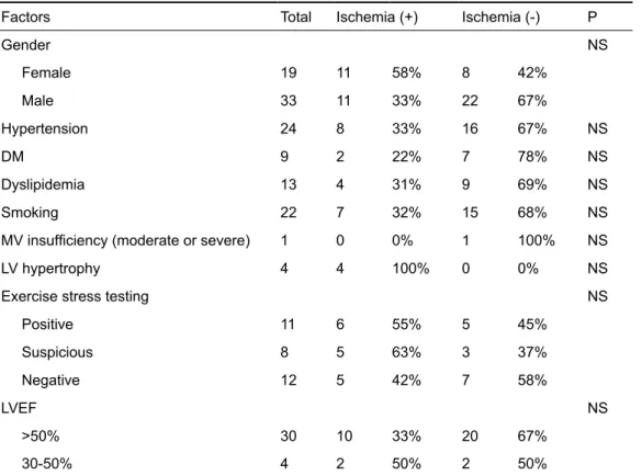
Table 1. Clinical, laboratory and demographic features of the patients and their statistical relationships with ischemia
Factors Total Ischemia (+) Ischemia (-) P
Gender NS Female 19 11 58% 8 42% Male 33 11 33% 22 67% Hypertension 24 8 33% 16 67% NS DM 9 2 22% 7 78% NS Dyslipidemia 13 4 31% 9 69% NS Smoking 22 7 32% 15 68% NS
MV insuffi ciency (moderate or severe) 1 0 0% 1 100% NS
LV hypertrophy 4 4 100% 0 0% NS
Exercise stress testing NS
Positive 11 6 55% 5 45% Suspicious 8 5 63% 3 37% Negative 12 5 42% 7 58% LVEF NS >50% 30 10 33% 20 67% 30-50% 4 2 50% 2 50%
MV: Mitral valve, LV: Left ventricle, LVEF: Left ventricular ejection fraction, NS: Non signifi cant (p>0.05) Table 2. Coronary angiography and myocardial perfusion scintigraphy fi ndings in 52 patients
Coronary artery lesions No of coronary artery Ischemia (+) Ischemia severity Ischemia (-) Mild Moderate severe
Normal 43 1 2% 1 42 98% Wall irregularity 13 3 23% 3 10 77% Nonstenotic plaque 24 7 30% 5 2 17 70% Stenotic (0-20%) 29 10 34% 7 3 19 66% Stenotic (20-50%) 17 7 41% 2 4 1 10 59% Total 126 28 22% 17 10 1 98 78%
Table 3. Coronary arteries having atherosclerotic lesions and ischemia
Affected coronary arteries of patients Total Ischemia (+) Ischemia (-)
RCA 8 4 50% 4 50%
Cx 8 1 13% 7 87%
LAD 10 4 40% 6 60%
RCA and LAD 9 5 56% 4 44%
RCA and Cx 3 1 33% 2 67%
LAD and Cx 8 5 63% 3 37%
All 6 2 33% 4 67%
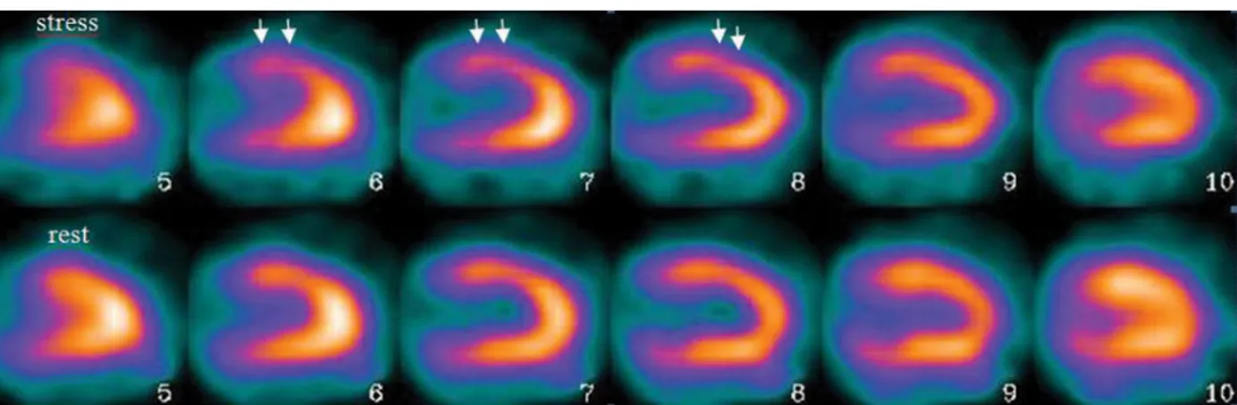
Figure 1. SPECT images of 49 years old woman in whom coronary angiography revealed nonstenotic plaque in LAD. On vertical long axis SPECT images, reversible perfusion defects refl ecting ischemia can be seen at anterior wall of the left ventricle (white arrows).
patients had some factors such as HT, DM, dyslipi-demia, cigarette smoking, LV hypertrophy or MV insuffi ciency that may cause myocardial ischemia. But statistical analysis showed that there was no signifi cant relationship between ischemia and these factors. Low subject number may contribute this result. Moreover, 6 ischemic patients didn’t have any of them. No one in this group had a history of any other known disease. In these circumstances we couldn’t explain ischemic changes by any reason other than endothelial dysfunction. Based upon this, we have thought that mild coronary atherosclerotic lesions can cause myocardial ischemia. But, we don’t have any data that whether ‘exercise-induced coronary spasm’ that may cause myocardial isch-emia was responsible for perfusion abnormalities at least in some of cases.
Verna et al.5 have studied 20 patients referred
for coronary angiography because of chest pain, abnormal exercise ECG, and abnormal myocardial perfusion fi ndings. In that study, patients were eval-uated by coronary anatomy and vasodilator capac-ity with IVUS and doppler fl ow veloccapac-ity measure-ments. They concluded that reversible perfusion defects seen on ‘single photon emission computed tomography’ (SPECT) images are often associated with angiographically unrecognized occult athero-sclerotic changes and an abnormal vasodilatation capacity of the coronary circulation. In our study, totally 83 coronary arteries (LAD, LCX or RCA) were evaluated and territories of 27 of them were found to be ischemic on MPS. We noticed that 17 of them (63%) were mildly, 9 (33%) were moderately
DISCUSSION
The atherosclerotic coronary lesions such as wall irregularity, nonstenotic plaque and mild coronary artery stenosis are normally expected not to cause myocardial ischemia during exertion. However, in some patients with normal coronary anatomy on CA ischemic changes can be revealed on MPS.9
Although many attenuation artifacts may cause such scintigraphic fi ndings, not every case can be explained as artifact. In fact, many condition such as DM, HT, cardiomyopathies, syndrom X, dys-lipidemia etc. may cause myocardial perfusion abnormalities due to endothelial or microvascular dysfunction.2,10-13
Many prior studies have emphasized the role of endothelial dysfunction as a potential cause of an-gina or abnormal functional results in patients with no signifi cant documented CAD. 14-20 Rodés-Cabau
et al. 3 have studied patients with angiographically
mild coronary stenosis (<50% diameter stenosis by quantitative coronary angiography) by IVUS, frac-tional fl ow reserve (FFR) measurements and MPS. They have concluded that plaque burden as deter-mined by IVUS might partly explain the presence of myocardial perfusion defects in cases of angio-graphically nonsignifi cant coronary lesions. How-ever, the high FFR values associated with these le-sions suggested that other mechanisms, such as en-dothelial/microvascular dysfunction, might also ac-count for perfusion abnormalities in these patients. In this study, we assessed totally 52 patients having mild artherosclerotic lesions and detected ischemia in 22 of them on SPECT images. Most of ischemic
and only 1 (4%) was severely ischemic. We found statistically poor correlation between lesion stage and ischemia severity (p=0.02, r=0.44). Mild isch-emic fi ndings on SPECT images may be deemed to be suspicious by some physicians, so ones may not report some scintigrapically mild ischemic fi ndings to increase specifi city of the procedure. Thus, the test results may vary from centers to centers.
A limitation of this study is that the myocar-dial ischemia caused by atherosclerotic lesions can be detected more accurately by PET imaging than MPS.21 Moreover; we didn’t perform fractional
fl ow reserve or doppler fl ow velocity measurements or any other test that evaluates endothelial/micro-vascular dysfunction to confi rm MPS fi ndings. We also didn’t do any other anatomical imaging tech-niques such as IVUS for coronary anatomy other than angiography.
In conclusion, based upon the study fi ndings, we suggest that insignifi cant atherosclerotic lesions (<50%) even at very early stages may cause myo-cardial ischemia in some of patients that can be de-tected easily by myocardial perfusion scintigraphy. Therefore, therapeutic intervention can be thought for this kind of the patients in case of having isch-emic fi ndings on MPS.
REFERENCES
1. Alqaisi F, Albadarin F, Jaffery Z, et al. Prognostic predictors and outcomes in patients with abnormal myocardial perfu-sion imaging and angiographically insignifi cant coronary artery disease. J Nucl Cardiol 2008;15:754-61.
2. Ammann P, Naegeli B, Rickli H, et al. Characteristics of pa-tients with abnormal stress technetium Tc 99m sestamibi SPECT studies without signifi cant coronary artery diameter stenoses. Clin Cardiol 2003;26:521-4.
3. Rodés-Cabau J, Candell-Riera J, Angel J, et al. Relation of myocardial perfusion defects and nonsignifi cant coronary lesions by angiography with insights from intravascular ul-trasound and coronary pressure measurements. Am J Car-diol 2005;96:1621-6.
4. Wiedermann JG, Schwartz A, Apfelbaum M. Anatomic and physiologic heterogeneity in patients with syndrome X: An intra-vascular ultrasound study. J Am Coll Cardiol 1995;25:1310-7.
5. Verna E, Ceriani L, Giovanella L, Binaghi G, Garancini S. “False-positive” myocardial perfusion scintigraphy fi nd-ings in patients with angiographically normal coronary arteries: Insights from intravascular sonography studies. J Nucl Med 2000;41:1935-40.
6. Erbel R, Ge J, Bockisch A, et al. Value of intracoronary ultra-sound and Doppler in the differentiation of angiographical-ly normal coronary arteries: A prospective study in patients with angina pectoris. Eur Heart J 1996;17:880-9.
7. Kemp HGJ. Left ventricular function in patients with the an-ginal syndrome and normal coronary arteriograms. Am J Cardiol 1973;32:375-6.
8. Beller GA, Zaret BL. Contributions of nuclear cardiology to diagnosis and prognosis of patients with coronary artery disease. Circulation 2000;101:1465-78.
9. Vrints CJ, Bult H, Hitter E, Herman AG, Snoeck JP. Impaired endothelium-dependent cholinergic coronary vasodilation in pa-tients with angina and normal coronary arteriograms. J Am Coll Cardiol 1992;19:21-31.
10. Egashira K, Inou T, Hirooka Y, Yamada A, Urabe Y, Takeshita A. Evidence of impaired endothelium-dependent coronary vasodilatation in patients with angina pectoris and normal coronary angiograms. N Engl J Med 1993;328:1659-64. 11. Quyyumi AA, Cannon RO, Panza JA, Diodati JG, Epstein
SE. Endothelial dysfunction in patients with chest pain and normal coronary arteries. Circulation 1992;86:1864-71. 12. Reis SE, Holubkov R, Lee JS, et al. Coronary fl ow velocity
response to adenosine characterizes coronary microvascu-lar func-tion in women with chest pain and no obstructive coronary disease. Results from the pilot phase of the Wom-en’s Ischemia Syndrome Evaluation (WISE) study. J Am Coll Cardiol 1999;33:1469-75.
13. Johnson BD, Shaw LJ, Buchthal SD, et al. Prognosis in women with myocardial ischemia in the absence of obstruc-tive coro-nary disease: Results from the National Institutes of Health-National Heart, Lung, and Blood Institute-Spon-sored Women’s Ischemia Syndrome Evaluation (WISE). Circulation 2004;109:2993-9.
14. Buchthal SD, den Hollander JA, Merz CN, Rogers WJ, Pep-ine CJ, Reichek N. Abnormal myocardial phosphorus-31 nuclear magnetic resonance spectroscopy in women with chest pain bu normal coronary angiograms. N Engl J Med 2000;342:829-35.
15. Ammann P, Naegeli B, Rickli H, et al. Characteristics of patients with abnormal stress technetium Tc-99m sestamibi SPECT studies without signifi cant coronary artery diameter stenosis. Clin Cardiol 2003;26:521-4.
16. Widlansky ME, Gokce N, Keaney JF,Vita JA. The Clinical Implications of Endothelial Dysfunction. J Am Coll Cardiol 2003;42:1149–60.
17. Picano E, Palinkas A, Amyot R. Diagnosis of myo-cardial ischemia in hypertensive patients. J Hypertens 2001;19:1177-83.
18. Fragasso G, Chierchia SL, Arioli F, et al. Coronary slow-fl ow causing transient myocardial hypoperfusion in patients with cardiac syndrome X: long-term clinical and functional prognosis. Int J Cardiol 2009;137:137-44.
19. Di Carli MF, Janisse J, Grunberger G, Ager J. Role of chronic hyperglycemia in the pathogenesis of coronary microvascular dysfunction in diabetes. Am Coll Cardiol 2003;16: 1387-93.
20. Camici PG. Coronary microvascular dysfunction in patients with cardiomyopathies. Circ Heart Fail 2008;1:150-2. 21. Husmann L, Wiegand M, Valenta I, et al. Diagnostic
ac-curacy of myocardial perfusion imaging with single photon emission computed tomography and positron emission to-mography: a comparison with coronary angiography. Int J Cardiovasc Imaging 2008;24:511-8.