DOI: 10.5455/annalsmedres.2019.10.607 2019;26(11):2507-12
The effectiveness of dual-magnetic controlled growth rods
in the distraction period in the treatment of early-onset
scoliosis
Sinan Erdogan1, Baris Polat2, Bahadir H. Gokcen3, Engin Carkci4, Tahsin Gurpinar4, Cagatay Ozturk3
1Baltalimani Bone and Joint Diseases Training and Research Hospital, Department of Orthopedics and Traumatology, Istanbul, Turkey 2Kyrenia University, Faculty of Medicine, Department of Orthopedics and Traumatology, Kyrenia, Cyprus
3Istinye University, Faculty of Medicine, Department of Orthopedics and Traumatology, Istanbul, Turkey 4Istanbul Training and Research Hospital, Department of Orthopedics and Traumatology, Istanbul, Turkey
Copyright © 2019 by authors and Annals of Medical Research Publishing Inc. Abstract
Aim: The aim of the present study is to evaluate our dual magnetic controlled growing rod practices in early-onset scoliosis in terms
of curve correction and control, and the effect on sagittal and coronal balance until definitive fusion surgery.
Material and Methods: Ten patients under 10 years of age who had a major curvature of more than 30 degrees with the diagnosis
of early-onset scoliosis and were operated on between 2014-2018 were included in the study. The mean age of the patients was 8.8 years (range, 7-10) and the mean follow-up was 26 months (range, 12-36). Magnetic rod was distracted at between 12-week intervals with a mean distraction per patient of 8.2 times (range, 6-12). Cobb angle, T1-T12 kyphosis angle, T1-S1 lengths, the coronal and sagittal balance were all evaluated.
Results: The mean pre-operative, post-operative and last follow-up Cobb angle results were 54°±16 (range, 40-88), 28°±14 (range,
10-55) and 30°±16 (range, 10-59), respectively (p<0.05). The mean pre-operative, post-operative and last follow-up T1-S1 lengths were 276 ± 53 mm (range, 170-365), 309±49 mm (range, 207-385) and 348±55 mm (range, 227-405), respectively (p<0.05). The mean pre-operative, post-operative and last follow-up kyphosis angle was 49°±18 (range, 21-74), 22°±8 (range, 10-39) and 21°±5 (range, 12-28), respectively (p<0.05). No complications were observed during the operation, outpatient distraction and at the last follow-up.
Conclusion: In our short-term follow-up, the dual-magnetic rod technique allowed the spinal growth and control of scoliosis and
lengthening.
Keywords: Early-onset scoliosis; growing-rod; magnetic rod; pediatric spine; scoliosis.
Received: 04.10.2019 Accepted: 05.11.2019 Available online: 02.12.2019
Corresponding Author: Baris Polat, Kyrenia University, Faculty of Medicine, Department of Orthopedics and Traumatology, Kyrenia,
Cyprus E-mail: [email protected]
INTRODUCTION
Early-onset scoliosis (EOS) is the common name given to deformities with a 10-degree or greater curvature of the spine associated with all etiologies seen before the age of 10 (1). Follow-ups, casting, bracing, traction, and surgical correction are among the treatment methods (2). The surgical treatment of early-onset scoliosis is challenging for orthopaedic surgeons. Definitive surgical treatments with a large fusion performed on small children with severe spinal deformity may result in severe pulmonary insufficiency, short trunk, and loss of growth (3). In order to prevent the occurrence of these problems, there is a need for treatment that allows the thorax to grow and is able to correct the deformity by reducing the length of hospital
stay and the number of surgeries and complications and to reduce the burden on the family (3).
In the last two decades, the growing rod system was significantly improved in the treatment of early-onset scoliosis. In 1962, Paul Harrington, the developer of the Harrington rod system, used instrumentation with fusionless distraction, which allowed spinal growth in children younger than 10 years (4). In the following period, Moe et al. popularized the fusionless and periodically-lengthening instrumentation that corrects the deformity using a hook- and a single-distraction rod, allowing the spine to grow (5).
The problems arising from the increasing use of conventional growing-rod techniques are the requirement
for repetitive surgical interventions, and the associated increased costs along with the development of autofusion, which complicates the definitive surgery. Sankar et al. described the ‘’law of diminishing returns”. They indicated that the spine would be harder and the amount of lengthening would decrease after repetitive surgeries. This hypothesis argues that autofusion of the spine results from repetitive surgeries, strong manual distraction and elongated instrumentation (6). Less invasive, more cost-efficient, magnetically-controlled growing rod systems, which do not require repetitive surgical interventions and have a lower risk of developing autofusion, have been developed because of these problems that arise from conventional methods (7). Takaso et al. first reported that the magnetic distraction systems could be used in spinal deformities (8). Many studies on the use of magnetic rods to eliminate the need for repetitive surgery and anaesthesia have been reported in the literature (9,10). The aim of the present study is to to evaluate our dual magnetic controlled growing rod practices in early-onset scoliosis in terms of curve correction and control, and the effect on sagittal and coronal balance until definitive fusion surgery.
MATERIAL and METHODS
In a single institution, 10 patients diagnosed with early onset scoliosis between 2014-2018 were operated by a single senior surgeon. This study received the approval of the institutional review board (RY-2019-10). All patients provided written informed consent. The study included patients who had a major curvature of 30 degrees or greater and a postoperative follow-up period of at least 12 months. Five patients (50%) had idiopathic scoliosis, three patients (30%) had congenital, and two patients (20%) had neuromuscular scoliosis. All patients underwent the same surgical procedure. Posteroanterior, lateral, right and left side bending radiographs were evaluated to determine the upper and lower stable vertebra of the curvature.
The patients were placed in the prone position on the spinal frame under general anaesthesia. Neuromonitorisation was used in all surgical procedures. After confirming the levels where proximal and distal vertebrae pedicle screws would be implanted with fluoroscopy, two separate subperiosteal dissections were carried out in these regions and a pedicle screw was implanted by preserving the facet joint structures. The number of screws used (range, 4-12) depended on the surgeon’s preference based on the patient’s weight and the degree of curvature. One screw per vertebra were instrumented in each of the upper and lower segments in one patient. Two screws per vertebra were instrumented in each of the upper and lower segment in seven patients. Three screws per vertebra were instrumented in each of the upper and lower segments in two patients. Sublaminar hooks were not used for any of the patients. Appropriate kyphosis and lordosis were given from the upper part of the rod and the lower part of the rod by a protecting actuator (5.5 mm thickness).
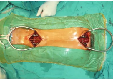
The magnetic rod was placed submuscularly between the proximal and distal screws without any additional incision (Figure 1). The magnetic rod was lengthened at 12-week intervals. In outpatient clinic conditions, the actuator portion of the magnetic rod was found with the help of a magnet while the patient was in the prone position, and the skin level was marked with a marker. The rod was lengthened through the actuator portion with the help of remote control by gently applying traction from the legs and under the armpit. A lengthening of 4 mm was obtained in each session. The radiological evaluation was repeated at the end of the lengthening procedure.
Figure 1. Intraoperative view of minimal invasive magnetically
controlled dual rod application.
Cobb angle, T1-T12 kyphosis angle, T1-S1 lengths, the coronal and sagittal balance were evaluated by the two authors during the preoperative (Figure 2), postoperative (Figure 3) and further follow-ups (Figure 4) of the patients. Two authors (S.E and B.P) assessed all images and measured all angles independently. The images were blinded and randomized. Where there was disagreement between the examiners, re-evaluation was performed until a consensus was reached.
Statistical Analysis
SPSS 15.0 for Windows (IBM Corporation, Chicago, IL, USA) was used for statistical analyses. The descriptive statistics were expressed as numbers and percentages for categorical variables. The mean and standard deviation were used as numerical variables for normally distributed data. The mean of T1-S1 length, Cobb angle, Kyphosis angle, Coronal and sagittal balance in the preoperative period, early postoperative period and the late postoperative period were analyzed with Repeated measures Anova with a Greenhouse-Geisser correction. Post-hoc tests using Bonferroni correction determined the differences among the pre-operative, early post-operative and late post-post-operative period. A p-value of <0.05 was considered to indicate significance. Inter-observer agreement was assessed using the ‘κ’ statistical test. A kappa value between 0.8 and 1 was considered perfect agreement.
Figure 2. Preoperative anteroposterior and lateral x-rays of 10-year-old male patient 1 showing an idiopathic scoliosis with 42 degrees of curvature.
Figure 3. Early postoperative anteroposterior and lateral x-rays of patient 1 showing a dual rod construct. The postoperative Cobb value was 18 degrees.
RESULTS
The mean age of the patients at the time of the first operation was 8.8 years (range, 7-10). The mean number of distractions per patient was 8.2 times (range, 6-12). Two rods were used in all patients. The mean number of
screws used was 8.5 ± 2.2 (range, 4-12). The mean follow-up was 26 months (range, 12-36).
Table 1. The mean pre-operative, post-operative and last follow-up radiological measurement
Preoperative PostoperativeInitial Last Follow-up p T1-S1 (mm) 276±53 309±49 348±55 <0.001* Cobb angle 54°±16 28°±14 30°±16 <0.001* Kyphosis angle 49°±18 22°± 8 21°±5 <0.001* Lumbar Lordosis angle 44°±11 33°±15 38°±11 0.061 Coronal balance (mm) 25±9 9±4 10±5 0.002* Sagittal balance (mm) -19±13 16±40 9±31 0.029*
* represents statistical significance
The mean preoperative Cobb angle was 54±16 degrees (range, 40-88), which was corrected to 28±14 degrees (range, 10-55) after the first surgery, and it was 30±16 degrees (range, 10-59) during the final follow-up. Considering the pre-operative and post-operative Cobb angles, it was stated that a statistically significant improvement was obtained (p <0.05), and this value was maintained until the last follow-up. The mean preoperative T1-S1 length was 276±53 mm (range, 170-365). The early postoperative T1-S1 length was 309±49 mm (range, 207-385). During the last follow-up, the T1-S1 length was evaluated to be 348±55 mm (range, 227-405). Given the pre-operative and final follow-up values, the mean lengthening obtained was 72±22 mm (range 40-120). The mean pre-operative kyphosis angle was 49±18 degrees (range, 21-74) while it was 22±8 degrees (range, 10-39) postoperatively and was found to be 21±5 degrees (range, 12-28) during the last follow-up.
Figure 4. 31. months follow-up anteroposterior and lateral x-rays
Considering the pre-operative and post-operative kyphosis angles, it was observed that there was a statistically significant decrease in kyphosis (p <0.05), and that patients developed hypokyphosis. The mean pre-operative, post-operative and last follow-up lumbar lordosis angles were 44°±11, 33°±15 and 38°±11, respectively (p>0.05). (Table 1).
The pre-operative coronal balance of the patients was 25±9 mm (range, 5-36), while it was found to be 9±4 mm (range, 2-17) post-operatively. The improvement in coronal balance was statistically significant (p <0.05), which was 10±5 mm (range, 3-19) during the final follow-up. There was no impairment in the coronal balance. The pre-operative sagittal balance was -19±13 mm (range, -40-6), while it was 16±40 mm (range, -27-89) mm postoperatively. It was measured to be 9±31 mm (range, -32-60) during the last follow-up. It was stated that a statistically significant change in sagittal balance was obtained (p <0.05) (Table 1). Interobserver agreement (k) was 0.96 for all assessments.
There were no complications during the operations and outpatient distraction. None of the patients in our series experienced complications such as autofusion, surgical wound site infection, implant loosening or failure until last follow-up.
DISCUSSION
An ideal system should allow the spine to grow from the first procedure and should be able to correct the curvature three-dimensionally (7). The conventional distraction-based growing rod technique, which allows normal growth, is the most commonly used technique today (11,12). The major problem of the conventional growing rod technique is the difficulty in the correction of final deformity caused by autofusion during the final fusion. This autofusion encountered during final fusion has a complex pathophysiology. The main factor causing this is the unnecessary dissection carried out in unfused vertebrae that will not be instrumented (9,13,14). Cahill et al. reported that 89% of the patients that they treated with the conventional growing rod method developed autofusion, and that the Cobb angle could be improved by 44% during the definitive fusion. They stated that they had to perform osteotomy on 7 patients due to autofusion (14). The magnetic rod system can overcome some of these complications inherent to conventional techniques. Autofusion may also occur in the magnetic rod system, but it is believed that non-invasive successive distractions reduce the likelihood of this phenomenon (9). None of the patients in our series experienced complications such as autofusion, surgical wound site infection, implant loosening, failure or major vascular injury during pedicle screw instrumentation (15). The treatments of all patients are still ongoing and no final fusion has been performed. Definitive fusion surgery has been planned and appointments have been given to all patients.
Rolton et al. indicated that the ratio of actual lengthening/ desired lengthening was not the same in their study conducted on a patient group whose surgical procedure was the magnetic rod system (16). Cheung et al. reported that the actual and expected rod distraction should be closely compared in magnetic rod system surgery (17). Ensuring the consistency between the planned distraction and the distraction occurring in vivo is one of the greatest challenges. The actual distraction extent cannot be evaluated without removing the rod and comparing it with the original length. Akbarnia et al. reported that 80% of the distraction they planned could be achieved in their study conducted on pig models (18). Rolton et al. found this value to be even lower (16). La Rosa et al. reported an inconsistency rate of 32% between the predicted and actual length (13). They stated that there might be a major difference between the predicted and actual lengthening due to overload if the distraction procedure is performed in the sitting position, and that success of the distraction could be improved by reducing the compression force on the magnet (actuator) and performing gentle traction under the armpit and from the pelvis in the lying position during the next distractions in order to overcome this problem (13). In our study, we also performed lengthening in the prone position by applying a slight traction in accordance with this procedure.
The duration between successive lengthenings mostly depends on the surgeon’s preference based on the patient’s age, growth potential, the flexibility of the curvature and diagnosis. The duration between the distractions in the conventional growing rod technique is between 6-9 months. Distractions in the magnetic rod system are performed noninvasively more frequently (9). In the literature, there is no consensus on the extent and timing of distraction (13). Although the frequency of lengthenings is not definitive, it is carried out more frequently than for classic rods. In our study, we performed 4 mm distractions during each session at 3-month intervals, and we are of the opinion that the effect of this period on controlled lengthening, patient follow-up and growth potential is sufficient.
Despite the success of the growing rod technique, the technique still has limitations in deformity correction, especially in the transverse plane. The system does not control the apex of the curvature directly but applies distraction from the lower and upper part of the spinal deformity (7). Acaroglu et al. indicated that the curvature could be controlled in the coronal and sagittal plane with fusionless distraction techniques in twelve children with progressive curvature whom they retrospectively followed up until the decision was made to apply definitive treatment without fusion, but that transverse plane deformities continued to increase (19). Another critical issue of the magnetic rod system is the difficulty of control in the sagittal profile due to bending problems in the actuator portion of the rod when it falls on the apex of scoliosis. Harrington-like 2-point distraction structures cause inevitable flattening in the thoracic
region. Such problems can be partially overcome with a smaller actuator (13). We observed a tendency towards hypokyphosis in the postoperative kyphosis values of the patients in our series. We are of the opinion that the system’s major problem is the postoperative development of hypokyphosis since we could not give curvature to the rod due to its magnetic field.
There are still discussions on proximal junctional kyphosis in growth-friendly procedures. It is thought that increased kyphosis might develop above the level of the last instrumented vertebrae depending on the increase in the non-instrumented area (9). None of the patients in our series developed junctional kyphosis. We believe that not applying distraction force on the screw by taking care to preserve the facet joint structure during surgery was effective in this regard.
We agree with Cheung et al. that the magnetic rod system exposes the patient to high-doses of radiation. We are of the opinion that it would be better to adjust the duration between lengthening periods and to take radiography only after rod lengthening in order to reduce the dose of radiation (13,17). In addition, it has been shown that measuring the amount of growth via Ultrasound is as reliable as X-ray in order to avoid the effects of radiation (20). There is currently no evidence of recurrent or major adverse effects due to repetitive distraction regarding the effects of electromagnetic fields on humans, especially for the local areas. Thus, the procedure could be considered relatively safe (9).
Implant-related complications such as rod breakages, rod malfunctions and implant pullout can occur in the MCGR technique. Choi et al. investigated the complications of 54 patients with magnetic growing rods in their multicentre retrospective research conducted in 2016 and found at least 1 complication in 21 (38.8%) of the 54 patients. 15 patients (27.8%) also underwent at least one revision surgery, while 6 patients (11.1%) developed rod fractures. Fixation problems arose in the proximal or distal screws of 7 patients (13%) (21). Dannawi et al. experienced rod fractures in 2 patients on whom they used a single rod. It was determined that the locking pin caused damage when the rod was examined by the manufacturer. They stated that the rod thickness was 4.5 mm in the two patients with rod fractures and that they used 5.5 mm rods on the other patients (22). In order to minimize the risk of implant-related complication, we used dual-rod, pedicule screw instead of hooks and up to 3 vertebral instrumentations in both the upper and lower segments in all our patients, and none of our patients experienced rod fracture.
Our study has several limitations, the most important of which is the relatively small sample size. Secondly, the follow-up period is comparably short. However, the lack of complications during the follow-up period is the strongest aspect of this paper, which might be the result of close follow-up and detailed questioning by the surgeons throughout the study, and tailored interventions for each of the patients in the study group.
CONCLUSION
During our distraction period of follow-up, the dual-magnetic rod technique allowed spinal growth as well as control of scoliosis and lengthening. Since the procedure is relatively novel in the area of orthopaedics in Turkey, and the collection of large case-series would require more time, the early findings of our study group could add to the current knowledge. Thus, the use of dual-magnetic controlled growth rods for the treatment of early-onset scoliosis is a safe and efficient protocol for the treatment of early-onset scoliosis in children, and could considered as the model of treatment among the conventional methods.
Competing interests: The authors declare that they have no competing interest.
Financial Disclosure: There are no financial supports.
Ethical approval: The study was approved by the Kyrenia University Scientific Research and Publication Ethics Committee (RY-2019-10). Sinan Erdogan ORCID: 0000-0002-8517-3925
Barıs Polat ORCID: 0000-0001-8229-6412
Huseyin Bahadir Gokcen ORCID: 0000-0002-5374-1166 Engin Carkci ORCID: 0000-0002-3918-4498
Tahsin Gurpinar ORCID: 0000-0002-8194-6492 Cagatay Ozturk ORCID: 0000-0003-3133-206X
REFERENCES
1. Eltorai AE, Fuentes C. Magnetic growth modulation in orthopaedic and spine surgery. Journal of Orthopaedics 2018;15:59-66.
2. Thompson W, Thakar C, Rolton DJ, et al. The use of magnetically-controlled growing rods to treat children with early-onset scoliosis: early radiological results in 19 children. Bone Joint J 2016;98:1240-7.
3. Rolton D, Richards J, Nnadi C. Magnetic controlled growth rods versus conventional growing rod systems in the treatment of early onset scoliosis: a cost comparison. Eur Spine J 2015;24:1457-61. 4. Harrington PR. Treatment of scoliosis: correction and
internal fixation by spine instrumentation. J Bone Joint Surg Am 1962;44:591-610.
5. Moe JH, Kharrat K, Winter RB, et al. Harrington instrumentation without fusion plus external orthotic support for the treatment of difficult curvature problems in young children. Clin Orthop Relat Res 1984;185:35-45.
6. Sankar WN, Skaggs DL, Yazici M, et al. Lengthening of dual growing rods and the law of diminishing returns. Spine 2011;36:806-9.
7. Li K, Zhao S, Wei X, et al. A preliminary study on a novel growth guidance rod system for early-onset scoliosis in a sheep model. Spine 2015;40:767-72.
8. Takaso M, Moriya H, Kitahara H, et al. New remote-controlled growing-rod spinal instrumentation possibly applicable for scoliosis in young children. J Orthop Sci 1998;3:336-40.
generation of growth-sparing techniques: preliminary clinical results of a magnetically controlled growing rod in 14 patients with early-onset scoliosis. Spine 2013;38:665-70.
10. Yılmaz B, Ekşi MŞ, Işik S, et al. Magnetically controlled growing rod in early-onset scoliosis: A minimum of 2-year follow-up. Pediatr Neurosurg 2016;51:292-6. 11. Akbarnia BA, Breakwell LM, Marks DS, et al. Dual
growing rod technique followed for three to eleven years until final fusion; the effect of frequency of lengthening. Spine 2008;33:984-90.
12. Korkmaz MF, Sevimli R, Selcuk EB, et al. Three-dimensional spinal deformity: Scoliosis. Medicine Science 2015;4:1796-808.
13. La Rosa G, Oggiano L, Ruzzini L. Magnetically controlled growing rods for the management of early-onset scoliosis: a preliminary report. J Pediatr Orthop 2017;37:79-85.
14. Cahill PJ, Marvil S, Cuddihy L, et al. Autofusion in the immature spine treated with growing rods. Spine 2010;35:1199-203.
15. Korkmaz MF, Erdem MN, Ozevren H, et al. Determining the optimal length and safety of pedicle screws in the T12 vertebra: A morphometric study. Cureus 2018;10:2156.
16. Rolton D, Thakar C, Wilson-MacDonald J, et al.
Radiological and clinical assessment of the distraction achieved with remotely expandable growing rods in early onset scoliosis. Eur Spine J 2016;25:3371-6. 17. Cheung KM, Cheung JP, Samartzis D, et al. Magnetically
controlled growing rods for severe spinal curvature in young children: a prospective case series. Lancet 2012;379:1967-74.
18. Akbarnia BA, Mundis GM, Salari P, et al. Innovation in growing rod technique: a study of safety and efficacy of a magnetically controlled growing rod in a porcine model. Spine 2012;37:1109-14.
19. Acaroglu E, Yazici M, Alanay A, et al. Three-dimensional evolution of scoliotic curve during instrumentation without fusion in young children. J Pediatr Orthop 2002;22:492-6.
20. Cheung JP, Bow C, Samartzis D, et al. Clinical utility of ultrasound to prospectively monitor distraction of magnetically controlled growing rods. Spine J 2016;16:204-9.
21. Choi E, Yazsay B, Mundis G, et al. Implant complications after magnetically controlled growing rods for early onset scoliosis: a multicenter retrospective review. J Pediatr Orthop 2017;37:588-92.
22. Dannawi Z, Altaf F, Harshavardhana NS, et al. Early results of a remotely-operated magnetic growth rod in early-onset scoliosis. Bone Joint J 2013;95:75-80.