Determination of ANAE and ACP-ase Positive Lymphocytes of
Peripheral Blood and Endometrium Tissues in Experimental
Hypothyroidism-Induced Rats
[1]Fatma ÇOLAKOĞLU
1 Hasan Hüseyin DÖNMEZ
2[1] This study was supported by Karamanoglu Mehmetbey University Scientific Research Projects (BAP) Coordinating Office
(Project no: 22-M-16)
1 Department of Nutrition and Dietetics, Faculty of Health Sciences, Karamanoglu Mehmetbey University, TR-70100
Karaman - TURKey
2 Department of Histology and embryology, Faculty of Veterinary Medicine, Selcuk University, TR-42075 Konya - TURKey
Article ID: KVFD-2018-20431 Received: 26.06.2018 Accepted: 13.11.2018 Published Online: 14.11.2018 How to Cite This Article
Çolakoğlu F, Dönmez HH: Determination of ANAe and ACP-ase positive lymphocytes of peripheral blood and endometrium tissues in experimental hypothyroidism-induced rats. Kafkas Univ Vet Fak Derg, 25 (2): 157-162, 2019. DOI: 10.9775/kvfd.2018.20431
Abstract
This study was aimed to provide information about the status of the immune system by revealing changes in peripheral blood leukocyte (PBL) percentages, ANAe- and ACP-ase(+) lymphocyte rates in peripheral blood (PB) and endometrium tissues of experimental hypothyroidism-induced rats. In this study, 15 healthy female Wistar Albino rats were used. Rats were fed through 4 weeks. The Group e (experimental, n=9) is group that were made hypothyroidism by intraperitoneal methimazole enjection for 2 weeks. Rats of the Group C (control, n=6) were untreated. In the 2nd and 4th weeks, ANAe- and ACP-ase(+) lymphocyte rates of the Group e were higher than Group C in PB. excepting eosinophil and basophil leukocyte rates, there was no statistical difference in the other PBL percentages in the both of weeks. In PB, while lymphocyte rate of the 4th week was no statistically different (P>0.05), it was found lower in Group C. There was no alteration in ANAe- and ACP-ase(+) lymphocyte rates of uterine tissue. As a result, whereas hypothyroidism caused significant alterations in PBL and T lymphocyte rates, the any marked changes was not observed in the uterine tissue.
Keywords: ACP-ase, ANAE, Endometrium, Hypothyroidism, Methimazole
Deneysel Hipotiroidizm Oluşturulan Ratlardaki Periferik Kan ve
Endometriyum Dokularında ANAE ve ACP-az Pozitif Lenfositlerin
Belirlenmesi
Öz
Bu çalışma, deneysel hipotiroidi oluşturulan ratların periferal kan lökosit (PKL) yüzdelerindeki, periferal kan ve endometriyum dokularındaki ANAe ve ACP-az (+) lenfosit oranlarındaki değişiklikleri açığa çıkararak bağışıklık sisteminin durumu hakkında bilgi vermeyi amaçlamaktadır. Çalışmada 15 sağlıklı dişi Wistar Albino rat kullanıldı. Ratlar 4 hafta boyunca beslendi. Grup D (deneysel, n=9) 2 hafta boyunca intraperitoneal methimazol enjeksiyonu ile hipotiroidizm oluşturulan gruptur. Grup K’nın (kontrol, n=6) ratları normal beslendi. İkinci ve dördüncü haftalarda, Grup D’nin periferal kandaki ANAe- ve ACP-az(+) lenfosit oranları Grup K’den yüksekti. eozinofil ve bazofil lökosit oranları hariç olmak üzere, her iki haftada da diğer PKL oranlarında istatistiksel bir fark yoktu. 4. haftadaki periferal kan lenfosit oranı istatistiksel olarak farklı bulunmazken (P>0.05), Grup K’de daha düşük bulundu. Uterus dokusunda ANAe- ve ACP-az(+) lenfosit oranlarında değişiklik görülmedi. Sonuç olarak, hipotiroidizmin PKL ve T lenfosit oranlarında önemli değişikliklere neden olurken uterus dokusunda herhangi belirgin bir değişikliğe yol açmadığı gözlendi.
Anahtar sözcükler: ACP-az, ANAE, Endometriyum, Hipotiroidizm, Methimazol
INTRODUCTION
Thyroid hormones play an important role not only in
metabolic disorders [1], but also in the development and
function of the immune and reproductive systems [2].
Hypothyroidism, one of the most common thyroid disorders
İletişim (Correspondence)
+90 338 2262185/4255 GSM: +90 533 3906020
[email protected]in humans, is insufficient production of thyroid hormones
by the thyroid gland [3]. This disorder can be arised as a
consequence of thyroid disfunction, impedement in mechanism that control thyroid function, or complication
during treatment of hyperthyroidism [4]. In studies showing
the effects of thyroid hormones on adaptive immunity have been seen that human hypothyroidism, as well as in rodents hypothyroidism induced pharmacologically and surgical, is associated with a decrease in thymic activity. Low concentrations of 3,3´,5-triiodo-L-thyronine (T3) and L-thyroxine (T4) can stimulate T cell proliferation including
cell-mediated immunity [5,6]. Activation of T lymphocyte
subtypes occur in severe hypothyroidism [7]. In clinical
cases of hypothyroidism the spontaneous migration of polymorph nuclear leukocytes (PMNL) was found to be
impaired [8]. Furthermore, the effects of hypothyroidism
are directly on gonadotropin and steroid hormones [9].
Hypothyroidism is one of the most obvious causes of infertility, menstrual disturbance, spontaneous recurrent abortion, and of stillbirths [10]. The effect of hypothyroidism
is more marked on the endometrium [11]. One of the most
complex tissues is endometrium. Because it undergoes many dynamic changes, such as cytokines, growth factors,
hormones and adhesion molecules [12,13]. Martinez et al.[14]
reported that T lymphocyte was major class of lymphocytes in uterine.
Alpha-naphthyl acetate esterase (ANAe) demonstration is a method, which is used distinct from each other of
T lymphocytes, B lymphocyte and monocytes [15] Acid
phosphatase (ACP-ase) demonstration is also a method and specific for cell populations in which the majority of
the T lymphocytes are formed in mammals [16].
This study was planned to provide information about the immune system by revealing changes in peripheral blood leukocyte (PBL) percentages, ANAe- and ACP-ase(+) lymphocyte rates in PB and endometrium tissues of experimentally hypothyroidised rats.
MATERIAL and METHODS
Research Material
ethic approval was obtained from Selcuk University experimental Medical Practice and Research Center (SUDAM) Animal experiments ethics Committee (2016/13). In this study, it was used 15 healthy female Wistar Albino rats (198-250 g), 12-14 weeks of age. Rats were caged individually on 12:12 h light-dark schedule at the room temperature (22±1°C), and fed with commercial rat food and water which were available ad bilitum.
Experimental Procedures
The rats were divided into two groups: animals from the first group (n=9) were made hypothyroid by intraperitoneal (IP) methimazole enjection (10 mg/kg/day) for 2 weeks as
per methods of Parija et al.[17] and Swann [18]. Animals in the
other group (n=6) were untreated control (C). In the 2nd
week, total serum T3 and T4 concentrations in PB which was taken from the lateral tail veins were determined using the ADVIA Centaur CP Immunoassay System (detection kits provided by Siemens). To see chronic effects on the tissues, the both of groups were made normal feeding
for 2 weeks. In the 4th week, the all rats were sacrificed by
cervical dislocation under general anesthesia with ketamin (10 mg/kg, IM) and ksilazin (5 mg/kg, IM).
Collection and Processing of Tissue Samples
In the 2nd and 4th weeks, from each blood samples, six
blood smears were prepared and fixed in a gluteraldehyde-acetone solution. Two smears were stained for each PBL
formula, ANAe and ACP-ase demonstrations [15]. Uterine
samples were fixed in formol calcium solution and the samples were incubated in 22 h formal sucrose solution and kept in 22 h Holt’s solution for enzyme demonstrations. Then, cryostat sections (12 μm) were taken from samples. These preparations were stained for ANAe and ACP-ase
demonstrations [19]. Both PB and uterine tissue samples were
stained with 1% methyl green (Merck) for counterstain. For PBL formula, blood smears were stained with May
Grünwald-Giemsa staining method [20].
Evaluation of the Stained Tissue Samples
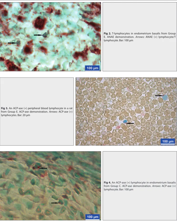
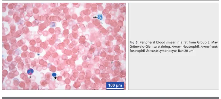
Respectively, as shown in Fig. 1 and Fig. 2, in the PB smears and uterine preparations which were made ANAe demonstration, there were dot-like reddish brown granules of ANAe (+) lymphocytes/T lymphocytes. Respectively, as shown in Fig. 3 and Fig. 4, in PB smears and uterine preparations lymphocytes containing one to three pinkish red cytoplasmic granules were considered to be ACP-ase (+). In each of the PB smears stained for ANAe and ACP-ase activity, 200 lymphocytes were counted and positivity rates were expressed as the percentage of counted cells. For PBL counts, 100 leukocytes were counted in a light microscope, and leukocyte formula were calculated (Fig.
5). In the uterine preparations, ANAe- and ACP-ase (+)
lymphocytes were counted in the total 0.1 mm2 area from
10 different uterine areas. Statistical Analysis
Comparison of parameters between groups of the 2nd and
4th weeks was analysed using Independent-Samples T test.
Differences in parameters between the 2nd and 4th weeks
within the groups was drawn using the Paired-Samples T test. Significance was set at P<0.05 [21].
RESULTS
Changes in T3 and T4 levels of groups after methimazole
treatment were given Table 1. In the 2nd and 4th weeks,
Group e when compared with Group C (P<0.05). According to the weeks within groups, it was statistically seen
that T3 level of 2nd week was lower than that of it in the
4th week (P<0.05) (Table 2). As shown in Table 3, in the
2nd and 4th weeks, the highest peripheral blood ANAe- and
ACP-ase (+) lymphocyte (Fig. 1 and Fig. 3, respectively) rates were determined in the Group e (P<0.05). In the
4th week, according to the PBL percentages (Fig. 5), the
eosinophil and basophil leukocyte rates were statistically lower in the Group e than that of the Group C (P<0.05). No statistically significant, it was observed that the Group e had higher lymphocyte rate than the Group C
(P>0.05). For both of weeks, there was no statistical difference in the other leukocytes between the groups (P>0.05). No the differences between two weeks within the groups are statistically significant (Table 4). In the endometrium, as shown in Table 5, while the ANAe(+) lymphocyte (Fig. 2) number in the Group C was 19.33/0.1
mm2, this rate was found 20.00/0.1 mm2 in the Group e.
There was no statistical difference between the groups (P>0.05). When the ACP-ase(+) lymphocyte (Fig. 4) number of the Groups C and e were examined, it was noted that there was no statistical difference between the groups (P>0.05).
Table 1. Serum T3 (pg/ml) ve T4 (ng/dl) levels ±Se
Groups FT3(2) FT4(2) FT3(4) FT4(4)
Control (n=6) 3.22±0.13a 2.35±0.06a 3.35±0.18a 2.39±0.14a
experimental (n=9) 2.83±0.21b 1.61±0.19b 2.12±0.15b 1.72±0.25b
a,b Values within a column with no common superscripts are significantly (P<0.05) different; FT3(2): 2nd week
serum T3, FT4(2): 2nd week serum T4, FT3(4): 4th week serum T3, FT4(4): 4th week serum T4
Table 2. Differences in parameters between the 2nd and 4th weeks within the groups (%)±Se
Week FT3 FT4
2nd 2.99±0.15a 1.90±0.15
4th 3.27±0.12b 2.00±0.14
a,b Values within a column with no common superscripts are significantly (P<0.05) different
Table 3. The proportions of peripheral bood lymphocyte and leukocyte in different weeks (%)±Se
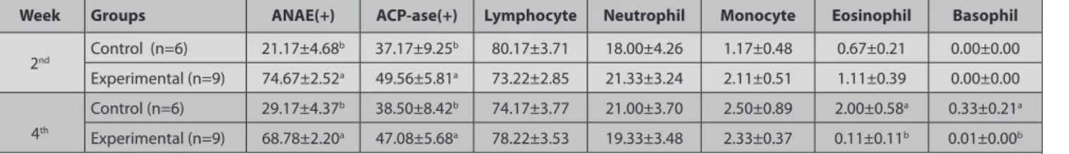
Week Groups ANAE(+) ACP-ase(+) Lymphocyte Neutrophil Monocyte Eosinophil Basophil
2nd Control (n=6) 21.17±4.68 b 37.17±9.25b 80.17±3.71 18.00±4.26 1.17±0.48 0.67±0.21 0.00±0.00 experimental (n=9) 74.67±2.52a 49.56±5.81a 73.22±2.85 21.33±3.24 2.11±0.51 1.11±0.39 0.00±0.00 4th Control (n=6) 29.17±4.37b 38.50±8.42b 74.17±3.77 21.00±3.70 2.50±0.89 2.00±0.58a 0.33±0.21a experimental (n=9) 68.78±2.20a 47.08±5.68a 78.22±3.53 19.33±3.48 2.33±0.37 0.11±0.11b 0.01±0.00b a,b Values within a column with no common superscripts are significantly (P<0.05) different
Fig 1. A peripheral blood T-lymphocyte in Group e. ANAe demonstration. Arrow: ANAe (+) lymphocyte/T lymphocyte. Bar: 20 μm
DISCUSSION
T3 and T4 hormones secreted by the thyroid gland are formed from a large prohormone molecule called as thyroglobulin and enter the cells by binding to the thyroid
hormone receptor α and β [22]. These thyroid hormones
which is necessary for sexual development and life regulate the steroid hormones secretion. Findings such as lipidode deficiency, impotence, menopause, excessive bleeding and menstrual irregularities are observed in
Fig 2. T-lymphocytes in endometrium basalis from Group
e. ANAe demonstration. Arrows: ANAe (+) lymphocyte/T lymphocyte. Bar: 100 μm
Fig 3. An ACP-ase (+) peripheral blood lymphocyte in a rat from Group e. ACP-ase demonstration. Arrows: ACP-ase (+) lymphocytes. Bar: 20 μm
Fig 4. An ACP-ase (+) lymphocyte in endometrium basalis from Group C. ACP-ase demonstration. Arrows: ACP-ase (+) lymphocyte. Bar: 100 μm
hypothyrodism. Although the most synthesized hormone in the thyroid gland is T4, the most effective is the T3 [23].
In humans, one of the most common thyroid disorders is hypothyroidism, which is defined as low levels of
thyroid hormones in the blood [3]. Deficiency of thyroid
hormone production causes serious abnormalities [1].For
establishing hypothyroidism in experimental animals, methimazole is frequently used in the treatment of human
hyperthyroidism [24]. In this study, the methimazole dose
and form of implementation given to the Group e was
determined from the results of previous studies [17,18].
ANAe is an enzyme that demonstrates T lymphocytes. It is also known that dot-like positivity is specific for T
lymphocytes [15]. ACP-ase is one of the lysosomal enzymes
in lymphocytes. Some investigators have demonstrated ACP-ase reactivity in human peripheral blood T
lympho-cytes [15,16]. In the direction of this information, we found
that T lymphocyte and ACP-ase (+) lymphocyte rates were higher in the Group e (P<0.05). This situation was explained that low serum T3 and T4 concentrations can stimulate T lymphocyte proliferation [7].
Although we did not statistically find any change in the other PBL excluding eosinophil and basophil leukocytes, we was observed that the lymphocyte ratio was higher in the Group e. This suggests that thyroid hormones have regulatory effects on immunological activity at the cellular
level [25]. There is also information that thyroid hormones
modify lymphocyte activity [26,27]. In severe hypothyroidism,
activation of T lymphocyte subclasses, reduction of natural killer cells and reduction of CD4 T lymphocyte responses are
seen [7]. Compared with healthy subjects, hypothyroidism
has been found to be impaired in the spontaneous migrations of polymorphonuclear leukocytes in clinical
cases [8]. Hypothyroidism is accompanied by spleen and
lymph node involution and decreases in humoral and
cellular immune responses [28].
As in other systemic mucosal tissues, uterine mucosa
normally contains T and B lymphocytes [29]. However, the
uterine tissue under the influence of hormonal changes is rearranged with the sexual cycle or the pregnancy.
Karaca et al.[30] reported that the distribution of ANAe (+) T
lymphocyte numbers in uterine tissues of pre-implantation period of goats was lower than that of non-pregnant animals. Akbulut et al.[15] reported significant reductions in
ANAe- and ACP-ase (+) lymphocyte numbers throughout the entire pregnancy in the decidua basalis region of endometrium. In the direction of the data obtained from this study, there was no change in terms of both ANAe- and ACP-ase(+) lymphocyte numbers and distribution in endometrium basalis. We can say that hypothyroidism has no any an effect on uterine in terms of lymphocyte number and distribution.
Table 4. Values of parameters between the 2nd and 4th weeks within the groups (%)±Se
Week ANAE(+) ACP-ase(+) Lymphocyte Neutrophil Monocyte Eosinophil Basophil
2nd 53.27±7.38 84.60±12.47 76.00±2.36 20.00±2.53 1.73±0.37 0.93±0.25 0.00±0.00
4th 52.93±5.59 89.60±4.61 76.60±2.57 20.00±2.48 2.40±0.40 0.87±0.34 0.13±0.91
No the differences between two weeks within the groups are statistically significant (P>0.05)
Fig 5. Peripheral blood smear in a rat from Group e. May Grünwald-Giemsa staining. Arrow: Neutrophil, Arrowhead: eosinophil, Asterisk: Lymphocyte. Bar: 20 μm
Table 5. The proportions of the ANAE(+) ve ACP-ase (+) lymphocytes in
endometrium basalis region of uterine (%) ± Se (number/0.1 mm2)
Week Groups ANAE(+) ACP-ase(+)
4th Control (n=6) 19.33±0.80 8.50±0.62
experimental (n=9) 20.00±0.71 8.00±0.44
Prevention of mental and developmental disorders is possible with a good understanding of the interaction between the endocrine and immune systems. The immune system is under the influences of many hormones. Routine control and treatment of thyroid gland functions at pre-pregnancy and throughout pre-pregnancy is an important factor for a healthy pregnancy. As a result of this study, hypothyroidism made significant changes both the peripheral blood T lymphocyte and leukocytes counts. Obtained findings may help doctors to evaluate the immunological status of hypothyroid women. Since the these techniques are simple, much cheaper, less time consuming applications, we suggest that it can be given as a laboratory service to assist of women in the early diagnosis of some gestational disorders. However, further studies should be planned in terms of our understanding more detailed of relationship between thyroid gland diseases and uterine.
As a result, whereas hypothyroidism caused significant alterations in PBL and T lymphocyte rates, the any marked changes was not observed in the uterine tissue.
A
cknowledgementWe thank to ‘‘Karamanoglu Mehmetbey University Scientific Research Projects (BAP) Coordinating Office’’, Project no: 22-M-16, for financial support.
REFERENCES
1. Cakic-Milosevic M, Korac A, Davidovic V: Methimazole induced hypothyroidism in rats: effets on body weight and histological characteristics of thyroid gland. Jugoslov Med Biohem, 23 (2): 143-147, 2004. DOI: 10.2298/JMH0402143C
2. Krassas GE: Thyroid disease and female reproduction. Fertil Steril, 74 (6): 1063-1070, 2000. DOI: 10.1016/S0015-0282(00)01589-2
3. Ladenson PW: Problems in the management of hypothyroidism. In, Braverman Le (ed): Diseases of the Thyroid. 161-176, Humana Press, Totawa, NJ, 2003.
4. Silva JE: Thyroid hormone control of thermogenesis and energy balance. Thyroid, 5, 481-492, 2005. DOI: 10.1089/thy.1995.5.481
5. Barreiro Arcos ML, Gorelik G, Klecha A, Genaro AM, Cremaschi GA: Thyroid hormones inrease inducible nitric oxide synthase gene expression from PKC-ζ in murine T lymphocytes. Am J Physiol Cell Physiol, 291 (2): C327-C336, 2006. DOI: 10.1152/ajpcell.00316.2005
6. Hodkinson CCF, Simpson EEA, Beattie JH, O’Conner JM, Campbell DJ, Strain JJ, Wallace JMW: Preliminary evidence of immune function modulation by thyroid hormones in healthy man and women aged 55-70 years. J Endocrinol, 202 (1): 55-63, 2009. DOI: 10.1677/JOe-08-0488 7. Volpe R: The immunomodulatory effects of antithyroid drugs are mediated via actions on tyhroid cells, affecting thyrocyte-signalling: A review. Curr Pharm Des, 7 (6): 451-460, 2001. DOI: 10.2174/ 1381612013397898
8. Hrycek A: Functional characterization of peripheral blood neutrophils in patients with primary hypothyroidism. Folia Biol, 39 (6): 304-310, 1993. 9. Stagnaro-Green A, Abalovich M, Alexander E, Azizi F, Mestman J, Negro R, Nixon A, Pearce EN, Soldin OP, Sullivan S, Wiersinga W: Guideliness of the American Thyroid Association for the diagnosis and management of the thyroid disease during pregnancy and postpartum.
Thyroid, 21 (10): 1081-1125, 2011. DOI: 10.1089/thy.2011.0087
10. Inuwa I, Williams MA: Morphometric study on the uterine horn and
thyroid gland in hypothyroid, and thyroxine treated hypothyroid rats. J
Anat, 188, 383-393, 1996.
11. Kirby JD, Jetton AE, Cooke PS, Hess RA, Bunick D, Ackland JF, Turek FW, Schwartz NB: Developmental hormonal profiles accompanying the neonatal hypothyroidism-induced increase in adult testicular size and sperm production in the rat. Endocrinology, 131, 559-565, 1992. DOI: 10.1210/endo.131.2.1639007
12. Sharkey A: Cytokines and implantation. Rev Reprod, 3, 52-61, 1998. 13. Salamonsen LA: Role of proteases in implantation. Rev Reprod, 4, 11-22, 1999.
14. Martinez CM, Buendia AJ, Sanchez J, Navarro JA: Immuno-phenotypical characterization of lymphocyte suppopulations of the uterus of non-pregnant and pregnant goats. Anat Histol Embryol, 34, 240-246, 2005. DOI: 10.1111/j.1439-0264.2005.00606.x
15. Akbulut B, Sur E, Okur DN: Determination of the AgNOR parameters, MN frequency ANAe and ACP-ase positivity of PBL in pregnants. Selçuk Tıp
Derg, 31 (4): 344-350, 2015.
16. Basso G, Cocito MG, Semenzato G, Pezzutto A, Zanesco L: Cytochemical study of thymocytes and T lymphocytes. Br J Haematol, 44 (4): 577-582, 1980. DOI: 10.1111/j.1365-2141.1980.tb08712.x
17. Parija SC, Mishra SK, Raviprakash V: Hypothyroid state reduces calcium channel function in 18-day pregnant rat uterus. Indian J Exp Biol, 44, 19-27, 2006.
18. Swann AC: Noradrenaline and thyroid function regulate (Na+-K+)
adenosine triphosphate independently in vivo. Eur J Pharmacol, 169 (2-3): 275-283, 1989. DOI: 10.1016/0014-2999(89)90025-3
19. Dönmez HH, Eken E, Beşoluk K, Sur E: The histological characteristics and localisation of ACP and ANAe positive lymphocytes in the eosophageal tonsil of the duck (Anas platyrhynchos). Avian Biol Res, 5 (1): 11-15, 2012. DOI: 10.3184/175815512x13264771062961
20. Demir R, Yılmazer S, Öztürk M, Üstünel İ, Demir N, Korgun ET, Akkoyunlu G: Histolojik Boyama Teknikleri Başvuru Kitabı. 267-269, Palme yayıncılık, Ankara, 2007.
21. SPSS: SPSS 21.0 for Windows. IBM SPSS Statistics, USA, 2012. 22. DeRuiter J: Thyroid hormone tutorial: the thyroid and thyroid hormones. In, DeRuiter J (ed): endocrine Pharmacotherapy Module: Thyroid, Summer, 1-16, 2001
23. Jameson JL, Weetman AP: Disorders of the thyroid gland. In, Jameson JL (ed): Harrison’s endocrinology. 62-69, The McGraw-Hill Companies, New york, 2010.
24. Homsanit M, Sriussadapom S, Vannasaeng S, Peerapatdit T, Nitiyanant W, Vichayanrat A: efficacy of single daily dosage of methimazole vs. propylthiouracil in the induction of euthyroidism. Clin
Endocrinol, 54, 385-390, 2001.
25. De Vito P, Incerpi S, Pederson JZ, Luly P, Davis FB, Davis PJ: Thyroid hormones as modulators of immune activities at the cellular level.
Thyroid, 21 (8): 879-890, 2011. DOI: 10.1089/thy.2010.0429
26. Keast D, Taylor K: The effect of tri-iodothyronine on the phytohaemaglutinin response of T lymphocytes. Clin Exp Immunol, 47, 217-220, 1982.
27. Ong ML, Malkin DG, Malkin A: Alteration of lymphocyte reactivities by thyroid hormones. Int J Immunopharmacol, 8, 755-762, 1986. DOI: 10.1016/0192-0561(86)90012-3
28. Klecha AJ, Genaro AM, Lysionek AE, Caro RA, Coluccia AG, Cremaschi GA: experimental evidence pointing to the bidirectional interaction between the immune system and the thyroid axis.
Int J Immunopharmacol, 22, 491-500, 2000. DOI:
10.1016/S0192-0561(00)00012-6
29. Herington JL, Bany BM: effect of the conceptus on uterine natural killer cell numbers and functionin the mouse uterus during decidualization.
Biol Reprod, 76, 579-588, 2007. DOI: 10.1095/biolreprod.106.056630
30. Karaca T, Yörük M, Uslu S, Uslu BA, Çetin Y: Preimplatasyon sürecindeki keçilerin dişi genital kanal organlarında plazma hücresi ve alfa naftil asetat esteraz pozitif lenfositlerin dağılımı. YYU Vet Fak Derg, 20 (2): 1-5, 2009.