Cenk YARDIMCI, Taylan ÖNYAY, Kamil Serdar İNAL, Birsen Deniz ÖZBAKIR, Ahmet ÖZAK
Ondokuz Mayıs University, Faculty of Veterinary Medicine, Department of Surgery, Samsun, Turkey.
Summary: Ectrodactyly is an exceptional congenital disorder which is seen at the distal part of the limbs and characterized with longitudinal bony and soft tissue clefts. These clefts can include digits, metacarpal bones, carpal joint or radius-ulna. A male, 18 week-old, Turkish Kangal dog was presented with the right forelimb lameness and inspectional abnormality with a long and wide cleft starting from the distal manus to the carpo-metacarpal joint. The long cleft between metacarpal bones was fixed with two lag screws after filling the gap with corticocancellous bone block and fusion podoplasty. Short term clinical results were favourable following the surgery, but long term clinical outcome was less favourable because the dog revealed mild lameness due to the erosion of skin surrounding the digital pads. Finally, this problem was solved by using pad protectors when walking on rough grounds.
Keywords: Congenital anomaly, dog, ectrodactyly, fusion podoplasty.
Kangal ırkı bir köpekte ektrodaktili’nin cerrahi sağaltımı
Özet: Ektrodaktili, bacağın distal kısmındaki yumuşak ve kemik dokuda uzunlamasına yarık ile karakterize, ender görülen konjenital bir hastalıktır. Bu yarık; parmakları, metakarpal kemikleri, karpal eklemi ve radius ulna’yı da kapsayabilir. Dört buçuk aylık, Kangal ırkı, erkek bir köpek sağ ön ayağında topallık ve dış bakıda belirlenebilen distalden başlayan ve karpometakarpal ekleme kadar uzanan, uzun ve geniş bir yarık şikayeti ile getirildi. Metakarpal kemikler arasındaki geniş yarık, kortikokansellöz blok kemik grefti ve füzyon podoplastiden sonra iki lag vidası ile stabilize edildi. Operasyonu takiben kısa dönem sonuçları olumlu iken, uzun dönem sonuçlarına bakıldığında, parmakların taban yastıklarının etrafındaki derinin erozyonundan dolayı hafif topallık gözlendi. Sonuç olarak bu problem de sert zeminde yapılan yürüyüşlerde koruyucu ped uygulaması ile çözüldü.
Anahtar sözcükler: Ektrodaktili, füzyon podoplasti, konjenital anomali, köpek.
Ectyrodactyly is an exceptional congenital anomaly which is seen at the distal part of limbs and characterized with longitudinal bony and soft tissue clefts (2, 9). These clefts can include digits, metacarpal bones, the carpal joint and radius-ulna (17). Splint-hand deformity, lobster claw deformity, hypodactyly and oligodactyly are considered synonymous to ectyrodactyly (2, 5).
The disorder has been reported in many of mammalian species such as dogs (3), cats (22), sheep (20), cattle (13), primates (16), humans (21), and tiger (19). In humans the condition can also be seen in combination with ectodermal dysplasia or cleft palate and main reason is usually genetic (9). Experimental studies have indicated that some teratogenic subtances (cadmium, etc.) can have inductory effect on ectrodactyly in laboratory animals (7). In dogs, ectrodactyly is generally unilateral and only two cases of bilateral formation have reported in veterinary literature until now (5, 17). Clinical view of the disease includes a total soft tissue cleft of different length between two separate segments which tend to split after weight-bearing (9). Radiologically, ectrodactyly is characterized as axial separation of the metacarpal and/or carpal bones
furthermore the cleft can reach up to the elbow joint. Also the ulna of the affected limb can be shorter, the elbow joint can be luxated or some bone segments can be absent (9, 17). Surgeons performed various interventions for ectrodactyly extending from soft tissue reconstruction techniques to euthanasia. In some of these reports, the condition was treated by carpal arthrodesis or carpometacarpal arthrodesis (9). The aim of present report is to describe the surgical management of a unilateral ectrodactyly first time in a Turkish Kangal dog.
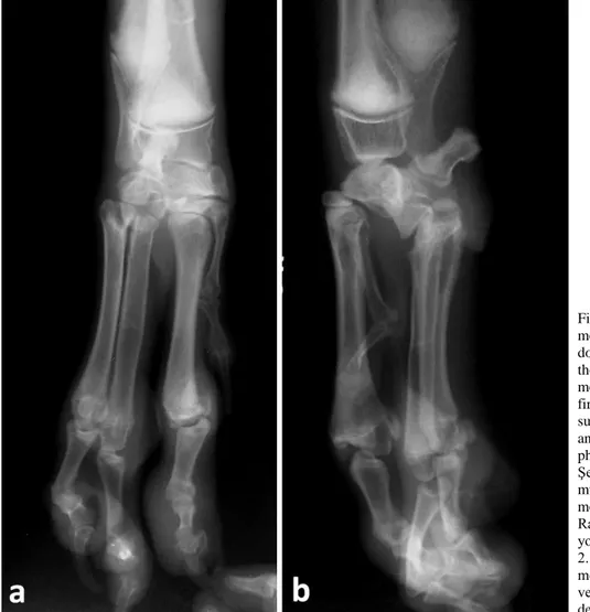
A male, 18 week old, Turkish Kangal dog was presented to the clinics of Ondokuz Mayıs University, Faculty of Veterinary Medicine with the right forelimb lameness. During clinical examination, the dog had moderate lameness on the right forelimb. An 11 cm long cleft starting from the distal manus to the carpo-metacarpal joint was observed (Figure 1a). The right foot pad did not touch the ground when the dog was in standing position. In addition, the lower limb deviated slightly to the medial, and a curvature deformity of nails was determined on the affected paw (Figure 1b). There was a mild valgus deviation in the carpal joint of affected limb.
Figure 1. Clinical view of the cleft at rest (a) and after load bearing (b) at the initial clinical examination. Şekil 1. Dinlenme anında (a) ve vücut ağırlığını taşıma sırasında (b) yarığının klinik görünümü.
Figure 2. Cranio-caudal (a) and mediolateral (b) radiographs of the dog at the initial examination. Note the absence of the 3rd carpal and
metacarpal bone with fusion of the first and second carpal bones. Lateral subluxation of 5th metacarpal bone,
and lateral deviation of all distal phalanges were also apparent. Şekil 2. Köpeğin ilk klinik muayenesindeki kraniokaudal (a) ve mediolateral (b) radyografileri. Radyografilerde 3.metakarpus’un yokluğu ve metakarpal kemiğin 1. ve 2. karpal kemikler ile füzyonu, 5. metakarpal kemiğin subluksasyonu ve tüm distal falanksların laterale deviasyonu.
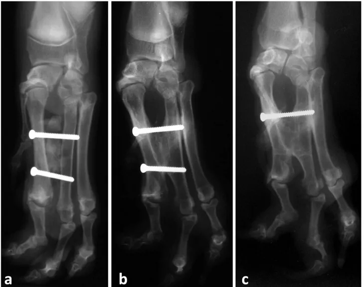
Figure 3. Cranio-caudal radiographs of the dog immediate (a), three months (b), and five months (c) after the operation. Note the deviation of the phalanges were worsened by the time.
Şekil 3. Operasyondan hemen sonra (a), operasyondan sonra 3. ay (b) ve 5. ay (c)’daki kraniokaudal radyografileri. Falanksların deviasyonundaki zaman içindeki artış net olarak gözlenmekte.
Upon radiographic evaluation, cranio-caudal radiographs demonstrated a 10 mm wide gap between the 2nd and 4th metacarpals, with the nonexistence of the 3rd
metacarpal bone (Figure 2a). In the right carpo-metacarpal joint, fusion of the 1st and 2nd carpal bones with the
absence of 3rd carpal bone were identified (Figure 2b).
Furthermore lateral subluxation of 5th metacarpal bone,
and lateral deviation of all distal phalanges were observed. As an additional radiographic finding, there was distinct malarticulation in the metacarpo-phalangeal joint of the second digit. Various levels of deviations were observed in all interphalangeal joints. In accordance with clinical and radiographic evaluations, the diagnosis was ectrodactyly.
The treatment consisted of two phases including bone reconstruction and fusion podoplasty. The skin of the manus over the 2nd and the 4th metacarpal bones was
incised parallel to each bone, starting at the distal
interphalangeal area. Both incisions were connected over the proximal side of the carpal joint forming an upside down “V” shape. The subcutaneous soft tissues were dissected using a periosteal elevator avoiding damage to the common digital artery and vein. During the surgery of the manus, a second surgeon performed the block ostectomy of the ipsilateral iliac crest and harvested the corticocancellous bone graft (3x1,5x1cm). After exposing the 2nd and 4th metacarpal bones, periostectomy of the
axial sides of both were performed. The bone graft was placed between the 2nd and 4th metacarpal bones, and fixed
with two 2,7mm lag screws bicortically (Figure 3a). Following reconstructive bone surgery, the upside down “V” sized defect was closed as an “I” shaped fusion podoplasty.
In the post-operative period, a soft padded bandage reinforced with a moulded palmar support was applied to the limb and the bandage was renewed every 3 days until
removal. After removing the bandage on postoperative 20th day, the owner was advised to refrain from rigorous
excercise. During the two months postoperatively, only leash walking was allowed. Due to the length of the metacarpal paw cleft, it was impossible to merge two parts of the soft pad. Initially, short term clinical and radiological results were favourable after the corrective surgery following bandage removal. A second surgery was proposed to the client but he refused it so the treatment was maintained as symptomatically. The animal had a very slight limp on the effected foot and shown no signs of pain while weight bearing or walking. The physical examination that was performed on the post operative 2nd month revealed medial rotation of the distal
phalanges and the excessive growth of nails (Figure 4a). Due to the skin contact of the distal digits to the ground instead of the foot pads, mild pododermatitis was developed (Figure 4b). This condition triggered slight pain and mild lameness while running on hard and rough ground. Three months after the surgery head of the distal screw caused mild serous discharge owing to pressure sore of the screw head on the medial side of the second metacarpal bone (Figure 3b). The distal screw was removed on postoperative 4th month but the proximal
screw was kept in place (Figure 3c). Postoperative 6th
month physical examination findings were in accordance
with the previous ones and the condition of the skin and the pain did not deteriorate further. Radiological examination demonstrated that the malarticulation occured after lateral subluxation of 5th metacarpal bone,
and lateral deviation of all distal phalanges has worsened. Long term clinical outcome was less favourable. The animal's limp has increased due to pain but the limb was still functional in terms of walking and weight bearing. The large foot pad still functioned as the primary contact to the ground, however the pads of the digits deviated medially due to the malformation hence the weight bearing surface became the skin. This phenomenon likely caused pain to the animal and it refused to apply its full body weight.
Ectrodactyly is generally described as the absence in the digital structures and the term is borrowed from the human malformation. Three parallel rays in embryonic development are responsible for the formation of the front limbs; the medial ray, which forms the radius, carpals and metacarpals of the first digit, the central ray, which forms the ulna, carpals and metacarpals of the 2nd digit and the
lateral ray which forms the rest of the digital bones and structures (6, 10). In our case, the 3rd carpal and metacarpal
and corresponding phalangeal bones were absent which suggests the developmental abnormality of the lateral ray. This finding was not in line with most of the previously
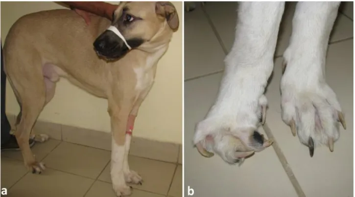
Figure 4. Clinical view of the dog two months after the surgery (a). Note the deviation of phalanges and mild pododermatitis of the right foot (b).
Şekil 4. Köpeğin operasyondan sonraki 2. aydaki klinik görünümü (a). Falanksların deviasyonu ve hafif pododermatisis net olarak görünmekte (b).
dog (2, 3, 4, 9, 11) however we could not find any accounts of a Turkish Kangal breed which is worth mentioning.
Each ectrodactyly case requires a different treatment approach as the condition has many diverse clinical appearances (5, 9,12). Milder cases may require no attention at all while more severe cases require surgical intervention. The treatment options that were presented in human literature are mostly incompatible in veterinary patients because of the functional differences of the limb (15). This is partially due to aiding the locomotion of the patient and partially to prevent deteriorationg conditions on the limb because of the malformation (23). Soft tissue reconstruction techniques may be sufficient to treat some cases but others may need bone reconstructive surgery and arthrodeses (1, 9, 18). The most severe cases may require amputation of the limb (2, 5, 8, 9, 17). Though our case might be considered a severe malformation, the client wanted the reconstructive surgery over amputation. As to the soft tissue reconstruction, fusion podoplasty is usually indicated in irreversible and non healing conditions of the digits but this case presented itself as the ideal candidate for the procedure as it allowed us to graft a corticocancellous bone graft instead of the 3rd metacarpal
bone. A previous study reported the fusion of two metacarpals via synostosis (17), we felt the presence of the bone graft would expedite the process and it provided a more controlled and limited area of fusion between the metacarpals while retaining mobility of the joint. Because there are many kinds of ectrodactyly the surgical intervention options are quite varied as well and the long term success of these options were suggested to be dependent on both the kind of malformation and the treatment used (9). Previous studies reported the changes on footpads to some extent due to the malformation (14,18). This might be worth mentioning because no matter how the deformity is repaired the footpad remains the weight bearing surface and the changes inflicted on it during surgical repair might have consequences like ours in long term clinical outcome.
As a result it can be concluded that; use of a corticocancellous block graft and fusion podoplasty can be an alternative treatment choice for the management of ectrodactyly in large breed dogs.
References
1. Andreoni A, Rytz U, Vannini R, et al. (2010): Ground
reaction force profiles after partial and pancarpal arthrodesis in dogs. Vet Comp Orthop Traumatol, 23, 1-6.
2. Barrand K (2004): Ectrodactyly in a West Highland white
terrier. J Small Anim Pract, 45, 315-318.
3. Bingel SA, Riser WH (1977): Congenital elbow luxation in
the dog. J Small Anim Pract,18, 445-456.
Bilateral ectrodactyly and spinal deformation in a mixed-breed dog. Can Vet J, 52, 47.
6. Entin MA (1976): Patterns of deformities in congenital
anomalies of the upper limb and their relation to the classification. Birth Defects Orig Artic Ser, 13, 231-241.
7. Feuston MH, Scott WJ (1985): Cadmium‐induced
forelimb ectrodactyly: A proposed mechanism of teratogenesis. Teratology, 32, 407-419.
8. Harasen G (2010): Surgical management of ectrodactyly in
a Siberian husky. Can Vet J, 51, 421.
9. Innes JF, McKee W, Mitchell R, et al. (2001): Surgical
reconstruction of ectrodactyly deformity in four dogs. Vet
Comp Orthop Traumatol, 14, 201-209.
10. Kanavel AB (1932): Congenital malformations of the
hands. Arch Surg, 25, 1-53.
11. Keller W, Chambers J (1989): Antebrachial metacarpal
arthrodesis for fusion of deranged carpal joints in two dogs.
J Am Vet Med Assoc, 195, 1382-1384.
12. Leighton R (1983): Surgical repair of a congenital defect
of the radius, ulna, and carpus in a dog. Mod Vet Pract, 41-44.
13. Leipold H, Huston K, Guffy M, et al. (1969): Ectrodactyly
in two beef calves. Am J Vet Res, 30, 1689.
14. Montgomery M, Tomlinson J (1985): Two cases of
ectrodactyly and congenital elbow luxation in the dog. J Am
Anim Hosp Assoc, 21, 781-785.
15. Montgomery RD, Milton JL, Mansfield PD, et al. (1989):
What is your diagnosis? Ectrodactyly. J Am Vet Med
Assoc, 194, 120-121.
16. Pearson K (1931): On the existence of the digital deformity-so-called "lobster-claw"-in the apes. Ann Eugen, 4, 339-340.
17. Pisoni L, Del Magno S, Cinti F, et al. (2014): Surgical
induction of metacarpal synostosis for treatment of ectrodactyly in a dog. Vet Comp Orthop Traumatol, 27,
166-171.
18. Pratschke K (1996): A case of ectrodactyly in a dog. Vet J, 49, 412-413.
19. Rahal SC, Volpi RS, Teixeira CR, et al. (2012):
Congenital deformity of the paw in a captive tiger: Case report. BMC Vet Res, 8, 98.
20. Ramadan R (1993): Hemimelia and ectrodactyly in a Najdi
sheep. Agri Practice, 14, 30-32.
21. Roelfsema N, Cobben J (1996): The EEC syndrome: A
literature study. Clin Dysmorphol, 5, 115-127.
22. Searle AG (1953): Hereditary split-hand in the domestic
cat. Ann Eugen, 17, 279-282.
23. Towle HA, Breur GJ (2004): Dysostoses of the canine and
feline appendicular skeleton. J Am Vet Med Assoc, 225,
1685-1692.
Geliş tarihi: 25.03.2016 / Kabul tarihi: 02.11.2016
Address for correspondence:
Assoc.Prof.Dr. Cenk Yardımcı Ondokuz Mayıs University,
Faculty of Veterinary Medicine, Department of Surgery, Samsun, Turkey.