Highly efficient nonradiative energy transfer
mediated light harvesting in water using aqueous
CdTe quantum dot antennas
Evren Mutlugun,
1Olga Samarskaya,
1Tuncay Ozel,
1Neslihan Cicek,
1Nikolai Gaponik,
2Alexander Eychmüller,
2and Hilmi Volkan Demir
1,*1Department of Physics, Department of Electrical and Electronics Engineering, UNAM — National Nanotechnology Research Center, Bilkent University, 06800, Ankara, Turkey
2
Physical Chemistry, TU Dresden, Bergstr.66b, 01062, Dresden, Germany *[email protected]
Abstract:
We present light harvesting of aqueous colloidal quantum dots to
nonradiatively transfer their excitonic excitation energy efficiently to dye
molecules in water, without requiring ligand exchange. These
as-synthesized CdTe quantum dots that are used as donors to serve as
light-harvesting antennas are carefully optimized to match the electronic structure
of Rhodamine B molecules used as acceptors for light harvesting in aqueous
medium. By varying the acceptor to donor concentration ratio, we measure
the light harvesting factor, along with substantial lifetime modifications of
these water-soluble quantum dots, from 25.3 ns to 7.2 ns as a result of their
energy transfer with efficiency levels up to 86%. Such nonradiative energy
transfer mediated light harvesting in aqueous medium holds great promise
for future quantum dot multiplexed dye biodetection systems.
©2010 Optical Society of America
OCIS codes: (230.5590) Quantum-well, -wire and -dot devices; (160.2540) Fluorescent and luminescent materials; (160.4670) Optical materials; (160.4760) Optical properties; (260.2160) Energy Transfer; (160.1435) Biomaterials
References and links
1. A. Georgi, C. Mottola-Hartshorn, A. N. Warner, B. Fields, and L. B. Chen, “Detection of individual fluorescently labeled reovirions in living cells,” Proc. Natl. Acad. Sci. U.S.A. 87(17), 6579–6583 (1990).
2. Y. Li, Y. T. H. Cu, and D. Luo, “Multiplexed detection of pathogen DNA with DNA- based fluorescence nanobarcodes,” Nat. Biotechnol. 23(7), 885–889 (2005).
3. K. Bacia, S. A. Kim, and P. Schwille, “Fluorescence cross-correlation spectroscopy in living cells,” Nat. Methods 3(2), 83–89 (2006).
4. C. Fang, A. Agarwal, K. D. Buddharaju, N. M. Khalid, S. M. Salim, E. Widjaja, M. V. Garland, N.
Balasubramanian, and D. L. Kwong, “DNA detection using nanostructured SERS substrates with Rhodamine B as Raman label,” Biosens. Bioelectron. 24(2), 216–221 (2008).
5. H. Tokudome, Y. Yamada, S. Sonezaki, H. Ishikawa, M. Bekki, K. Kanehira, and M. Miyauchi,
“Photoelectrochemical deoxyribonucleic acid sensing on a nanostructured TiO2 electrode,” Appl. Phys. Lett. 87(21), 213901 (2005).
6. B. O’Reagen, and M. Grätzel, “A low-cost, high-efficiency solar cell based on dye-sensitized colloidal TiO2 films,” Nature 353(6346), 737–740 (1991).
7. S. L. Li, K. J. Jiang, K. F. Shao, and L. M. Yang, “Novel organic dyes for efficient dye-sensitized solar cells,” Chem. Commun. (Camb.) 26(26), 2792–2794 (2006).
8. E. Mutlugün, S. Nizamoglu, and H. V. Demir, “Highly efficient nonradiative energy transfer using charged CdSe/ZnS nanocrystals for light-harvesting in solution,” Appl. Phys. Lett. 95(3), 033106 (2009).
9. W. W. Yu, E. Chang, R. Drezek, and V. L. Colvin, “Water-soluble quantum dots for biomedical applications,” Biochem. Biophys. Res. Commun. 348(3), 781–786 (2006).
10. S. Kim, and M. G. Bawendi, “Oligomeric Ligands for Luminescent and Stable Nanocrystal Quantum Dots,” J. Am. Chem. Soc. 125(48), 14652–14653 (2003).
11. M. Grabolle, M. Spieles, V. Lesnyak, N. Gaponik, A. Eychmüller, and U. Resch-Genger, “Determination of the Fluorescence Quantum Yield of Quantum Dots: Suitable Procedures and Achievable Uncertainties,” Anal. Chem. 81(15), 6285–6294 (2009).
12. N. Gaponik, D. V. Talapin, A. L. Rogach, K. Hoppe, E. V. Shevchenko, A. Kornowski, A. Eychmüller, and H. Weller, “Thiol-Capping of CdTe nanocrystals: An alternative to organometallic synthetic routes,” J. Phys. Chem. B 106(29), 7177–7185 (2002).
#124054 - $15.00 USD Received 9 Feb 2010; revised 23 Apr 2010; accepted 28 Apr 2010; published 7 May 2010
13. A. L. Rogach, T. Franzl, T. A. Klar, J. Feldmann, N. Gaponik, V. Lesnyak, A. Shavel, A. Eychmüller, Y. P. Rakovich, and J. F. Donegan, “Aqueous synthesis of thiol-capped CdTe nanocrystals: State-of-the-art,” J. Phys. Chem. C 111(40), 14628–14637 (2007).
14. T. Förster, “Zwischenmolekulare Energiewanderung und Fluoreszenz,” Ann. Phys. 437(1-2), 55–75 (1948). 15. A. R. Clapp, I. L. Medintz, J. M. Mauro, B. R. Fisher, M. G. Bawendi, and H. Mattoussi, “Fluorescence
resonance energy transfer between quantum dot donors and dye-labeled protein acceptors,” J. Am. Chem. Soc. 126(1), 301–310 (2004).
16. D. M. Willard, and A. Van Orden, “Quantum dots: Resonant energy-transfer sensor,” Nat. Mater. 2(9), 575–576 (2003).
17. I. L. Medintz, A. R. Clapp, F. M. Brunel, T. Tiefenbrunn, H. T. Uyeda, E. L. Chang, J. R. Deschamps, P. E. Dawson, and H. Mattoussi, “Proteolytic activity monitored by fluorescence resonance energy transfer through quantum-dot-peptide conjugates,” Nat. Mater. 5(7), 581–589 (2006).
18. S. Sadhu, and A. Patra, “Composition effects on quantum dot-based resonance energy transfer,” Appl. Phys. Lett. 93(18), 183104 (2008).
19. P. S. Chowdhury, P. Sen, and A. Patra, “Optical properties of CdS nanoparticles and the energy transfer from CdS nanoparticles to Rhodamine 6G,” Chem. Phys. Lett. 413(4-6), 311–314 (2005).
20. T. Pons, I. L. Medintz, M. Sykora, and H. Mattoussi, “Spectrally resolved energy transfer using quantum dot donors: Ensemble and single-molecule photoluminescence studies,” Phys. Rev. B 73(24), 245302 (2006). 21. E. Alphandery, L. M. Walsh, Y. Rakovich, A. L. Bradley, J. F. Donegan, and N. Gaponik, “Highly efficient
Förster resonance energy transfer between CdTe nanocrystals and Rhodamine B in mixed solid films,” Chem. Phys. Lett. 388(1-3), 100–104 (2004).
22. X. Y. Wang, Q. Maa, Y. B. Lia, B. Li, X. G. Su, and Q. H. Jin, “Studies on fluorescence resonance energy transfer between dyes and water-soluble quantum dots,” Can. J. Anal. Sci. Spectrosc. 50, 141–146 (2005). 23. J. Li, F. Mei, W. Y. Li, X. W. He, and Y. K. Zhang, “Study on the fluorescence resonance energy transfer
between CdTe QDs and butyl-rhodamine B in the presence of CTMAB and its application on the detection of Hg(II),” Spectrochimica Acta Part A 70(4), 811–817 (2008).
24. Q. Chen, Q. Ma, Y. Wan, X. Su, Z. Lin, and Q. Jin, “Studies on fluorescence resonance energy transfer between dyes and water-soluble quantum dots,” J. Biolumin. Chemilumin. 20(4-5), 251–255 (2005).
25. A. R. Clapp, I. L. Medintz, and H. Mattoussi, “Förster resonance energy transfer investigations using quantum-dot fluorophores,” Chem. Phys. Chem 7(1), 47–57 (2006).
26. J. R. Lakowicz, Principles of Fluorescence Spectroscopy (New York: Springer,2006).
27. V. K. Komarala, A. L. Bradley, Y. P. Rakovich, S. J. Byrne, Y. K. Gun’ko, and A. L. Rogach, “Surface plasmon enhanced Förster resonance energy transfer between the CdTe quantum dots,” Appl. Phys. Lett. 93(12), 123102 (2008).
28. S. Chanyawadee, R. T. Harley, M. Henini, D. V. Talapin, and P. G. Lagoudakis, “Photocurrent Enhancement in Hybrid Nanocrystal Quantum-Dot p-i-n Photovoltaic Devices,” Phys. Rev. Lett. 102(7), 077402 (2009).
1. Introduction
Today dye molecules are widely used in molecular biology. They offer several benefits
including good biocompatibilty, high quantum yield, and reasonable photostability. Some of
their common applications include biolabeling, biodetection, and spectroscopy [1–3]. Also,
their small size of only a few nanometers allows for their use in molecular-level detection
systems (e.g., in double stranded deoxyribonucleic acid (DNA), which is not possible with
larger size biotags [4, 5]). Despite these unique advantages, organic dyes, suffer one main
drawback that they cannot be efficiently pumped over a wide spectral range beyond their
characteristically narrow absorption spectra (for example, see Fig. 1a for Rhodamine B). This
limits their use in some important applications that require broader spectral operation
including spectral multiplexing (in which various dyes can simultaneously be pumped using a
single source). Also for dyes, spectral extension of their optical absorption is important in
photovoltaic applications where dyes are used as sensitizers [6]. The dye sensitized solar cells
require a wide spectral response for an enhanced efficiency, but broadening of the absorption
spectra of dyes generally necessitates complicated chemical modifications [7].
To address these problems, by using semiconductor quantum dot nanocrystals,
nonradiative Förster-type resonance energy transfer (FRET) can be employed to enable light
harvesting at optical wavelengths beyond the absorption range of dye molecules, and thus to
effectively extend their absorption. Such nanocrystals feature high-efficiency, Gaussian-like
distributed emission along with high tunability of absorption/emission characteristics, which
make them good candidates as donors for light harvesting, as we have shown in our previous
work [8]. However, the solubility of such donor quantum dots in aqueous media is a critical
issue to provide biocompatibility and enable biological applications [9]. To be dissolved in
water, the ligand exchange of nanocrystals is an alternative, but this comes at a cost of
#124054 - $15.00 USD Received 9 Feb 2010; revised 23 Apr 2010; accepted 28 Apr 2010; published 7 May 2010
significantly decreased quantum efficiency [10]. On the other hand, aqueous CdTe quantum
dots provide as-synthesized water solubility and high quantum yield [11], and their synthesis
has already been studied and well established [12, 13]. For these reasons, aqueous CdTe is
one of the best candidates as light-harvesting antenna in water. However, such as-synthesized
aqeuous quantum dots have not been investigated or demonstrated for light harvesting in
water to date, although this is of ciritical importance for spectrally extended bioimaging and
biolabeling applications.
Nonradiative transfer of the electronic excitation energy from electronically excited donor
molecules to optically luminescent acceptor molecules in close proximity was first discussed
by Theodor Förster in 1948 [14]. Till date FRET has been extensively studied in different
FRET pairs of dyes and nanocrystals for various applications [15–20] (also including CdTe
nanocrystals [21–25]). Although these previous reports have demonstrated FRET mechanism
using such a large variety of FRET pairs, light harvesting based on FRET using aqueous
nanocrystals has not been reported. In the previous work of our group, light harvesting of
organic nanocrystals synthesized in apolar solvents was investigated; this, however,
undesirably came at the cost of requiring ligand exchange. Avoiding the need for the ligand
exchange, the light harvesting factor of as-synthesized aqueous nanocrystals and their
systematic tuning and control in aqueous medium for light harvesting have not been reported.
In this paper, different than the prior works of our group and the others, we propose and
demonstrate light harvesting of aqueous colloidal CdTe quantum dots to nonradiatively
transfer their excitonic excitation energy efficiently to dye molecules in water, and present
systematic tuning and control of their light harvesting activity in aqueous medium, without the
need for ligand exchange. We investigated the effects of Förster radius of these aqueous
nanocrystals on modifying their lifetimes and controlling their light harvesting factor in water.
We studied the operation of these CdTe nanocrystal donors, serving as optical antennas for
acceptor Rhodamine B molecules, with their steady state photoluminescence (SSPL)
spectroscopy and further investigated and analyzed their significantly modified
photoluminescence decay kinetics for light harvesting with their time resolved
photoluminescence (TRPL) spectroscopy.
With acceptor to donor (A/D) concentration ratio varied in water, we controlled the light
harvesting factor of the donor CdTe quantum dots, with their substantial lifetime
modifications as a result of the nonradiative energy transfer with high efficiency levels (up to
86%). We further analyzed the controlled change in the lifetime of the acceptor molecules and
investigated the resulting trend of increasing energy transfer efficiency versus decreasing light
harvesting enhancement of the acceptor emission with the increased A/D concentration ratio,
discussing the fundamental tradeoffs and practical feasibility of nonradiative energy transfer
assisted light harvesting operation with reasonable efficiency and enhancement.
2. Experimental characterization, analyses, and results
In this work we colloidally synthesized aqueous CdTe quantum dots in two different sizes to
study the effect of Förster radius on the energy transfer efficiency and light harvesting
activity. Our synthesis procedure follows the method previously described in detail in
references 11 and 12. In our synthesis, 4.59 g of Cd(ClO
4)
2x6H
2O is dissolved in 0.5 l of
Milli-Q water in 1 l three-neck reaction flask. 1.33 g of TGA is added to the mixture, which
turns into milky appearance. The pH of this mixture is then increased to 11.8 – 12.0 by
dropwise addition of NaOH upon vigorous stirring. After this step, the reaction mixture
becomes clear or slightly turbid. To prepare tellurium precursor, 0.8 g of Al
2Te
3is transferred
into a small three-neck flask in the glove box and then deaerated by passing Ar for 50-60 min
in the setup. 10 ml of deaerated 0.5 M H
2SO
4is slowly poured into Al
2Te
3lumps to produce
H
2Te gas, which is carried by a slow Ar flow and bubbled through the mixture containing
cadmium precursor for 40-50 min. The resulting red-black mixture is refluxed at 100°C to
obtain the desired nanocrystal size. The reaction mixture is then cooled to room temperature
and filtered. The CdTe quantum dots are finally separated by size selective precipitation.
#124054 - $15.00 USD Received 9 Feb 2010; revised 23 Apr 2010; accepted 28 Apr 2010; published 7 May 2010
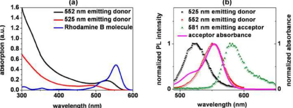
Figure 1a shows the optical absorption spectra of these differently sized CdTe quantum
dot donors carefully chosen by size selection, along with that of Rhodamine B acceptor
molecules in water. Figure 1b depicts the photoluminescence spectra of these CdTe quantum
dots selectively chosen to emit at the peak emission wavelengths of 525 nm and 552 nm
(corresponding to 2.04 nm and 2.98 nm in size, respectively), presented here along with the
emission and absorption spectra of the acceptor dye molecules alone to show the spectral
overlap. Here the absorbance measurements were taken using Cary UV-VIS
spectrophotometer and the photoluminescence measurements were carried out using Cary
Eclipse fluorescence spectrophotometer.
Fig. 1. (a) Absorbance spectra of the two differently sized aqueous CdTe nanocrystal quantum dots (emitting at 552 and 525 nm) together with that of the Rhodamine B dye molecules. (b) Normalized photoluminescence spectra of our aqueous CdTe quantum dots (donors) selectively chosen to emit at the peak wavelengths of 525 nm and 552 nm, along with the emission and absorption spectra of the Rhodamine B molecules (acceptors). The donors emitting at 552 nm provide a better spectral match to the electronic structure of these acceptors.
Förster radius, which is defined as the distance between the donor and acceptor molecules
yielding a 50% efficient energy transfer, is given by Eq. (1)
2 4 1/6
0
0.211(
D( ))
R
κ
n Q J
−λ
=
(1)
where
κ
2is the dipole orientation factor; n is the refractive index of the medium;
D
Q
is the
quantum efficiency of the donor;
J
( )
λ is the overlap integral of the donor emission spectrum
and the acceptor absorption spectrum; and
R
ois expressed in terms of Angstroms (Å) [26].
Figure 1b is used to calculate the spectral overlap integrals ( )
J
λ , which leads to 5.5 × 10
15and 9.2 × 10
15for the donor quantum dots emitting at 525 nm and 552 nm, respectively.
Subsequently, the quantum efficiencies of the donor molecules are experimentally measured
to be 10% and 54% for 525 nm emitting and for 552 nm emitting dots, respectively using
Rhodamine 6G as the reference dye. This difference in the quantum efficiency is commonly
observed for this type of nanocrystals; their quantum efficiencies increase with increasing
size, which is inevitable for such only core structures. Using Eq. (1), the Förster radii
calculated are
R
o=
4.7 and
R
o=
6.7 nm, for 525 nm and 552 nm quantum dots, respectively.
This difference in the Förster radius comes both from the spectral overlap and quantum
efficiency difference. Based on this observation, the quantum dot donors emitting at 552 nm
are found to have a wider Förster interaction range and thus to be a better optimized match to
the acceptor dyes, compared to 525 nm emitting dots,
2.1 Steady State Photoluminescence (SPPL) measurements
To observe FRET, we first performed SSPL measurements (with optical excitation at 375 nm)
while adding controlled amounts of acceptor molecules into the aqueous donor solution, thus
changing A/D ratio in increments. Figure 2a presents SSPL spectra for our CdTe quantum
#124054 - $15.00 USD Received 9 Feb 2010; revised 23 Apr 2010; accepted 28 Apr 2010; published 7 May 2010
dots emitting at 552 nm used as the donors and Fig. 2b shows the results of the same
measurements repeated using 525 nm emitting dots as the donors, both starting with the same
concentration levels and changing A/D concentration ratio in an identical manner in water. In
this steady state characterization, optical excitation is chosen at 375 nm to be consistent with
that of the time resolved measurements that use a 375 nm laser diode pump. It is worth noting
that our donor molecules are optically well excited at 375 nm, while this excitation
wavelength is out of the absorption range of the acceptor molecules (Fig. 1a). Here the
concentrations of the donors (without acceptors) and acceptors (without donors) used in Fig.
1a correspond to the same starting points of Fig. 2 before adding the acceptor molecules into
the aqueous donor solution. As A/D concentration ratio is increased, we clearly observe
simultaneously the quenching of donor emission and the enhancement of acceptor emission in
increments.
Fig. 2. SSPL spectra taken by adding controlled amounts of dye acceptors into the aqueous donor solution using CdTe quantum dots emitting at the peak wavelength of (a) 552 nm and (b) 525 nm. The legends show the corresponding A/D concentration ratios (A/D = 1.8–152.8). (Note that these PL intensity levels are measured using the same arbitrary units and that they are presented using the scales as indicated on their plots, for clear visibility.)
2.2 Time Resolved Photoluminescence (TRPL) measurements
To better understand the emission kinetics, we also performed and analyzed TRPL
measurements, again by adding controlled amounts of acceptors into the aqueous donor
solution (and changing A/D concentration ratios) in identical increments, both for 552 nm and
525 nm quantum dot donors. TRPL measurements were conducted using PicoQuant FluoTime
200 time resolved spectroscopy system with a fixed laser diode head at 375 nm wavelength
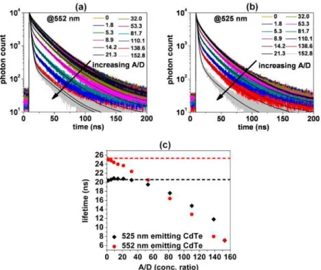
having pulse widths of <70 ps . Figure 3 shows the evolution of photon count decay over time,
parameterized with respect to the varied A/D concentration ratios, using 552 nm emitting
quantum dots (given in Fig. 3a) and 525 nm emitting ones (given in Fig. 3b) at the
corresponding donor emission wavelengths. Each decay curve is also shown together with its
corresponding numerical exponential fits, which exhibit good fitting to the experimental data.
These measurements and numerical analyses are performed at the corresponding peak
emission wavelengths of the donor nanocrystals since there is an insignificant overlap
between the donor and acceptor emission spectra at these peak wavelengths (Fig. 1b), which
makes the analysis safe (with no detectable crosstalk between the emission of donor and
acceptor molecules). The comparison of their donor photoluminescence decay lifetimes is also
presented as a function of A/D concentration ratio in Fig. 3c. Here we clearly observe
increasingly faster photoluminescence decay of donors. We also see that the donor lifetimes
diverge away more from the starting baseline of only donors and are shortened further more
for 552 nm emitting quantum dots (with its lifetimes modified from 25.3 ns to 7.2 ns) than
those of 525 nm dots (with its lifetimes modified from 20.4 ns to 7.1 ns). This is because 552
nm emitting CdTe quantum dots are better optimized to match Rhodomine dye molecules and
thus serve as better light-harvesting antennas to these dyes.
#124054 - $15.00 USD Received 9 Feb 2010; revised 23 Apr 2010; accepted 28 Apr 2010; published 7 May 2010
Fig. 3. TRPL measurements of donor molecules taken by adding controlled amounts of dye acceptors into the aqueous donor solution, using CdTe quantum dots emitting at the peak wavelength of (a) 552 nm and (b) 525 nm, all shown together with their corresponding numerical fits, and along with a comparative analysis of the donor photoluminescence decay lifetimes both for 552 and 525 nm emitting dots as a function of A/D concentration ratio (c). In the last plot, the red (black) dotted baseline represents the lifetime of only donors of 552 nm (525 nm) emitting dots, without any acceptors in the mixture.
Furthermore, we also performed TRPL characterization and analyses at the acceptor
emission wavelengths. The peak emission wavelength of the acceptor is 581 nm, where there
is a weak tail component of the donor emission. Therefore, in addition to the peak wavelength
581 nm, we performed all of the measurements and lifetime analyses also at 605 nm where
there is no detectable donor emission, for safe comparison. This allowed us to make sure the
effect of this tail overlap of the donor is insignificant. The evolution of photon count lifetimes
at 581 nm and 605 nm are given as a function of time together with their numerical fits for
552 nm emitting quantum dots in Fig. 4a and for 525 nm emitting quantum dots in Fig. 4b.
Both of their comparative lifetime analyses are given in Fig.s 4c and 4d. Due to the energy
feeding as a result of FRET process, we see that the acceptor photoluminescence decay
lifetimes are increased compared to the baseline of only acceptors, which is consistent with
the previous literature [27, 28]. Using 552 nm emiting donors, we observe the lifetime of the
acceptor molecule increases from 1.68 ns to 23.24 ns. As a function of the increasing A/D
concentration ratio, since the rate of the enhancement on the emission of acceptor molecule
decreases for larger A/D (Fig. 5b), the modifed lifetimes also converge towards the baseline.
Regardless of the analyses conducted at either of the wavelengths (581 nm or 605 nm), we
observe the same trend of the acceptor lifetime modifications, again with a stronger
modification for the better optimized light-harvesting 552 nm emitting CdTe quantum dots in
these experiments. All of the lifetime analysis results are also listed in Table 1-VII along with
their amplitudes A
iand
χ
2, chi-square values. The intensity weigted lifetime,
τ
intis calculated
through Eq. (2)
2 int i i/
i i i iA
A
τ
= ∑
τ
∑
τ
(2)
#124054 - $15.00 USD Received 9 Feb 2010; revised 23 Apr 2010; accepted 28 Apr 2010; published 7 May 2010
whereas the amplitude wighted lifetime,
τ
ampis calculated using Eq. (3).
/
amp i i i
i i
A
A
τ
= ∑
τ
∑
(3)
Fig. 4. TRPL measurements of acceptor molecules while varying the A/D concentration ratio, shown along with their numerical fits using (a) 552 nm and (b) 525 nm emitting quantum dots and comparative analysis of the acceptor photoluminescence decay lifetimes for emission (c) at 581 nm (acceptor peak with a weak donor tail) and (d) at 605 nm (strong acceptor tail with no donor tail as a function of A/D concentration ratios. In both plots, the dashed baseline represents the lifetime of only acceptors without any donors.
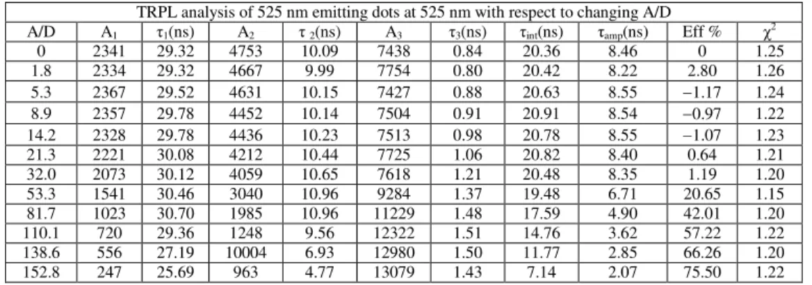
Table 1. TRPL measurement analysis (at 525 nm) of the 525 nm emitting donors varying the A/D concentration ratio.
TRPL analysis of 525 nm emitting dots at 525 nm with respect to changing A/D
A/D A1 τ1(ns) A2 τ 2(ns) A3 τ3(ns) τint(ns) τamp(ns) Eff % χ2
0 2341 29.32 4753 10.09 7438 0.84 20.36 8.46 0 1.25 1.8 2334 29.32 4667 9.99 7754 0.80 20.42 8.22 2.80 1.26 5.3 2367 29.52 4631 10.15 7427 0.88 20.63 8.55 −1.17 1.24 8.9 2357 29.78 4452 10.14 7504 0.91 20.91 8.54 −0.97 1.22 14.2 2328 29.78 4436 10.23 7513 0.98 20.78 8.55 −1.07 1.23 21.3 2221 30.08 4212 10.44 7725 1.06 20.82 8.40 0.64 1.21 32.0 2073 30.12 4059 10.65 7618 1.21 20.48 8.35 1.19 1.20 53.3 1541 30.46 3040 10.96 9284 1.37 19.48 6.71 20.65 1.15 81.7 1023 30.70 1985 10.96 11229 1.48 17.59 4.90 42.01 1.20 110.1 720 29.36 1248 9.56 12322 1.51 14.76 3.62 57.22 1.22 138.6 556 27.19 10004 6.93 12980 1.50 11.77 2.85 66.26 1.20 152.8 247 25.69 963 4.77 13079 1.43 7.14 2.07 75.50 1.22
#124054 - $15.00 USD Received 9 Feb 2010; revised 23 Apr 2010; accepted 28 Apr 2010; published 7 May 2010
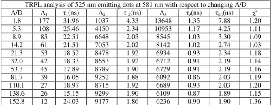
Table 2. TRPL measurement analysis (at 581 nm) of the 525 nm emitting donors varying the A/D concentration ratio.
TRPL analysis of 525 nm emitting dots at 581 nm with respect to changing A/D
A/D A1 τ1(ns) A2 τ 2(ns) A3 τ3(ns) τint(ns) χ2 1.8 177 31.96 1037 4.33 13648 1.35 7.88 1.20 5.3 108 25.46 4150 2.34 10953 1.17 4.25 1.11 8.9 85 22.51 6648 2.05 8545 1.03 3.30 1.09 14.2 61 21.51 7053 2.02 8142 1.02 2.74 1.03 21.3 53 18.52 8478 1.92 6934 0.93 2.34 1.18 32.0 42 18.33 8653 1.92 6712 0.91 2.19 1.14 53.3 45 17.89 8789 1.90 6729 0.91 2.19 1.16 81.7 39 16.05 9252 1.88 6092 0.86 2.03 1.19 110.1 27 18.97 8715 1.92 6689 0.93 2.03 1.20 138.6 26 15.15 9299 1.90 6109 0.87 1.89 1.15 152.8 12 24.03 9177 1.86 6236 0.90 1.90 1.36
Table 3. TRPL measurement analysis (at 605 nm) of the 525 nm emitting donors varying the A/D concentration ratio.
TRPL analysis of 525 nm emitting dots at 605 nm with respect to changing A/D
A/D A1 τ1(ns) A2 τ2(ns) A3 τ3(ns) τint(ns) χ2 1.8 128 46.63 404 9.03 14116 1.46 11.29 1.16 5.3 117 26.16 5379 2.23 9774 1.07 4.62 1.17 8.9 98 21.87 7350 2.02 7983 0.99 3.39 1.19 14.2 51 23.24 7585 2.00 7815 0.98 2.73 1.07 21.3 57 23.50 7200 2.05 8094 1.02 2.88 1.06 32.0 44 16.98 8977 1.93 6397 0.90 2.15 1.23 53.3 45 17.29 8895 1.90 6614 0.86 2.16 1.22 81.7 38 17.00 9089 1.92 6397 0.88 2.09 1.21 110.1 29 18.90 9230 1.91 6061 0.88 2.07 1.20 138.6 27 15.80 9512 1.89 5789 0.83 1.92 1.25 152.8 13 17.37 9138 1.86 6186 0.88 1.79 1.18
Table 4. TRPL measurement analysis (at 552 nm) of the 552 nm emitting donors varying the A/D concentration ratio.
TRPL analysis of 552 nm emitting dots at 552 nm with respect to changing A/D
A/D A1 τ1(ns) A2 τ2(ns) A3 Τ3(ns) τint(ns) τamp(ns) Eff. % χ2
0 2524 36.75 6271 15.85 4265 1.26 25.26 1.12 0 1.12 1.8 2605 35.90 6016 15.61 4662 1.21 25.02 1.13 3.90 1.13 5.3 1572 41.03 6062 15.95 5216 1.57 24.81 1.17 12.83 1.17 8.9 2151 35.78 4973 15.35 6317 1.27 24.40 1.14 20.63 1.14 14.2 2212 34.70 4567 14.76 6742 1.32 23.99 1.11 25.14 1.11 21.3 2063 34.85 4498 14.92 6827 1.42 23.67 1.12 26.55 1.12 32.0 1561 34.80 3657 14.87 8501 1.46 22.44 1.14 41.63 1.14 53.3 967 35.42 2418 14.83 10387 1.56 20.51 1.13 58.55 1.13 81.7 521 34.78 1201 13.95 12215 1.62 16.38 1.15 74.03 1.15 110.1 320 34.61 720 12.74 13095 1.63 12.86 1.17 80.54 1.17 138.6 348 25.09 3560 2.78 10949 1.33 8.01 1.23 85.23 1.23 152.8 269 25.79 4194 2.65 10499 1.28 7.16 1.15 86.08 1.15
#124054 - $15.00 USD Received 9 Feb 2010; revised 23 Apr 2010; accepted 28 Apr 2010; published 7 May 2010
Table 5. TRPL measurement analysis (at 581 nm) of the 552 nm emitting donors varying the A/D concentration ratio.
TRPL analysis of 552 nm emitting dots at 581 nm with respect to changing A/D
A/D A1 τ1(ns) A2 τ2(ns) A3 τ3(ns) τint(ns) χ2 1.8 1566 35.80 3515 15.18 8940 1.41 23.23 1.12 5.3 1042 34.53 2124 14.24 10977 1.44 20.67 1.10 8.9 774 34.15 1518 13.53 12159 1.46 18.64 1.12 14.2 508 34.86 1080 13.33 12809 1.48 16.38 1.16 21.3 486 32.94 913 12.23 13030 1.50 14.85 1.12 32.0 425 31.51 755 10.77 13304 1.49 13.05 1.14 53.3 278 31.41 543 9.88 13606 1.51 10.36 1.14 81.7 237 28.43 568 6.27 13723 1.48 7.96 1.12 110.1 209 25.08 4409 2.48 10629 1.18 6.05 1.05 138.6 181 24.82 5258 2.32 9634 1.13 5.51 1.06 152.8 167 24.72 4266 2.44 10793 1.19 5.22 1.05
Table 6. TRPL measurement analysis (at 605 nm) of the 552 nm emitting donors varying the A/D concentration ratio
TRPL analysis of 552 nm emitting dots at 605 nm with respect to changing A/D
A/D A1 τ1(ns) A2 τ2(ns) A3 τ3(ns) τint(ns) χ2 1.8 701 42.56 1591 15.37 11970 1.46 23.28 1.13 5.3 548 40.07 1320 14.82 12481 1.51 19.85 1.11 8.9 478 36.82 971 13.21 13060 1.52 16.85 1.15 14.2 342 37.02 763 13.08 13383 1.53 14.63 1.14 21.3 306 33.70 594 10.39 13481 1.50 12.05 1.14 32.0 300 33.09 596 10.16 13407 1.53 11.55 1.17 53.3 255 27.88 897 4.70 13627 1.43 7.99 1.10 81.7 225 25.53 3185 2.73 11804 1.26 6.47 1.09 110.1 161 24.90 5866 2.28 9180 1.11 5.13 1.05 138.6 134 23.86 6681 2.16 8412 1.04 4.42 1.08 152.8 119 25.71 5840 2.25 9138 1.09 4.55 0.98
Table 7. TRPL measurement analysis (at 581 and 605 nm) of the 581 nm emitting acceptors varying the A/D concentration ratio.
TRPL analysis of 581 nm emitting Rhodamine B molecules at 581 nm
A1 τ1(ns) A2 τ2(ns) A3 τ3(ns) τint(ns) χ2
−17 0.001 8277 1.98 7105 1.006 1.68 1.88
TRPL analysis of 581 nm emitting Rhodamine B molecules at 605 nm
A1 τ1(ns) A2 τ2(ns) A3 τ3(ns) τint(ns) χ2
10225 1.89 25883 0.53 −22115 0.45 1.69 1.23
2.3 Light harvesting analyses and remarks
Fig.s 3 and 4 demonstrate clearly the effect of Förster radius on the lifetime modifications of
the donor and acceptor molecules. For further analyses, we also calculate energy transfer
efficiency and light harvesting enhancement of the acceptor emission. The energy transfer
efficiency is extracted from the amplitude weighted donor lifetime,
τ
ampin the presence and
absence of the acceptor molecules using Eq. (4)
1
DA.
FRET Dτ
η
τ
= −
(4)
#124054 - $15.00 USD Received 9 Feb 2010; revised 23 Apr 2010; accepted 28 Apr 2010; published 7 May 2010
Here
τ
DAis the ampitude weighted lifetime of donors in the presence of acceptors and
τ
Dis
that of donors in the absence of acceptors [26].
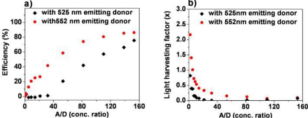
Figure 5a reveals the comparison of efficiency levels extracted from TRPL measurements.
Here we observe that the energy transfer efficiency increases with the increased A/D
concentration ratio, as the donors find more acceptors around them to transfer more of their
excitation energy. Tuning the A/D concentration ratios and using better optimized 552 nm
emitting quantum dot donors, we observe a maximum energy transfer efficiency of 86%,
which is obtained at an A/D concentration ratio of 152.8 in our experiments. This comparison
shows that the efficiency levels are higher using 552 nm emitting quantum dots than those of
525 nm emitting ones.
To show the effect of nonradiative energy transfer mediated light harvesting on the
emission enhancement of the acceptor molecules, we further compute the light-harvesting
factor for the acceptor emission (Fig. 5b). These calculations are carried out through fitting
SSPL measurements (in Fig. 2) of the donor quantum dots to a Gaussian distribution and
comparing the overall emission (donor + acceptor mixture) with the emission of only
acceptors (corresponding to the same concentration of acceptor molecules used in each A/D
concentration point). In these calculations, the tail overlap of the donor emission on the
acceptor emission is also considered, and any possible contribution from the tail (although
weak) is also subtracted. Here we observe that the relative enhancement factor of the acceptor
emission is decreased with the increased A/D concentration ratio, because the acceptors
increasing in number find fewer donors around them to be fed via nonradiative energy
transfer, which indicates a tradeoff between the efficiency and light harvesting factor.
Also, we again observe that 552 nm emitting aqueous CdTe quantum dots are better
light-harvesting antennas in water for Rhodamine B dye molecules in comparison to those quantum
dots emitting at 525 nm. In the light of these experiments and analyses, such light harvesting
is possible; however, one needs to consider the tradeoff between efficiency and enhancement
factor to choose an operating point. These results also indicate that nonradiative energy
transfer assisted light harvesting may enable quantum dot multiplexed dye biodetection
systems, on which we are currently working.
Fig. 5. Comparison of (a) FRET efficiencies and (b) enhancement of the acceptor emission, using 552 nm and 525 nm emitting CdTe quantum dot donors, as a function of the A/D concentration ratio.
3. Conclusion
In conclusion, as a proof-of-concept demonstration, we have presented nonradiative energy
transfer based light harvesting of aqueous colloidal CdTe quantum dot antennas for dye
molecules in water. Our experiments show that these quantum dots used as donors need to be
carefully optimized to match Rhodamine B used as acceptors. In our experiments, we have
observed strong lifetime modifications of these CdTe quantum dots from 25.3 to 7.2 ns. We
have demonstrated the energy transfer efficiency tuning up to 86% as the acceptor-donor
#124054 - $15.00 USD Received 9 Feb 2010; revised 23 Apr 2010; accepted 28 Apr 2010; published 7 May 2010
concentration ratio is varied. These experiments indicate that nonradiative energy transfer
mediated light harvesting using aqueous quantum dots leads to enhanced emission of dye
molecules in water at wavelengths beyond the absorption range of the dyes. One should also
note that a good operating point in the A/D concentration ratio for a specific donor-acceptor
pair has to be set to provide both reasonably high efficiency and high light harvesting of the
acceptor emission. This nonradiative energy transfer assisted light harvesting holds great
potential for future quantum dot multiplexed biological and optoelectronic applications.
Acknowledgements
This work is supported by EU-FP7 Nanophotonics4Energy NoE, BMBF TUR 09/001, and
TUBITAK EEEAG 106E020, 107E297, 107E088, 109E002, and 109E004. HVD
acknowledges support from ESF-EURYI and TUBA-GEBIP, and EM, from
TUBITAK-BIDEB.
#124054 - $15.00 USD Received 9 Feb 2010; revised 23 Apr 2010; accepted 28 Apr 2010; published 7 May 2010