TÜRK DO
Ğ
A VE
FEN DERG
İ
S
İ
Bingöl Üniversitesi Fen Bilimleri Enstitüsü
tarafından yayımlanmaktadır
Published by Institute of Science of Bingol
Bingöl Üniversitesi Fen Bilimleri Enstitüsü tarafından yayımlanmaktadır
Published by Institute of Science of Bingol University
Yıl/Year: 2015 Cilt/Volume: 4 Sayı/Number: 2
http://dergipark.ulakbim.gov.tr/tdfd/
Tr. Doğa ve Fen Derg. − Tr. J. Nature Sci. 2015 Vol. 4 No. 2
Bingöl Üniversitesi Fen Bilimleri Enstitüsü Adına Sahibi Owner on behalf of Institute of Science of Bingol University
Doç. Dr. İbrahim Yasin ERDOĞAN Fen Bilimleri Enstitüsü Müdürü
Director of Institute of Science
Editör/Editor Yrd. Doç. Dr. Özgür ÖZGÜN
Yazışma Adresi/Correspondence Adress Bingöl Üniversitesi Fen Bilimleri Enstitüsü Müdürlüğü 12000 Bingöl
Tel/Phone: +90 (426) 215 00 72 Faks/Fax: +90 (426) 216 00 34
Yayın Türü/Publication Type Bilimsel Dergi
Scientific Journal
DANIŞMA KURULU/ADVISORY BOARD
Prof. Dr. Ali Sayil ERDOĞAN Bingöl Üniversitesi
Prof. Dr. Erkan BOYDAK Bingöl Üniversitesi
Prof. Dr. Fahrettin YAKUPHANOĞLU Fırat Üniversitesi
Prof. Dr. Fehim FINDIK Sakarya Üniversitesi
Prof. Dr. Hüsamettin BULUT Harran Üniversitesi
Prof. Dr. İbrahim TÜRKOĞLU Bingöl Üniversitesi
Prof. Dr. İskender DEMİRKOL Bingöl Üniversitesi
Prof. Dr. Latif KELEBEKLİ Ordu Üniversitesi
Prof. Dr. Lütfi BEHÇET Bingöl Üniversitesi
Prof. Dr. Mehmet ÇİFTCİ Bingöl Üniversitesi
Prof. Dr. Nurullah SARAÇOĞLU Bingöl Üniversitesi
Prof. Dr. Orhan KURT Ondokuz Mayıs Üniversitesi
Prof. Dr. Turgay ŞENGÜL Bingöl Üniversitesi
Prof. Dr. Yusuf ZEREN Mersin Üniversitesi
Doç. Dr. Abdullah MART Bingöl Üniversitesi
Doç. Dr. Ahmet ÇETİN Bingöl Üniversitesi
Doç. Dr. Alaaddin YÜKSEL Bingöl Üniversitesi
Doç. Dr. Ali Rıza DEMİRKIRAN Bingöl Üniversitesi
Doç. Dr. Asım KAYGUSUZ İnönü Üniversitesi
Doç. Dr. Bekir BÜKÜN Harran Üniversitesi
Doç. Dr. Ecevit EYİDURAN Iğdır Üniversitesi
Doç. Dr. Hamit Özkan GÜLSOY Marmara Üniversitesi
Doç. Dr. Hasan KILIÇ Bingöl Üniversitesi
Doç. Dr. Hisamettin DURMAZ Harran Üniversitesi
Doç. Dr. Hüseyin VAPUR Çukurova Üniversitesi
Doç. Dr. İbrahim Yasin ERDOĞAN Bingöl Üniversitesi
Doç. Dr. İlkay ŞİŞMAN Sakarya Üniversitesi
Doç. Dr. Kağan KÖKTEN Bingöl Üniversitesi
Doç. Dr. Murat SOYLU Bingöl Üniversitesi
Doç. Dr. Mustafa KOYUN Bingöl Üniversitesi
Doç. Dr. Mustafa YAZGAN İstanbul Teknik Üniversitesi
Doç. Dr. Muttalip ÇİÇEK Dicle Üniversitesi
Doç. Dr. Nezir YILDIRIM Bingöl Üniversitesi
Doç. Dr. Orhan YILMAZ Yüzüncü Yıl Üniversitesi
Doç. Dr. Ramazan MERAL Bingöl Üniversitesi
Doç. Dr. Ramazan SOLMAZ Bingöl Üniversitesi
Yrd. Doç. Dr. Halil ŞİMŞEK Bingöl Üniversitesi
Yrd. Doç. Dr. Mücahit PAKSOY Kahramanmaraş Sütçü İmam Üniversitesi
Yrd. Doç. Dr. Ömer AKGÖBEK Harran Üniversitesi
Yrd. Doç. Dr. Özgür ÖZGÜN Bingöl Üniversitesi
Yrd. Doç. Dr. Sabri YURTSEVEN Harran Üniversitesi
Yrd. Doç. Dr. Tahir AKGÜL Sakarya Üniversitesi
TEŞEKKÜR/ACKNOWLEDGEMENT
Bu sayıda yer alan çalışmaların yazarlarına ve değerli zamanlarını ayırarak görüş ve önerileri ile katkıda bulunan hakemlerimize teşekkür eder, saygılar sunarız.
Tr. Doğa ve Fen Derg. − Tr. J. Nature Sci. 2015 Vol. 4 No. 2
İÇİNDEKİLER/CONTENTS
The various biological activities of Rheum ribes extract on different types of cell
Musa Tartik, Ekrem Darendelioglu, Gurkan Aykutoglu, Giyasettin Baydas
1
Ekonomik bir yöntemle hazırlanan tek faz -Fe
2O
3ve -Fe
2O
3nanopartiküllerinin karakterizasyonu ve
elektrokimyasal biyosensör uygulaması
Güllüzar Demir, İbrahim Y. Erdoğan
8
Yüksek gerilim izolatörlerinde yüzey kaçak akımlarının ANFIS yöntemiyle hesaplanması
Ersin Turan, Muhsin Tunay Gençoğlu, Abdulkerim Karabiber
16
An investigation of microstructure of the worn surfaces on drum in brake systems
Muzaffer Erdoğan, İbrahim Yavuz, Ali Erçetin
21
Composition and antioxidant capacity of the essential oils of Alyssum pateri Nyár subsp. prostratum (Nyár)
Dudley (Brassicaceae)
Mehmet Kadir Erdogan, İbrahim Halil Geçibesler, Lütfi Behçet
25
Ekmeklik buğday genotiplerinde erken kuraklığa dayanıklılığın ölçütü olarak fide kalımı
Sinan Bayram, Ali Öztürk, Murat Aydın
30
Resveratrol attenuates diabetic nephropathy by modulating organic transporters and heat shock proteins
in streptozotocin-induced diabetic rats
Oguzhan Ozdemir, Zeynep Tuzcu
36
Erzurum ve çevresinde yetiştirilen bazı domates çeşitlerinin (Lycopersicon esculentum) besinsel bileşimi
İhsan Güngör Şat, Nurhan Turhan
44
In vitro antioxidant activities of various solvent extracts from Tanacetum balsamita L. subsp. balsamita
Mehmet Kadir Erdogan, Gıyasettin Baydas
49
Yapay zekâ tekniklerinin inşaat mühendisliği problemlerinde kullanılması üzerine bir çalışma
Mehrzad Mohabbi Yadollahi, Ahmet Benli
55
Döküm endüstrisinde kurumsal kaynak planlama yazılımlarının kullanımının değerlendirilmesi
Didem Güleryüz Çilingir, Murat Çolak
60
Hayvansal ürünlerde dioksinler
Alper Baran, Ahmet Erdoğan
66
Tr. Doğa ve Fen Derg. − Tr. J. Nature Sci. 2014 Vol. 3 No. 1özgü 01
Tr. Doğa ve Fen Derg. − Tr. J. Nature Sci. 2015 Vol. 4 No. 2
The various biological activities of Rheum ribes extract on different
types of cell
Musa Tartik
1, Ekrem Darendelioglu
1, Gurkan Aykutoglu
1, Giyasettin Baydas*
2Abstract
The worldwide acceptance to use traditional herbs for medicinal remedies is a common event in recent times. Herbal plants have positive benefits rather than the side effects of marketing drugs. Therefore, numerous traditional plants are used to prevent various types of cancer, diabetes and viral diseases. These findings provided an attention in herbal medicine to discover rational and natural drugs and isolate biologically active compounds.
Rheum ribes (R. ribes) is most popular herbal plant to obtain raw drug materials. Its fresh stems, leaves and petioles are
commonly consumed as crude drugs in Asian countries. This study was designed to examine the biological activities of ethanol extract of Rheum ribes root (EERR) such as antioxidant activity on human umbilical vein endothelial cells (HUVECs), anticancer effectiveness in prostate cancer cells (PC3) and antimicrobial activity against several microorganisms. Moreover, the some active chemical compounds were determined in EERR.
The purpose of the present study was planned to evaluate the activity of EERR on the induction of oxidative cell injury in PC3 and HUVECs. Treatment of HUVECs with EERR decreased hydrogen peroxide (H2O2) induced reactive oxygen species
(ROS) overproduction and lipid peroxidation (LPO) levels. Furthermore, EERR caused an increase on productions of ROS and malondialdehyde (MDA) in PC3 cells and significantly decreased cell viability. Besides, the antibacterial and antifungal analysis showed that EERR is active against Staphylococcus aureus, Enterobacter aerogenes, Escherichia coli and Saccharomyces
cerevisiae. It was concluded that EERR has both pro-oxidant ability against PC3 cells and antioxidant activity on HUVECs.
Keywords: HUVECs; LPO; PC3; Rheum ribes; ROS
Rheum ribes ekstraktının farkli hücre tipleri üzerindeki biyolojik etkileri
ÖzetSon yıllarda, geleneksel olarak kullanılan şifalı bitkilerin tedavi amaçlı kullanımı bütün dünya tarafından kabul görmektedir. Bu tür bitkiler piyasadaki çok sayıda ilacın yan etkilerine rağmen birçok pozitif etkiye sahiptirler. Bu yüzden sayısız şifalı bitki; diyabet, çeşitli kanser türleri ve viral hastalıklarda tedavi amacıyla kullanılmaktadır. Bu bulgular, birçok araştırmacının dikkatini şifalı bitkilere yönelterek, doğal ve oranlı ilaçlar keşfetmeye teşvik etmiştir.
Rheum ribes (ışkın) da bu şifalı bitkilerden olup, ham maddeleri tedavi amaçlı kullanılmaktadır. Anadolu ve Asya’nın çeşitli
ülkelerinde gövdesi, yaprakları ve sapları taze ya da pişirilmiş olarak tüketilmektedir. Bu çalışma Rheum ribes kök etanol ektraktinin (EERR) biyolojik aktivitelerini belirlemek için tasarlanmıştır. Bu çalışmada insan umblikal ven endotel hücresi (HUVEC)’te antioksidan, prostat kancer hücresi (PC3)’de anti-kanser etkisi ve bazı mikroorganizmalarda ise antimikrobiyal aktivitesine bakıldı. Ayrıca ekstraktın içindeki 4 farklı aktif fitokimyasalın miktarıda belirlendi.
Bu çalışmanın amacı; oksidatif stres açısından indüklenmiş HUVEC ve PC3 hücrelerinde EERR’ın aktivitesini değerlendirmektir. Alınan sonuçlara göre, hidrojen peroksit (H2O2) ile indüklenmiş HUVEC’ler EERR ile muamele edildiğinde,
hücrelerde artmış olan reaktif oksijen türleri (ROS) ve lipid peroksidasyonu (LPO) seviyelerini düşürdüğü görüldü. Ayrıca aynı dozların PC3 hücrelerinde ise ROS ve LPO’yu artırdığı ve buna bağlı olarak hücre canlılığını düşürdüğü gösterildi. Diğer taraftan, EERR’ın Staphylococcus aureus, Enterobacter aerogenes, Escherichia coli ve Saccharomyces cerevisiae organizmalarına karşı antimikrobiyal aktivitesinin olduğu belirlendi. Bunlara ek olarak, EERR’ın içinde; chrysin, narinegenin ve quarcetin’nin varlığı ve miktarları tespit edildi. Sonuç olarak, EERR’ın PC3 hücrelerinde pro-oksidant bir etki ile hücreyi öldürdüğü, ama HUVEC’lerde ise antioksidan bir ajan olarak hücreyi koruduğu saptandı.
Anahtar Kelimeler: HUVECs; LPO; PC3; Rheum ribes; ROS
Abbreviations: Caffeic Acid Phenethyl Ester (CAPE); Human Umbilical Vein Endothelial Cell (HUVEC); Hydrogen Peroxide (H2O2); Lipid Peroxidation (LPO); Malondialdehyde (MDA); luria broth (LB); Microgram/mililiter (µg/ ml); Micromolar (µM); Nitric Oxide (NO); Reactive Oxygen Species (ROS); Prostate Cancer Cells (PC-3 cells); Thiobarbituric Acid Reactive Substances (TBARS); Water Soluble Tetrazolium-1 (WST-1); yeast extract peptone dextrose (YPD); Ethanol Extract of Rheum Ribes Root (EERR).
1 Bingol University, Faculty of Science and Literature, Department of Molecular Biology and Genetics, 12000 Bingol, Turkey 2
Bingol University Rectorate, Bingol University, 12000 Bingol, Turkey
* Corresponding author E-mail: [email protected]; Tel.: (+90) (426) 216-0001 (3400); Fax: (+90) (426) 215-1020
M. Tartik et al. The various biological activities of Rheum ribes extract on different types of cell
Tr. Doğa ve Fen Derg. − Tr. J. Nature Sci. 2015 Vol. 4 No. 2ö zgü 02
1. Introduction
Treatment of chronic infectious and several hard-curable diseases such as cancer, diabetes and Alzheimer with available drugs are inadequate to present consistent solutions in modern medicine remedies. In addition, increasing studies revealing the side effects of marketing drugs being chemically produced [1–3] and also the resistance that many pathogenic microorganisms improve against commercial antibiotics [4,5]. Recently, these findings provided an attention in herbal medicine to discover the basis for rational drug of natural origin and isolate biologically active compounds to treat diseases. In fact that, nature has a balance of doses that is crucial to be alive. The importance of dose was expressed many years ago by Paracelsus "All things are poison and nothing is without poison; only the dose makes a thing not a poison." [6]
In recent years, there is a common worldwide acceptance to use traditional herbs that have been preferred since ancient times as a medicinal drug. Herbal plants have positive benefits such as easier accessibility and lower side effects to control and prevent various types of cancer, diabetes and viral diseases [7]. Therefore, there are a number of singly prepared or combined herbal drugs to use for medicinal remedies [8,9].
Rheum species from Polygonaceae family are also
important herbal plants to be used in pharmacological research. Especially, Rheum ribes (R. ribes) is most popular one to obtain the raw materials of crude drugs in Asian countries [10]. Its fresh stems, leaves and petioles are commonly consumed as a raw and cooked vegetable in Lebanon, Iran and eastern Turkey [11]. R. ribes is locally named as ‘‘ıskın, rhurbarb, rivas and ribes’’ in daily languages.
R. ribes is a wild perennial vegetable species growing in
sub regions of North and Central Asia. Its wild forms mostly found between 1800 and 2800 altitude rocky countryside of Iran and Anatolia. Additionally, R. ribes is the only Rheum specie geographically adapting in Turkey [12]. Moreover, it can compatible with extreme dark, arid and very clayish acidic or alkaline soils conditions and also stay alive at freezing cold weather [13].
Conventionally, R. Ribes has been used as a diuretic remedy for urinary inflammation in Turkey [14] but also eaten as soothing food and mood modifier in Iran [15]. It has been also consumed for hypertension, obesity, haemorrhoids and stomachic problems [16]. Furthermore, the numerous of studies have shown various biological effects of R. ribes root, stem, leaf, seed and flower in different solvent fractions. Its root, stalk, leaves and seed extracts have exhibited significant antimicrobial activities against clinical gram negative pathogens and Bacillus subtilis and
Enterobacter aerogenes [17,18]. Besides that, antiviral
activity of R. ribes were tested and resulted in high activity against Herpes simplex virus [19]. Trichomonas vaginalis is a pathogenic bacteria causing Trichomoniasis that is the most common sexually transmitted diseases in woman. Recently bacteria have built up drug resistance, so looking for new effective remedies. The study of Naemi et al. [20] demonstrated that the flower extract of R. ribes can be used as an anti-trichomonas agent by human [20].
Processed R. ribes root can control blood glucose level in diabetic patients by significant reduction [21,22]. In addition, Uyar et al. [23] displayed that its ethyl acetate fraction has an antioxidant capability and cytotoxic properties against HL-60 cells [23]. R.ribes is also strong antioxidant to decrease oxidative damage [24]. Pyrocatechol and quercetin [22] are potential antioxidants which provide important phenolic constituents in ethanol extract of Rheum
ribes root (EERR) [25].
The objective of present study was planned to evaluate the activity of EERR on the induction of oxidative cell injury in prostate cancer cells (PC3) and human umbilical vein endothelial cells (HUVECs), therefore, changes in lipid peroxidation (LPO) and reactive oxygen species (ROS) generation were measured and the survival rate of treated-PC3 was determined to investigate anticancer activity. Moreover, EERR induced DNA fragmentation was detected to examine the relation of its cytotoxicity with apoptosis. In addition to that, EERR was phytochemically studied by HPLC-MS/MS (Exactive Plus mass spectrometer) to determine the correlation of antitumor activity with the active compounds. It was also aimed to evaluate antifungal and antibacterial activity of EERR to examine various biological properties of plant.
2. Experimental Procedures 2.1. Materials
Trichloro acetic acid (TCA) (PubChem CID:6421), 2',7'-Dichlorofluorescein diacetate (PubChem CID:53705683), tris (PubChem CID:6503), sodium chloride (PubChem
CID:5234), ethylenediaminetetraacetic acid (EDTA)
(PubChem CID:6049), 2-thiobarbituric acid (TBA)
(PubChem CID:2723628) and malondialdehyde bis
(PubChem CID:67147) were purchased from Merck Company (Germany).
2.2. Preparation of Extract
R. ribes was collected from the rocky countryside of
Karlıova, Bingol, Turkey. The 6 g of R. ribes root was extracted with 150 ml ethanol by shaking and extraction at room temperature. Then, EERR were filtered with Whatman No. 1 filter paper. The filtrates were evaporated with vacuum incubator and later then the prepared ethanol extract was stored under a dry condition.
2.3. Cell Culture
PC3 were used to assess the anticancer effect of EERR on PC3 cells. Cells were purchased from American Type Culture Collection (ATCC, USA). Cells were cultured in RPMI 1640 complete media containing 10 % fetal bovine serum and 1 % penicilin (1000 IU/ml)-streptomycin (10 g/ml) solution in humidified 5 % CO2 incubator. Cells were
subcultured every two days and used between 5-7 passages. HUVECs were supplied by Eskişehir Anatolia University, Plant Drug and Scientific Research Center. Cells were cultured in endothelial growth media containing growth supplements, fetal bovine serum, human fibroblast growth factor (hFGF), human epithelial growth factor (hEGF),
hydrocortisone, gentamicin-amphotericin-B, vascular
endothelial growth factor (VEGF) and ascorbic acid (Lonza, USA). Cells were grown in humidified incubator with 5 % CO2 and used at 5-10 passages.
2.4. Cell Cytotoxicity Assay
The effects of EERR on PC3 cell cytotoxicity were monitored using WST-1 cell proliferation assay kit (Clontech Laboratories, USA). PC-3 cells were pre-cultured, and 5x10³ of harvested cells were seeded in 96-well plate.
Cells were treated with various dose of EERR; 60, 125, 250, 500 and 1000 µg/ml. Later, 5 µl of WST-1 reagent was added to each well and then 4h incubation, absorbance was measured for each well at 450 nm (reference: 630 nm) by Spectra Max Plus 384 Microplate Reader (Molecular
M. Tartik et al. The various biological activities of Rheum ribes extract on different types of cell
Devices, USA). The EERR spectrum was blanked to the samples.
2.4.1. Experimental Grouping
PC3 cells were divided into two groups. In control group, cells were treated with just complete growth media. In EERR group, cells were treated with two of best cytotoxic doses (500 and 1000 µg/ml) of EERR that were determined in cell cytotoxicity assay. Then two groups were cultured in complete media for 24h.
HUVECs were divided into four groups. There was no agent in control group. 400 µM H2O2 (oxidative stress
inducing agent) [26] was used in positive control group. In EERR group, cells were incubated with 400 µM H2O2 and
500, 1000 µg/ml ethanol extract of R. ribes root for 24h. 2.5. Reactive Oxygen Species Assay
ROS generation was measured using 2’, 7’-Dichlorodihydrofluorescin diacetate (DCFH-DA). PC-3 cells were treated as in experimental grouping part (2.4.1). 5 µM of DCFH-DA was added to 1x 10⁶ cells for each group. Then, cells were incubated at 37 oC for one hour.
Fluorescence intensity of each group was measured with a spectrofluorometer (Perkin-Elmer LS-55, USA) using exitation and emmission wavelengths of 485 nm and 525 nm. Fluorescence intensity was expressed as relative fluorescence unit for each sample [27].
ROS was measured in HUVECs using same method as in PC3 cells. HUVECs cells were treated as in experimental grouping part (2.4.1).
2.6. Lipid Peroxidation Assay
LPO levels for experimental groups (2.4.1) were assayed by measuring the amount of thiobarbituric acid reactive substances (TBARS) formed from malondialdehyde (MDA) resulted from lipid peroxidation reactions. Cells were harvested and centrifuged at 2500 rpm for 2 minutes. After that, 250 µl trichloroacetic acid (70% w/v) and 1 mL of thiobarbituric acid (0.8% w/v) were added to cells and incubated at 95 oC for 30 minutes in boiling water bath. Later, cells cooled on ice for 5 minutes and centrifuged at 15000 rpm for 10 minutes. The supernatants were taken and measured at 532 nm in 96-well plate by Spectra Max Plus 384 Microplate Reader (Molecular Devices, USA). Malondialdehyde bis was used as a standard. MDA levels was calculated and expressed as µM for each sample [28]. LPO assayed in HUVECs using same method as in PC3 cells. HUVECs cells were treated as in experimental grouping part (2.4.1).
2.7. DNA Fragmentation Assay
DNA fragmentation was observed by agarose gel electrophoresis. PC3 cells were cultured and treated as stated in experimental grouping part (2.4.1). After treatment, the cells were centrifuged and washed in PBS. The kit extraction protocol (G-Dex, Belgium) was taken as a guide to isolate DNA. The fragmentation of nucleosomal DNA was analyzed by running on 1.5% agarose gel in Tris-Acetate-EDTA (TAE) with Ethidium Bromide. The DNA bands were imagined using a GelDoc EZ (Bio Rad, USA). 2.8. The Exactive Plus Mass Spectrometer (HPLC– MS/MS) Analysis
The 1000 ppm stock solution of phenolics compounds; caffeic acid phenethyl ester (CAPE), quercetin, chrysin and
naringenin were prepared in methanol. The calculated volumes of each standard stock solution were taken to prepare a 10 ppm mix solution. Then it was diluted to derive 0.050, 0.100, 0.250, 0.500 and 1.000 ppb concentrations. Calibration curve of each phenolic compounds were plotted with regard to their various concentration volumes. Hypersil gold column (50×4.6 mm, 1.9 μm particle size) was preferred in chromatographic separation (Thermo Fisher Scientific). (A) Aquatic phase with 1% formic acid and (B) acetonitrile with 1% formic acid were mobile phases of experimental part. During that process, the multi-step gradient mobile phase flowed with 0.3 ml/min rate and 20 μL from each sample was injected. The multi-step gradient programme was followed as 0.0 –1.0 min, 65-70% B; 1.0– 3.0 min, 70-85% B; 3.0–5.0 min, 85–90% B; 5.0–8.0 min, 85-90; 8.0-10.0 min 95% B.
The Exactive plus orbitrap was used as mass spectrometry to arrange ESI source. Xcalibur, Tune and Chromeleon data softwares (ThermoFinnigan, San Jose, CA, USA) were carried out to control device and process data acquisition. The instrument was performed in positive-ion mode, with spray voltage set at 3.50 kV, sheath gas flow 40, auxiliary gas flow 10 (au) and the ESI capillary temperature 320 °C. The full-mass scan covered the mass detection from m/z 250 to 310. The instrument determined and measured amount of present compounds in accordance with mass
ranges, exact mass and specific retention time.
Quantifications were calculated according to the accurate mass search technique (±5 ppm) by comparison the specific retention times and precise mass of present standards [29]. The accurate masses of sample standards were found by
searching in “http://planetorbitrap.com/ and
www.mzcloud.org” database. 2.9. Antimicrobial Activity Test
The EERR was tested for antifungal activity aganist
Saccharomyces cerevisiae and antibacterial capability
against various pathogenic organisms; Staphylococcus
aureus, Enterobacter aerogenes and Escherichia coli. The
cells were measured at specific absorbance for each microorganism (mostly 600 nm) with Spectra Max Plus 384 Microplate Reader (Molecular Devices, USA). The pure culture of microorganism were prepared in liquid solution of LB broth for bacteria and yeast extract peptone dextrose (YPD) for yeast in rotary shaker with 110 rpm at 32 ºC overnight. The 96 well plates were used to test antimicrobial activity and each well initially consisted of 8.0 x 10⁶ cells. Three columns of well plate was used; the first one included growing medium with EERR but without microorganism as a negative control and blank, the second one was for positive
antibiotic (Streptomycin+Amphycylin) control with
microorganism and the final one was for various dose of extract treated to microorganism. After that, cells were incubated in rotary shaker with 110 rpm at 32 ºC for 12 h. 2.10. Statistical Analysis
Statistical analysis was made GraphPad Prism 5.01 software.and comparable data groups were evaluated by one-way ANOVA Newman-Keuls Post-Hoc Test; p<0.05 was considered significant.
3. Results
3.1 Effects of EERR on PC3 cell cytotoxicity
We further evaluated the cytotoxic effects of various concentrations of EERR using WST-1 assay. Different concentrations showed significant cytotoxic effect on PC3
M. Tartik et al. The various biological activities of Rheum ribes extract on different types of cell
Tr. Doğa ve Fen Derg. − Tr. J. Nature Sci. 2015 Vol. 4 No. 2özgün 03
cells in dose-dependent manner for 24h. When compared to control cells, five different doses of EERR decreased cell viability (Fig.1A, p<0.001).
The EERR caused DNA fragmentation was also determined to understand hallmark of death reason. Two doses of EERR were chosen according to cell viability results in Fig. 1A. Both doses displayed a clear DNA fragmentation effect when compare to untreated control cells (Fig. 2A).
3.2 Antioxidant capacity of EERR on HUVECs
To investigate antioxidant capacity of EERR on HUVECs were induced by H2O2. The ROS and MDA levels
were measured in EERR treated HUVECs inducing with H2O2. There is a significant difference between control and
treated cells in accordance with various doses of EERR (Fig. 2A and 2C).
Fig. 1. The cytotoxicity and DNA fragmentation analysis of EERR. (A) The cell viability results of treatment PC3 with the various doses of EERR for 24 h. (B) The two best cytotoxic doses of EERR was treated to PC3 to determine DNA fragmentation effects. No extract was added to control cells. (Data were expressed as mean±SD with three independent experiments. (*** p<0.001 vs cont; n=3)
Fig. 2. The consequences of EERR on PC3 and HUVEC cells as an anticancer and antioxidant agent. (A) Total ROS production of EERR in H2O2 induced HUVEC for 24h. (B) Graph showing ROS generation of various doses of EERR in PC-3 cells for 24h.
(C) LPO results of two best cytotoxic doses of EERR in H2O2 induced HUVEC for 24h. (D) Graph showing TBARS results of
two best doses of EERR .in PC3 cells 24h. No extract was added to control cells. (Data were expressed as mean±SD with three independent experiments. (* p< 0.05 vs cont, ** p<0.01 vs cont and *** p<0.001 vs cont, ap<0.05 vs H2O2 and bp<0.01 vs H2O2,
n=3)
M. Tartik et al. The various biological activities of Rheum ribes extract on different types of cell
3.3. Effects of EERR on LPO levels and ROS production in PC3
In order to evaluate whether EERR could affect ROS and LPO generations in PC3 cells, we measured ROS and MDA concentrations in EERR treated PC3 cells. As in Fig. 2B, fluorescence intensity shows ROS generation in each sample. According to results, EERR induced ROS production in PC3 cells. When compared to control cells 500 and 1000 µg/ml of EERR significantly increased ROS in dose-dependent manner (p<0.01 and p<0.001 respectively).
MDA concentrations measured as a result of LPO. The EERR induced LPO in dose-dependent manner in PC3 cells for 24h. Comparing to control cells, 500 and 1000 µg/ml EERR increased MDA level significantly (p<0.05 and p<0.01) as seen Fig.2D.
3.4 HPCL-MS/MS analysis of phytochemical constituents in EERR
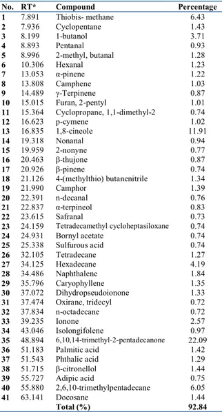
The phytochemical analysis of EERR represented in Table.1. The HPLC-MS-MS analysis resulted in the
persence of some important natural products such as flavonoids and phenolics. A kind of flavonoid or phenolic products mostly displayed clear intensity as [M +H]+ ions in the positive mode.
The experimental results are demonstrated the presence of chrysin, narinegenin and quarcetin as an example of flavonoids and phenolics. These important natural compounds were determined in extract of EERR by HPLC-MS-MS analysis. Their percentage in sample of EERR were calculated and indicated in Table 1.
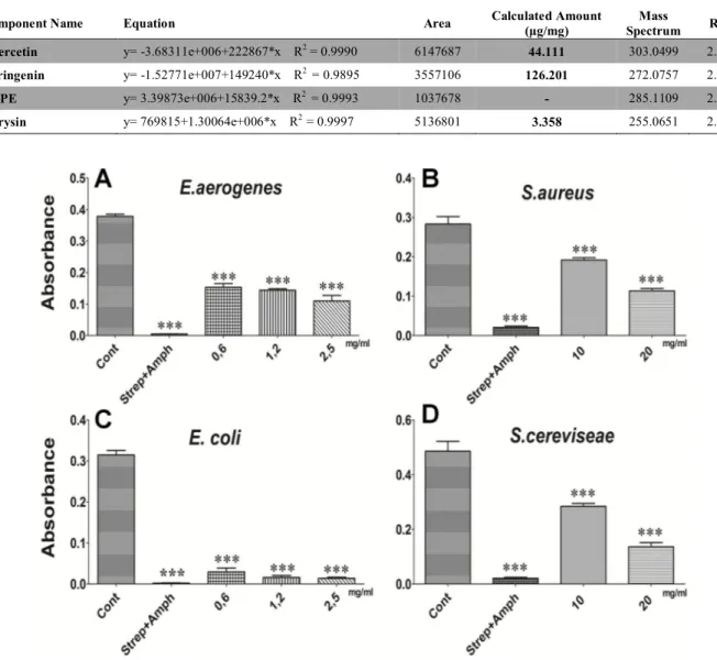
3.5 Effects of EERR on bacteria and yeast
To evaluate antimicrobial activity of EERR, the various doses of it were analyzed against different bacteria and yeast. Fig. 3 shows that antimicrobial capability of minimum inhibition concentration (MIC) doses on microorganisms. The results in Fig. 3A, 3B and 3C indicate that various EERR doses have good antibacterial activity against E.
aerogenes, S. aureus and E. coli. Moreover, it should be
active against S.cereviseae in higher dose (Fig. 3D).
Table 1. Chemical compounds detected in EERR
Component Name Equation Area Calculated Amount (µg/mg) Mass Spectrum RT Quercetin y= -3.68311e+006+222867*x R2 = 0.9990 6147687 44.111 303.0499 2.20 Naringenin y= -1.52771e+007+149240*x R2 = 0.9895 3557106 126.201 272.0757 2.08 CAPE y= 3.39873e+006+15839.2*x R2 = 0.9993 1037678 - 285.1109 2.08 Chrysin y= 769815+1.30064e+006*x R2 = 0.9997 5136801 3.358 255.0651 2.36
Fig. 3.The antimicrobial activity of EERR on growth rate of several bacteria and yeast cells. Cells were treated with various doses of EERR and same amounts of antibiotic mix as a positive control. (A) The antibacterial activity of EERR on gram-negative bacteria, E. aerogenes, (B) gram-positive bacteria, S. aureus and (C) gram-gram-negative bacteria, E. coli for various doses for 12 h. (D) The antifungal activity of EERR on S. cereviseae for various doses for 12 h. Data are presented as means ±SEM (n=3). ***p<0.001 and *p<0.05 Cont, antibiotic mix and various doses of EERR
M. Tartik et al. The various biological activities of Rheum ribes extract on different types of cell
Tr. Doğa ve Fen Derg. − Tr. J. Nature Sci. 2015 Vol. 4 No. 2ö zgü 06
4. Discussion
The entire biological activity of R. ribes and underlying mechanisms by which it might be effective are yet unclarified, although a number of biological properties of it have been revealed by testing on different model organisms. In the current study, we indicated that treatment of PC3 cells with the EERR decreased cell proliferation. We also found that the pro-oxidant activity of EERR in PC3 cells, which upregulated the ROS generations and LPO. It is exactly known that ROS and LPO are highly reactive to decide cell destiny [30,31]. On the other hand, the antioxidant activity of R. ribes has been previously reported [24,25]. We revealed that treatment of H2O2 induced HUVECs with the
various doses of EERR provided a decrease in ROS and MDA levels. Therefore, it might be suggested that EERR lead to pro-oxidant activity in cancer cell, in addition to its antioxidant ability in epithelial cells
There are limited number of studies about antitumor activity of R. ribes to reveal cytotoxicity and apoptotic effects of its root extract [23]. Furthermore, a report shown that emodin component from Rheum species induced apoptosis in human lung squamous carcinoma by activating mitochondrial pathway [32]. We infer that EERR may decrease cell viability due to increasing intracellular ROS and LPO levels.
Herein, we partially assessed mechanisms by which EERR can cause death of PC3 cells. Therefore, our results further displayed that DNA were fragmented by the various doses of EERR in PC3 cells. The DNA fragmentation is accepted a highlight of apoptosis [33]. We suggest that EERR might induce DNA fragmentation as a result of apoptosis.
As known, the quantitative and qualitative
phtyochemical screening of chemical constituents in natural plant extracts provide essential information for discovery of novel drugs. At the present study we analyzed the ingredient of EERR to determine some important phenolic and flavonoids including chrysin, naringenin, quarcetin and CAPE. All of these phytochemical chemicals have both immune modulator and anticancer ability. It is definitely elicited that the antioxidant activity and c.ytotoxicity capability have a linear correlation with phenolic and flavonoid concentrations [34,35]. It can be said that they may contribute and determine effectiveness of the biological activities of EERR.
Naringenin is a type of flavonoid that behaves as antioxidant, immune modulator, free radical scavenger and anticancer agent to regulate cell health [36-38]. Chrysin, is also a well-known natural flavone, has been clarified to be an antioxidant and anticancer agent in numerous pharmacological actions. It is commonly known as a wide distributed major dietary flavonol and revealed as a potential antioxidant, anti-proliferative and pro-apoptotic agent [39]. These findings together with our results strongly suggest that phenolic and flavonoid context of R. ribes might lead its bioactivities.
In the present study, we have also examined the antimicrobial activity of EERR in various bacteria cultures;
E. aerogenes, S. aureus and E. Coli. We have suggested that
EERR has antimicrobial activity, similar to studies investigated antibacterial activity of R. ribes [17,40]. Moreover, we also demonstrated that EERR inhibited
S.cereviseae growth at high doses as antifungal agent, unlike
a study argued that it is ineffective against yeast [18]. A plenty of researches clearly displayed that the different part of R. ribes plant are active against several pathogens [20,40]. However, it is not clear yet to explain the dose-dependent effectiveness of R. ribes against different type of microorganisms. Our findings were fairly shown that
the low doses of EERR strongly decreased growth rate of E.
aerogenes and E. Coli. Otherwise, the high doses of it were
just active against S. Aureus and S.cereviseae.
In conclusion, various biological properties of EERR have been revealed in present study. We observed that ERRR increases ROS and LPO levels which might inhibit the cell viability of PC3 as a pro-oxidant. Furthermore, the DNA of PC3 cells were fragmented with treatment of EERR. Additionally, we demonstrated that it has protective effects against H2O2 induced HUVECs by decreasing
overproduction of ROS and LPO. The chemical constituents of EERR were elicited to determine amount of active compounds related to its biological activities such as chrysin, naringenin and quarcetin. In addition, the antimicrobial activity of EERR was displayed against several microorganisms. Therefore, the present results suggest that EERR has potential to be a basis for anticancer, antimicrobial and antioxidant agents.
Conflict of interest
The authors declare that there are no conflicts of interest.
References
[1] Xu R., Wang Q. Large-scale automatic extraction of side effects associated with targeted anticancer drugs from full-text oncological articles. J. Biomed. Inform., 55, 64–72, 2015.
[2] Terada T., Noda S., Inui K. Management of dose variability and side effects for individualized cancer pharmacotherapy with tyrosine kinase inhibitors. Pharmacol. Ther., 152, 125–134, 2015.
[3] Tokajuk A., Krzyżanowska-Grycel E., Tokajuk A., Grycel S., Sadowska A., Car H. Antidiabetic drugs and risk of cancer. Pharmacol. Reports, 67(6), 1240– 1250, 2015.
[4] Laxminarayan R., Duse A., Wattal C., Zaidi A. K. M., Wertheim H. F. L., Sumpradit N., Vlieghe E., Hara G. L., Gould I. M., Goossens H., Greko C., So A. D., Bigdeli M., Tomson G., Woodhouse W., Ombaka E., Peralta A. Q., Qamar F. N., Mir F., Kariuki S., Bhutta Z., Coates A., Bergstrom R., Wright G. D., Brown E. D., Cars O. Antibiotic resistance—the need for global solutions. Lancet Infect. Dis., 13(12), 1057–1098, 2013.
[5] Rodríguez-Rojas A., Rodríguez-Beltrán J., Couce A., Blázquez J. Antibiotics and antibiotic resistance: A bitter fight against evolution. Int. J. Med. Microbiol., 303 (6), 293–297, 2013.
[6] Paracelsus T. Die dritte Defension wegen des Schreibens der neuen Rezepte. Septem Defensiones. 1538.
[7] Vermani K,. Garg S. Herbal medicines for sexually transmitted diseases and AIDS. J. Ethnopharmacol., 80(1), 49–66, 2002.
[8] Tahir L., Khan N. Antibacterial potential of crude leaf, fruit and flower extracts of tagetes minuta l. J. Public Heal. Biol. Sci., 1(3), 74–78, 2012.
[9] Mohanta B., Chakraborty A., Sudarshan M., Dutta R. K., Baruah M. Elemental profile in some common medicinal plants of India. Its correlation with traditional therapeutic usage. J. Radioanal. Nucl. Chem., 258(1), 175–179, 2003.
M. Tartik et al. The various biological activities of Rheum ribes extract on different types of cell
[10] Kashiwada T., Nonaka Y., Nishioka G., Yamagishi I. Galloyl and hydroxycinnamoyl glucoses from Rhubarb. Phytochemistry, 27, 1473–1477, 1988. [11] Krishnaiah D., Sarbatly R., Nithyanandam R. A
review of the antioxidant potential of medicinal plant species. Food Bioprod. Process., 89(3), 217–233, 2011.
[12] Cullen J., Flora of Turkey and The East Aegean Islands. Edinbg. Univ. Press, 2, 268–269, 1966. [13] Türkmen Ö., Çirka M., Şensoy S. Initial evaluation of
a new edible wild rhubarb species (Rheum ribes L.) with a modified weighted scaling ındex method. Pakista. J. Bio. Sci., 8(5), 763–765, 2005.
[14] Cakilcioglu U., Turkoglu I. An ethnobotanical survey of medicinal plants in Sivrice (Elazığ-Turkey). J. Ethnopharmacol., 132(1), 165–175, 2010.
[15] Sayyah M., Boostani H., Pakseresht S., Malayeri A. Efficacy of hydroalcoholic extract of Rheum ribes L. in treatment of major depressive disorder. J. Med. Plants Res., 3(8), 573–575, 2009.
[16] Abu-Irmaileh B. E., Afifi F. U. Herbal medicine in Jordan with special emphasis on commonly used herbs. J. Ethnopharmacol., 89 (2), 193–197, 2003. [17] Sedigheh B., Bazzaz F., Khajehkaramadin M. In vitro
antibacterial activity of rheum ribes extract obtained from various plant parts against clinical ısolates of gram-negative pathogens.IJPR., 2, 87–91, 2005. [18] Alan Y., Erbil N., Digrak M. In vivo antimicrobial
activity of Rheum ribes ekstracts obtained from various plant parts from Turkey. 1(4), 23–29, 2012. [19] Hudson J. B., Lee M. K., Sener B., Erdemoglu N.
Antiviral activities in extracts of turkish medicinal plants. Pharm. Biol., 38(3), 171–175, 2000.
[20] Naemi F., Asghari G., Yousofi H., Yousefi H. A., Chemical composition of essential oil and anti trichomonas activity of leaf , stem , and flower of Rheum ribes L . extracts. AJP., 4(3), 191–199, 2014. [21] Hamzeh S., Farokhi F., Heydari R., Manaffar R.
Renoprotective effect of hydroalcoholic extract of Rheum ribes root in diabetic female rats. AJP., 4(6), 392–401, 2014.
[22] Adham A. N., Naqishbandi A. M. HPLC analysis and antidiabetic effect of Rheum ribes root in type 2 diabetic patients. Zanco J. Med. Sci., 19(2), 2015. [23] Uyar P., Evaluation of in vitro antioxidative, cytotoxic
and apoptotic activities of Rheum ribes ethyl acetate extracts. J. Plant Sci. (Science Publ. Group), 2(6), 339, 2014.
[24] Yildirim I., Kutlu T., Takim K. Comparison of antioxidant activity of Rheum ribes fruits and seed methanolic extracts against protein oxidation and lipid peroxidation. Pakistan J. Biol. Sci., 18, (5), 232–239, 2015.
[25] Öztürk M., Aydoğmuş-Öztürk F., Duru M. E., Topçu G. Antioxidant activity of stem and root extracts of Rhubarb (Rheum ribes): An edible medicinal plant. Food Chem., 103(2), 623–630, 2007.
[26] Hou X., Tong Q., Wang W., Xiong W., Shi C., Fang J. Dihydromyricetin protects endothelial cells from hydrogen peroxide-induced oxidative stress damage
by regulating mitochondrial pathways. Life Sci., 130, 38–46, 2015.
[27] Shen H., Shi C., Shen Y. Detection of elevated reactive oxygen species level in cultured rat hepatocytes treated with aflatoxin B1. Free Radic. Biol. Med., 21(2), 139–146, 1996.
[28] Smith M. T., Thor H., Hartzell P., Orrenius S. The measurement of lipid peroxidation in isolated hepatocytes. Biochem. Pharmacol., 31, 19–26, 1982. [29] Medana C., Carbone F., Aigotti R., Appendino G.,
Baiocchi C. Selective analysis of phenolic compounds in propolis by HPLC-MS/MS. Phytochem. Anal., 19(1), 32–39, 2008.
[30] Sena L. A., Chandel N. S. Physiological Roles of Mitochondrial Reactive Oxygen Species. Mol. Cell, 48(2), 158–167, 2012.
[31] Panieri E., Gogvadze V., Norberg E., Venkatesh R., Orrenius S., Zhivotovsky B. Reactive oxygen species generated in different compartments induce cell death, survival, or senescence. Free Radic. Biol. Med., 57, 176–187, 2013.
[32] Lee H.Z., Effects and mechanisms of emodin on cell death in human lung squamous cell carcinoma. Br. J. Pharmacol., 134(1), 11–20, 2001.
[33] Tong W. M., Cortes U., Wang Z. Q. Poly(ADP-ribose) polymerase: a guardian angel protecting the genome and suppressing tumorigenesis. Biochim. Biophys. Acta, 1552(1), 27–37, 2001.
[34] Hassan L. E. A., Ahamed M. B., Majid A. S., Baharetha H. M., Muslim N. S., Nassar Z. D., Majid A. M. Correlation of antiangiogenic, antioxidant and cytotoxic activities of some Sudanese medicinal plants with phenolic and flavonoid contents. BMC Complement. Altern. Med., 14(1), 406, 2014.
[35] Gregoris E., Stevanato R. Correlations between polyphenolic composition and antioxidant activity of Venetian propolis. Food Chem. Toxicol., 48(1), 76– 82, 2010.
[36] Hatkevich T., Ramos J., Santos-Sanchez I., Patel Y. M. A naringenin-tamoxifen combination impairs cell proliferation and survival of MCF-7 breast cancer cells. Exp. Cell Res., 27, 1–9, 2014.
[37] Fouad A., Albuali W. H., Zahran A., Gomaa W. Protective effect of naringenin against gentamicin-induced nephrotoxicity in rats. Environ. Toxicol. Pharmacol., 38(2), 420–9, 2014.
[38] Kumar S. P., Birundha K., Kaveri K., Devi K. T. R. Antioxidant studies of chitosan nanoparticles containing naringenin and their cytotoxicity effects in lung cancer cells. Int. J. Biol. Macromol., 78, 1–9, 2015.
[39] Gardi C., Bauerova K., Stringa B., Kuncirova V., Slovak L., Ponist S. Quercetin reduced in flammation and increased antioxidant defense in rat adjuvant arthritis. 583, 150–157, 2015.
[40] Kazemi R., Parsa M. Antimicrobial potential of root , stalk and leaves extracts of Rheum ribes. 3(1), 10–13, 2014.
Tr. Doğa ve Fen Derg. − Tr. J. Nature Sci. 2014 Vol. 3 No. 1özgü 08
Tr. Doğa ve Fen Derg. − Tr. J. Nature Sci. 2015 Vol. 4 No. 2
Ekonomik bir yöntemle hazırlanan tek faz -Fe
2
O
3
ve -Fe
2
O
3
nanopartiküllerinin
karakterizasyonu
ve
elektrokimyasal
biyosensör uygulaması
Güllüzar Demir
1, İbrahim Y. Erdoğan*
1Özet
-Fe2O3 ve -Fe2O3 nanopartikülleri etkin bir kimyasal proses temeline dayanan pratik ve ekonomik bir yol kullanılarak
hazırlandı. Nanopartiküller, X-ışını kırınımı (XRD), atomik kuvvet mikroskopi (AFM), enerji dağılımlı X-ışını spektroskopi (EDX), fotolüminesans (PL) spektroskopi, Fourier dönüşümlü infrared (FT-IR) spektroskopi ve dönüşümlü voltametri (CV) teknikleri kullanılarak karakterize edildi. -Fe2O3’ten -Fe2O3’e dönüşüm esnasında nanopartiküllerin faz ve kristal boyut
değişimleri FTIR, XRD, AFM ve PL teknikleri kullanılarak incelendi. -Fe2O3 ve -Fe2O3 faz için kristal boyutları sırasıyla
9’dan 12 nm’ye değişir. Alfa ve gama faz Fe2O3 partiküllerinin XRD desenleri sırasıyla yüksek tercihli (1 0 4) ve (3 1 1)
yönelimi gösterdi. Örneklerin EDX analizleri Fe ve O’nin yaklaşık 2:3 atomik oranla yapıda bulunduklarını doğrular. -Fe2O3 ve
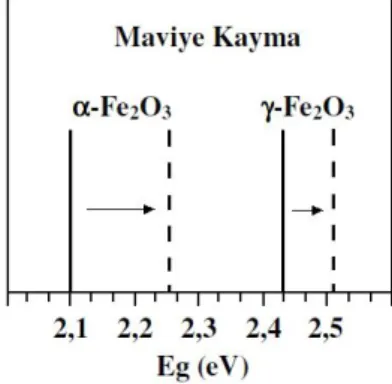
-Fe2O3 nanopartiküllerinin kuantum sınırlama etkisi PL ölçümleri sonucu doğrulandı. Hacmine göre yüzey alanının yüksek
olması sonucu -Fe2O3 ve -Fe2O3 nanopartikülleri, ileri optiksel ve elektriksel nanocihazlarda bulunan sensörlerdeki
uygulamalar için ideal adaylardır. Sonuçlar, -Fe2O3 ve -Fe2O3 nanopartiküllerinden yapılan sensörlerin oda sıcaklığında
fizyolojik çözeltilerdeki hidrojen peroksitin biyosensitivitesi için yüksek duyarlılık sergilediğini gösterir. Anahtar Kelimeler: Fe2O3; nanopartikül; elektrokimya; kuantum sınırlama etkisi; biyosensör uygulaması
The characterization and electrochemical biosensor application of single
phase -Fe
2O
3and -Fe
2O
3nanoparticles prepared by an economical method
Abstract
Nanoparticles of the -Fe2O3 and -Fe2O3 were prepared using a practical and economical route based on an efficient
chemical process. Nanoparticles were characterized by X-ray diffraction (XRD), atomic force microscopy (AFM), energy dispersive X-ray spectroscopy (EDX), photoluminescence (PL) spectroscopy, Fourier transform infrared (FT-IR) spectroscopy, and cyclic voltammetry (CV). Phase and crystallite size variations of nanoparticles during -Fe2O3 to -Fe2O3 transformation
were examined using FTIR, XRD, AFM, and PL techniques. The crystal sizes vary for the -Fe2O3 and -Fe2O3 phase from 9 nm
to 12 nm, respectively. XRD patterns of alpha and gamma phase Fe2O3 particles obtained highly preferential (104) and (311)
orientation, respectively. EDX analyses of samples confirm that Fe and O are present in approximately 2:3 atomic ratio. The quantum-confined effect of the -Fe2O3 and -Fe2O3 nanoparticles are confirmed by the PL measurements. The -Fe2O3 and
-Fe2O3 nanoparticles with the high surface-to-volume ratio are ideal candidates for applications in the sensors, as well as in
advanced optical and electric nanodevices. The results demonstrate that the sensors made of the -Fe2O3 and -Fe2O3
nanoparticles exhibit high sensitivity for biosensing of hydrogen peroxide in a physiological solution at room temperature. Keywords: Fe2O3; nanoparticle; electrochemistry; quantum confined effect; biosensor application
1. Giriş
Fe2O3, birçok endüstriyel uygulamada kullanılan çok
önemli bir malzemedir. Fe2O3 farklı dört farklı kristal
yapısına sahiptir. Bu kristal yapılardan uygulama alanı bulanlar -Fe2O3 ve -Fe2O3 kristal yapılarıdır. Diğer kristal
yapıları (-Fe2O3 ve -Fe2O3) kararsız olduklarından
aaaaaaaaaaaaaaaaaaaaaaaaaaaaaaaaaaaaaaaaaaaaaaaaaaaaaa aaaaaaaaaaaaaaaaaaaaaaaaaaaaaaaaaaaaaaaaaaaaaaaaaaaaaa aaaaaaaaaaaaaaaaaaaaaaaaaaaaaaaaaaaaaaaaaaaaaaaaaaaaaa aaaaaaaaaaaaaaaaaaaaaaaaaaaaaaaaaaaaaaaaaaaaaa
uygulama alanı bulamazlar. -Fe2O3, hematit olarak bilinen
rombohedral faz demir oksit olup, bir n-tipi yarı iletkendir (Bant aralığı enerjisi, Eg 2,1 eV) ve çevre koşullarında termodinamik olarak en kararlı demir oksit fazıdır. Hematit, gaz sensörü, manyetik kayıt ortamları, lityum-iyon pilleri, korozyon önleyici, sensör, su arıtma ve pigmentleri kapsayan birçok alanda kapsamlı uygulamalara sahiptir [1-4]. -Fe2O3, maghemit olarak bilinen kübik spinel faz demir
oksit olup, bir n-tipi yarı iletkendir (Eg 2,43 eV) ve çevre 1 Bingöl Üniversitesi, Fen Edebiyat Fakültesi, Kimya Bölümü, 12000 Bingöl,
Türkiye
*Sorumlu yazar E-posta: [email protected]
Demir ve Erdoğan, Ekonomik bir yöntemle hazırlanan tek faz -Fe2O3 ve -Fe2O3 nanopartiküllerinin karakterizasyonu ve …
koşullarında termodinamik olarak yarı kararlı (-Fe2O3’ten
sonra en kararlı) demir oksit fazıdır. Maghemit magnetik kayıt ve bilgi depolama sistemleri, katalizör, gaz sensörleri, biyosensörler ve magnetoptik cihazlarda uygulamalara sahiptir [5-9].
Son yıllarda, -Fe2O3 ve -Fe2O3 gibi metal oksit
nanoyapılar, hem teorik yönleri hem de onların nanocihazlardaki potansiyel uygulamaları sebebiyle kayda değer bir ilgi uyandırmıştır [10-12]. Birçok malzemenin, özellikle yarıiletken malzemelerin optik, yapısal, magnetik, elektronik ve katalitik özellikleri, nanometre ölçeğine inildiğinde boyutlara bağımlı hale gelir [13-15]. Nanoteknoloji, malzemelerin yapısal, optik, elektronik, katalitik, fiziksel ve kimyasal özelliklerini malzemenin boyutlarını nano seviyelerde ayarlayarak kontrol edebilme işidir. Yani, nanoteknolojide belirli bir malzeme, boyutları nano skalada kısıtlanarak, değiştirilerek ve ayarlanarak birçok farklı maddenin özelliklerine sahip olabilir. Böylece tek bir madde, çok farklı maddelerin özelliklerini gösterebilir hale getirilmektedir. Geleneksel bulk materyallerle karşılaştırıldığında geniş yüzey alanlı ve kuantum sınırlama etkisine sahip düşük boyutlu nano skala materyaller, daha farklı ve gelişmiş elektronik, optik, magnetik ve katalitik özellikler sergiler. Bu özellikler birçok alanda çok sayıda gelişmelere, yeniliklere sebep olmaktadır [16-19].
Farklı organik ve inorganik bileşiklerin nano boyutlarda
hazırlaması için yapılan çalışmalar neticesinde,
nanomalzemelerin bulk hallerine göre değiştirilebilen ve ayarlanabilen ilgi çekici çoklu özellikler kazanabildikleri görülmüştür [20-22]. Bunun sonucu olarak malzemelerin nano boyutlarda hazırlanması üzerine araştırmalar her geçen gün daha da artmaktadır. Magnetik nanopartiküller klinik teşhisler için biyomedikal uygulamalarda ve tümörlerin magnetik rezonans görüntülenmesi, magnetik destekli ilaç tedavisi, kanser tedavileri gibi canlı dokulardaki uygulamalarda yeni bir teknoloji olarak ortaya çıkmaktadır. Özellikle küresel olarak biçimlendirilmiş demir oksitlerin kullanımı onların üstün kimyasal ve termal kararlılığı, sertliği, toksik ve radyoaktif olmayışından dolayı biyomedikal alanda muazzam bir artış göstermektedir. [23-26].
Bu çalışmada, -Fe2O3 ve -Fe2O3 yarıiletken
nanopartikülleri, geliştirdiğimiz basit, hızlı ve ekonomik bir kimyasal yöntem kullanılarak hazırlandı. Bu çalışma ile hem asidik ortamda demir iyonlarının direk oksidasyonu sağlanmış hem de bazik ortamda elde edilen demir oksitlere göre çok daha saf ve homojen demir oksit nanopartikülleri hazırlanmıştır. Bu ekonomik yöntem ile hazırlanan Fe2O3
nanopartikülleri literatürde benzer yöntemlerle elde edilenlere göre daha küçük boyutlarla üretilebilir ve hidrojen
peroksitin hassas olarak saptanarak analizini
gerçekleştirmek için uygun bir potansiyele sahip olabilir. 2. Materyal ve Metot
Çalışmalarımızın tamamında çözücü olarak 18 Mohm cm’den daha yüksek kalitede deiyonize su ve saf etanol kullanıldı. Çözeltiler, Merck firmasının yüksek saflıktaki (%99,99) kimyasallarının deiyonize saf suda ve saf etanolde çözülmesi suretiyle hazırlandı. Hazırlanan çözeltilerin pH’ları ortama ilave edilen asit, baz ya da tampon çözelti ile istenilen değere ayarlandı ve bu değerler pH metre yardımıyla okundu. Çeşitli asitlerden elektrot oluşturulması işleminde yararlanıldı. Fe(NO3)3.9H2O ve Na2EDTA
(C10H14N2Na2O8) kullanılan kimyasallardır. Elektrokimyasal
çalışmalarımızda; elektrokimyasal analizör olarak CHI tipi işlemsel yükselteçli potansiyostat, çalışma elektrotu olarak altın disk elektrot, demir oksit modifiye altın disk elektrot,
referans elektrot olarak Ag/AgCl elektrot, karşıt elektrot olarak da platin tel elektrot kullanıldı. Deiyonize su kullanılarak hazırlanan Fe2O3 nanopartiküllerinden oluşan
süspansiyon (100 mg/mL) 30 dakika boyunca ultrasonik karıştırıcıda bekletildikten sonra altın elektrot yüzeyine düzgün ve homojen bir şekilde kaplandı ve kullanılmadan önce kurutuldu. Çalışmalarımızda malzemelerin PL spektrumları, Shimadzu RF-5301 PC Spektroflorofotometre ile IR bölge spektrumları ise Pelkin Elmer Spectrum 100 FT-IR spektrometre ile incelendi. Optiksel özelliklerin incelenmesinde cam ve kuvartz küvetler, IR özelliklerinin incelenmesinde KBr diskler kullanıldı. Morfoloji ve boyutun
belirlenmesinde Park Systems atomik kuvvet
mikroskobundan yararlanıldı. Malzemelerin kristal yapısı ve boyut analizleri XRD yardımıyla belirlendi. Kristalizasyon çalışmalarımızda, λ = 1,5405 Å (0,154 nm) dalga boyuna sahip Cu-Kα kaynaklı Rigaku–2200 D/Max XRD cihazından yararlanıldı.
3. Tartışma ve Bulgular
Hem -Fe2O3 hem de -Fe2O3 yarıiletken
nanopartikülleri, yeni geliştirdiğimiz basit, hızlı ve ekonomik bir kimyasal yöntem kullanılarak hazırlandı. Bu yöntem demir oksit partiküllerinin sanayide üretilmesinde kullanılan yöntemlere göre oldukça basit ve ekonomik olmakla birlikte karmaşık cihaz ve sistemlere de ihtiyaç duymaz. Bu sayede demir oksit nanopartikülleri basit bir laboratuvar ortamında kolaylıkla hazırlanabilir. Literatürde Fe2O3 partiküllerinin kimyasal yöntemlerle hazırlanmasında
genellikle birçok basamak üzerinden işlem
gerçekleştirilmektedir. Öncelikle demir iyonu sağlayacak bir demir tuzu, NaOH, NH3 gibi bir baz kullanılmak suretiyle
Fe(OH)3 halinde çöktürüldükten sonra çeşitli işlemlerle
Fe3O4 üzerinden Fe2O3 partiküllerinin sentezi
gerçekleştirilmektedir (Eşitlik 1). Fe3+ + OH- Fe(OH)
3 Fe3O4 Fe2O3 (1)
Bu çalışmalar sonucu elde edilen Fe2O3 partikülleri hem
-Fe2O3 hem de -Fe2O3 fazlarını içermekle birlikte
bünyesinde çeşitli demir hidroksit türlerini de
barındırmaktadır. Bu duruma bazik ortamda çalışmanın sebep olduğu bilinmektedir. Biz çalışmalarımızda asidik ortamda çalışarak bu dezavantajı ortadan kaldırdık. Bazik ortamda demir iyonlarını oksitlemek için baz ilavesi ve farklı süreçler gereklidir. Geliştirdiğimiz bu yeni teknik sayesinde hem asidik ortamda demir iyonlarının direk oksidasyonu sağlanmış hem de bazik ortamda elde edilen demir oksitlere göre çok daha saf demir oksit nanopartikülleri hazırlanmıştır. Burada amaç, tek faza sahip, homojen -Fe2O3 ve -Fe2O3 nanopartiküllerini hazırlamak
ve istenildiği zaman bir fazdan ötekine geçişi sağlayacak basit bir yol geliştirmektir. Ayrıca elde edilen nanoyapıların kristal yapıda olması önemlidir. Çalışmalarımızda kristal yapıların bazik ortama göre asidik ortamda çok daha yüksek verimle hazırlanabildiğini gördük. Bazik ortamda hazırlanan malzemeler, kristal yapıdan çok amorf yapıya geçme eğilimi gösterdi. Bunun sonucu olarak çalışmalarımızı büyük çoğunlukla asidik ortamlarda gerçekleştirdik. Yaptığımız çalışmalar sonucunda elde ettiğimiz -Fe2O3 ve -Fe2O3
nanopartikülleri literatürde elde edilenlere göre daha küçük boyutlara sahiptir. Geleneksel bulk demir oksitlerle karşılaştırıldığında geniş yüzey alanlı ve kuantum sınırlama etkisine sahip düşük boyutlu demir oksit nanopartikülleri, daha farklı ve gelişmiş elektronik, optik, magnetik ve katalitik özellikler sergiler. Bu çalışmada, -Fe2O3 ve
-Fe2O3 nanopartikülleri aşağıda ayrıntısı verilen çok basit ve
Demir ve Erdoğan, Ekonomik bir yöntemle hazırlanan tek faz -Fe2O3 ve -Fe2O3 nanopartiküllerinin karakterizasyonu ve …
Tr. Doğa ve Fen Derg. − Tr. J. Nature Sci. 2015 Vol. 4 No. 2özgün 110
olarak Fe(NO3)3.9H2O tuzu kullanıldı. Derişimi 0,1 M
olacak şekilde tartılan Fe(NO3)3.9H2O, 25 mL deiyonize
suda çözüldü (pH5). Bu çözelti üzerine ayrı bir kapta hazırlanan 25 mL 0,1 M EDTA çözeltisi ilave edildi. Daha sonra bu karışım üzerine saf etanol çözeltisinden 150 mL (suyun yaklaşık 3 katı) ilave edildi. Bu karışım bir magnetik karıştırıcılı ısıtıcıda 300 rpm’de karıştırmak suretiyle 30 dakika 80 C’de ısıtıldı (Eşitlik 2). Daha sonra çözelti soğutulmadan dekantasyon yöntemiyle ısıya dayanıklı porselen bir kap içine alındı ve dipte kalan çökelti atıldı. Böylece sadece çözeltiye dağılmış daha küçük boyutlara sahip partiküller alınırken dibe çökmüş daha büyük partiküller ayrılmış oldu. Porselen kaba alınan çözelti karışımı hemen bir fırına yerleştirilerek 250 C’de 2 saat tutuldu. Fırından çıkarılan kap oda sıcaklığında soğumaya bırakıldı. Daha sonra 5000 rpm’de 5 dakika santrifüj işlemine tabi tutularak partiküller ayrıldı. Sonra santrifüj kullanılarak çökelti 1 defa destile su ile 3 defa da saf etanol ile yıkandı. Son olarak laboratuvar ortamında, oda sıcaklığında kendi haline bırakarak kurutuldu. Bu şekilde hazırlanan (Eşitlik 3) toz halindeki çökelti kahverengidir ve saf -Fe2O3 nanopartiküllerinden oluşur.
Fe(NO3)3.9H2O Fe(NO3)3 (2)
4Fe(NO3)3 2-Fe2O3 + 12NO2(g) + 3O2(g) (3)
Yukarıda anlatılan yöntemle hazırlanan -Fe2O3
nanopartikülleri sadece ısıl bir işlem kullanılarak -Fe2O3
nanopartiküllerine dönüştürüldü. Bunun için fırına alınan -Fe2O3 nanopartikülleri, fırının sıcaklığı dakikada 5 C
arttırılmak suretiyle (5 C/dak) 700 C’ye çıkarıldı ve 1 saat bu sıcaklıkta tutuldu. Daha sonra fırından çıkarılan partiküller kendi haline bırakılarak oda sıcaklığına gelinceye kadar soğutuldu. Böylece hazırlanan toz kırmızı renge sahiptir ve saf -Fe2O3 nanopartiküllerinden oluşur (Eşitlik
4).
-Fe2O3 (kahverengi) -Fe2O3 (kırmızı) (4)
Yarıiletken Fe2O3, oldukça yön bağımlı bir yapıya
sahiptir ve büyüme yönü istenildiği şekilde
sınırlandırılabilir. Bunun sonucu olarak kolaylıkla Fe2O3
partiküllerinin boyutu küçültülebilir ve Fe2O3
nanopartikülleri hazırlanabilir. Burada, kullanılan EDTA
(etilendiamintetraasetikasit)’nın etkisiyle kalıp
kullanılmadan yarıiletken nanopartiküllerin üretimi söz konusudur. EDTA’nın açık formülü Şekil 1’de verilmiştir.
Şekil 1. EDTA ligantının açık formülü
Burada EDTA hem yüzey aktif madde hem dağıtıcı (disperse edici) hem de bir yardımcı yükseltgeyici olarak görev alır. Böylece partiküllerin bir araya gelmesi (agglomerasyonu) önlenerek oldukça yüksek yüzey alanına sahip Fe2O3 nanopartikülleri meydana gelir. Kahverengi
-Fe2O3 nanopartiküllerinden kırmızı -Fe2O3
nanopartiküllerinin ısıl işlemle eldesi esnasında sıcaklık dereceli olarak 700 C’ye çıkarılmalıdır. Yaptığımız çalışmalarda en uygun ısıl prosedürün fırının sıcaklığını dakikada 5 C arttırmak olduğu bulunmuştur. Isıtma işlemi aşama aşama yapıldığı takdirde faz geçişleri için kristal düzlemlerinin dönüşümlerinin tamamlanması mümkün olmaktadır. Böylece saf faz demir oksit eldesi mümkün
olmaktadır. Fırının sıcaklığının direk olarak 700 C’ye çıkarılması durumunda elde edilen yapının -Fe2O3 ve
-Fe2O3’in bir karışımı olduğu görülmüştür. Demirin
oksidasyonu bazik ortamda baz ilavesiyle
gerçekleştirilmektedir.
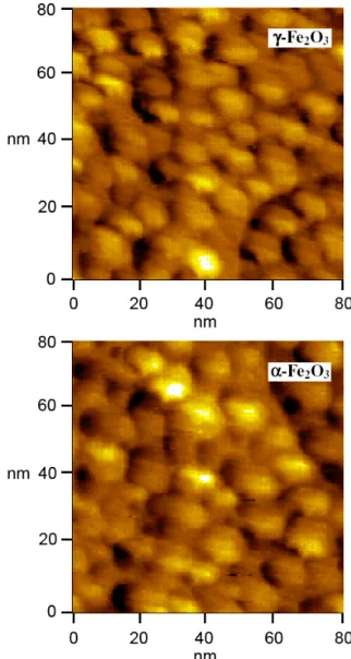
Yarıiletken Fe2O3 nanopartiküllerinin morfolojileri AFM
tekniğinden yararlanılarak belirlendi. Şekil 2, pH’sı yaklaşık
5 olan asidik ortamda hazırlanan demir oksit
nanopartiküllerinin boyutlarını ve şekillerini gösteren AFM görüntüleridir.
Şekil 2. -Fe2O3 ve -Fe2O3 nanopartiküllerinin AFM
görüntüleri
Bu AFM görüntüleri hem -Fe2O3 hem de -Fe2O3
nanopartiküllerinin taneciklerinin küresel yapıda ve oldukça homojen (tek tip) boyutlara sahip olduğunu göstermektedir. Kimyasal olarak hazırlanan -Fe2O3 nanopartikülleri
ortalama 10 nm boyutlara sahip iken, -Fe2O3’ün aşamalı bir
prosedürle ısıl işleme tabi tutulması sonucunda hazırlanan -Fe2O3 nanopartikülleri ortalama 14 nm boyutlara sahiptir.
Bu durum, -Fe2O3’den -Fe2O3’e faz değişimi neticesinde
tanecik boyutunun arttığını göstermektedir. Böylece, faz değişimi ile nanopartiküllerin şekillerinde herhangi bir
değişiklik olmaz. Bununla birlikte faz değişimi
nanopartiküllerin boyutlarının değişmesine sebep olur. Bu sonuç literatürle uyumlu olup, -Fe2O3’ün -Fe2O3’den daha
küçük partikül boyutuyla elde edilebildiği tezini doğrulamaktadır.
Demir ve Erdoğan, Ekonomik bir yöntemle hazırlanan tek faz -Fe2O3 ve -Fe2O3 nanopartiküllerinin karakterizasyonu ve …
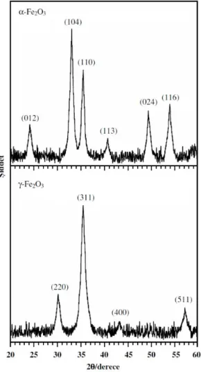
Demir oksit nanopartiküllerinin faz dönüşümleri ve kristal yapıları XRD tekniğinden yararlanılarak incelendi. Kafes sabitleri a 0,5036 ve c 1,3749 nm olan, hematit olarak bilinen rombohedral faz -Fe2O3 ve kafes sabitleri a
0,5036 nm olan, maghemit olarak bilinen kübik spinel faz -Fe2O3 nanopartiküllerinin XRD difraktogramları Şekil 3’te
verilmiştir.
Şekil 3. -Fe2O3 ve -Fe2O3 nanopartiküllerinin XRD
difraktogramları
Kimyasal olarak hazırlanan -Fe2O3 nanopartiküllerinin
XRD difraktogramlarında 2 ölçeğinde 24,1, 33,2, 35,6, 40,9, 49,5 ve 54,1’de difraksiyon pikleri gözlendi. Bu pikler sırasıyla, -Fe2O3’ün (0 1 2), (1 0 4), (1 1 0), (1 1 3),
(0 2 4) ve (1 1 6) yönelimlerine karşılık gelir. -Fe2O3
nanopartiküllerinin XRD difraktogramlarında 2 ölçeğinde 30,2, 35,6, 43,3 ve 57,3’de difraksiyon pikleri gözlendi. Bu difraksiyon pikleri sırasıyla, -Fe2O3’ün (2 2 0), (3 1 1),
(4 0 0) ve (5 1 1) yönelimlerine karşılık gelir. XRD sonuçlarından görüldüğü üzere her iki difraktogramda, birbirinden tamamen farklı piklere yani, birbirinden tamamen farklı difraksiyon desenlerine sahiptir. Hem
-Fe2O3 nanopartiküllerinin hem de -Fe2O3
nanopartiküllerinin difraksiyon desenleri saf faza sahiptir ve difraktogramlarda başka herhangi bir türe ait bir pik yoktur. Bunun sonucu olarak uyguladığımız bu yöntemle -Fe2O3
ve -Fe2O3 partikülleri tamamen saf ve homojen faz ile elde
edilebilir. XRD sonuçları, -Fe2O3 nanopartiküllerinin
düzlemler arası mesafesi, d = 0,270 nm olan (1 0 4) yönelimini, -Fe2O3 nanopartiküllerinin ise düzlemler arası
mesafesi, d = 0,252 nm olan (3 1 1) yönelimini tercih ettiğini gösterdi. Yani, -Fe2O3 nanopartiküllerinin kristal
düzlemleri arasındaki mesafe -Fe2O3 nanopartiküllerinden
büyüktür. Bu durum -Fe2O3 nanopartiküllerinin boyutunun
-Fe2O3 nanopartiküllerinin boyutundan daha büyük
olduğunu gösterir. Bu XRD sonuçları ile AFM sonuçları oldukça iyi bir uyum içindedir. Karşılaştırma amacıyla -Fe2O3 nanopartiküllerinin XRD sonuçları faz demir okside
ait 33-0664 numaralı JCPDS-ICDD kartı ile -Fe2O3
nanopartiküllerinin XRD sonuçları ise faz demir okside ait 39-1346 numaralı JCPDS-ICDD kartı ile karşılaştırılmış olup sonuçta elde edilen verilerin bu kartlarla mükemmel bir uyum içinde olduğu görülmüştür.
XRD sonuçlarından yararlanılarak, Scherrer eşitliği (Eşitlik 5) kullanılarak partikül boyutu (D) belirlenebilir.
cos K D (5) Burada D partikül boyutu (nm), K Scherrer sabiti (0,9),
kullanılan x-ışınının dalga boyu (0,154 nm), pik yarı yüksekliğinin tam genişliği (FWHM), pikin difraksiyon açısıdır. XRD verilerinde elde edilen piklerden ve Scherrer eşitliğinden yararlanılarak -Fe2O3 nanopartiküllerinin
boyutunun 9 nm, -Fe2O3 nanopartiküllerinin boyutunun ise
12 nm olduğu belirlenmiştir. Bu veriler AFM verileriyle iyi bir uyum içerisindedir. Hem XRD hem de AFM sonuçları faz dönüşümü gerçekleştiğinde partikül boyunun değiştiğini göstermektedir. Partikül boyutu, ’dan ’ya faz değişimi gerçekleştiğinde artmaktadır.
Demir oksit nanopartikülleri için -Fe2O3 ve -Fe2O3 faz
dönüşümleri ve yapı tayinleri XRD’nin yanı sıra FT-IR
yöntemiyle de incelendi. Şekil 4, demir oksit
nanopartiküllerine ait FT-IR spektrumlarıdır.
Şekil 4. -Fe2O3 ve -Fe2O3 nanopartiküllerinin FT-IR
spektrumları
Bu spektrumlar, -Fe2O3 ve -Fe2O3 faz oluşumlarını
doğrular. -Fe2O3 nanopartiküllerine ait IR spektrumu, 448
ve 572 cm-1’de iki adet pike sahiptir. Bu pikler, faz Fe2O3’deki karakteristik Fe-O bağ gerilme titreşimine aittir.
-Fe2O3 nanopartiküllerine ait IR spektrumu ise, 461, 580 ve
635 cm-1’de üç adet pike sahiptir. Bu pikler, faz Fe2O3’deki karakteristik Fe-O bağ gerilme titreşimine aittir.
-Fe2O3, -Fe2O3’e göre daha fazla titreşim bölgesine sahip
olup bu durum literatürle uyumludur. -Fe2O3 faza ait pikler,
-Fe2O3 faza ait piklere göre daha yüksek dalga sayısına ve
daha yüksek enerjiye sahip bölgededir. Bu durum, daha
küçük partikül boyutuna sahip olan -Fe2O3
nanopartiküllerinin titreşmesi için daha yüksek enerjili IR ışınına ihtiyaç duymasından ileri gelir. Bu sonuç, XRD verileriyle uyumludur.