CD8 Lineage-specific Regulation of Interleukin-7 Receptor
Expression by the Transcriptional Repressor Gfi1
*
□SReceived for publication, May 8, 2012, and in revised form, July 23, 2012 Published, JBC Papers in Press, August 3, 2012, DOI 10.1074/jbc.M112.378687
Davinna L. Ligons‡1, Ceren Tuncer§1, Brett A. Linowes‡, Izzet Mehmet Akcay§2, Sema Kurtulus§2,3, Emre Deniz§2, Belkis Atasever Arslan§4,5, Safak Isil Cevik§2, Hilary R. Keller‡, Megan A. Luckey‡, Lionel Feigenbaum¶, Tarik Möröy储, Tulin Ersahin**, Rengul Atalay**, Batu Erman§‡‡1,6, and Jung-Hyun Park‡1,7
From the‡Experimental Immunology Branch, National Cancer Institute, Bethesda, Maryland 20892, the§Biological Sciences and
Bioengineering Program, Faculty of Engineering and Natural Sciences, Sabanci University, Orta Mahalle, Üniversite Caddesi No. 27, Tuzla, Istanbul 34956, Turkey, the¶Science Applications International Corporation-Frederick, National Cancer Institute–Frederick
Cancer Research and Development Center, Frederick, Maryland 21702, the储Institut de Recherches cliniques de Montréal,
Département de microbiologie et immunologie, Université de Montréal, Montréal (Québec) H2W 1R7, Canada, the **Department of Molecular Biology and Genetics, Bilkent University, Bilkent, Ankara 06800 Turkey, and the‡‡Sabanci University, Nanotechnology Research and Application Center, SUNUM, Istanbul 34956, Turkey
Background:Expression of the IL-7R␣gene is up-/down-regulated during T/B-lymphocyte development.
Results:IL-7R␣gene transcription is repressed by the transcription factor Gfi1, specifically in CD8⫹T-lymphocytes.
Conclusion:Treatment by dexamethasone down-regulates Gfi1, which contributes to glucocorticoid receptor mediated up-regulation of IL-7R expression.
Significance:The mechanism by which the IL-7R gene gets turned on and off during development is a critical issue in biology.
Interleukin-7 receptor␣(IL-7R␣) is essential for T cell sur-vival and differentiation. Glucocorticoids are potent enhancers of IL-7R␣expression with diverse roles in T cell biology. Here we identify the transcriptional repressor, growth factor inde-pendent-1 (Gfi1), as a novel intermediary in glucocorticoid-in-duced IL-7R␣up-regulation. We found Gfi1 to be a major inhib-itory target of dexamethasone by microarray expression profiling of 3B4.15 T-hybridoma cells. Concordantly, retroviral transduction of Gfi1 significantly blunted IL-7R␣up-regulation by dexamethasone. To further assess the role of Gfi1 in vivo, we generated bacterial artificial chromosome (BAC) transgenic mice, in which a modified Il7r locus expresses GFP to report Il7r gene transcription. By introducing this BAC reporter transgene into either Gfi1-deficient or Gfi1-transgenic mice, we document
in vivo that IL-7R␣transcription is up-regulated in the absence
of Gfi1 and down-regulated when Gfi1 is overexpressed.
Strik-ingly, the in vivo regulatory role of Gfi1 was specific for CD8ⴙ, and not CD4ⴙT cells or immature thymocytes. These results identify Gfi1 as a specific transcriptional repressor of the Il7r gene in CD8 T lymphocytes in vivo.
A critical issue in biology is the mechanism by which genes get turned on and off during development and differentiation. Because IL-7 receptor (IL-7R) proteins provide critical survival signals to developing lymphocytes, the expression of the Il7r gene that encodes the IL-7R␣ receptor protein is tightly regu-lated at different stages of T and B lymphocyte development and precisely timed to stages when selection and programmed cell death occur in the immune system (1–3). The expression of IL-7R␣ follows an on-off-on pattern in the thymus at the CD4⫺CD8⫺double negative (DN),8CD4⫹CD8⫹double posi-tive (DP), and CD4⫹CD8⫺or CD4⫺CD8⫹single positive (SP) stages, respectively (4). Thus, developmental cues during thy-mocyte differentiation control IL-7R␣ expression. During CD8⫹memory cell generation in the peripheral immune sys-tem, Il7r gene expression again correlates with developmental outcome, in that long-lived memory cell precursors up-regulate IL-7R␣ expression and short-lived CD8⫹ cells lose IL-7R␣ expression (5). Notably, up-regulated IL-7R␣ expression is not sufficient to drive long-lived memory CD8⫹T cell generation, even though IL-7R␣ up-regulation clearly marks progenitors of this T cell subset (6, 7). Importantly, the differentiation signals that match IL-7R␣ expression to CD8 T cell fate remain unknown.
*This work was supported, in whole or in part, by a National Institutes of
Health (NIH) grant from the Intramural Research Program, NCI, Center for Cancer Research.
□S
This article containssupplemental Figs. S1–S5.
1These authors contributed equally to this work.
2Supported by the Scientific and Technological Research Council of Turkey
scholarships.
3Present address: Div. of Immunobiology, Cincinnati Children’s Hospital
Medical Center, University of Cincinnati College of Medicine, Cincinnati, OH 45229.
4Supported by the Turkish National Academy of Science post-doctoral
fellowship.
5Present address: Department of Electroneurophysiology, Uskudar
Univer-sity, Istanbul, Turkey.
6Supported by TUBITAK 1001 Grant 109T315, National Institutes of
Health-FIRCA Grant 1R03TW008208, and National Institutes of Health-GRIP Grant 1R01TW007270. To whom correspondence may be addressed: FENS, Sabanci University, Istanbul, Turkey. Tel.: 483-9530; Fax: 90-216-483-9550; E-mail: [email protected].
7To whom correspondence may be addressed: Exp. Immunol. Branch, NCI,
National Institutes of Health, Bldg. 10, Rm. 5B17, Bethesda, MD 20892. Tel.: 301-451-7641; Fax: 301-496-0887; E-mail: [email protected].
8The abbreviations used are: DN, double negative; DP, double positive; SP,
single positive; GR, glucocorticoid receptor; Dex, dexamethasone; BAC, bacterial artificial chromosome; IRES, internal ribosome entry site; ZF, zinc finger; LIP, lymphopenia-induced homeostatic proliferation; TCR, T cell receptor; GABP, GA-binding protein; Gfi1, growth factor independent-1.
at BILKENT UNIVERSITY on November 9, 2017
http://www.jbc.org/
In T cells, IL-7R␣ expression is thought to be primarily reg-ulated at the transcriptional level through an array of nuclear factors whose expression is also tightly controlled during devel-opment and activation. Several transcription factors that con-trol Il7r gene expression have been identified. The promoter of
Il7rcontains binding sites for the PU.1 transcription factor, which is necessary for the IL-7R␣ expression in developing B cells (8, 9). The same site is occupied in T cells by another ETS family transcription factor, GABP (10). Promoter occupancy by these factors likely prevents CpG methylation of promoter sequences and subsequent down-regulation of expression in mature T cells (11). Additionally, in human thymopoiesis, Notch may be complementing these ETS family proteins by acting through a conserved RBP-Jk/CSL binding site close by in the promoter of the Il7r gene (12). Therefore, down-regulation of Notch expression at the DP stage may be causative of the complete loss of Il7r gene transcription in murine DP thymo-cytes. Also, the potential role of microRNAs acting on the Il7r gene locus, specifically at the DP stage has not been addressed and needs to be tested. Furthermore, the zinc finger protein Gfi1, for which a regulatory role was originally proposed in T cells and more recently confirmed in pro-B cells, was shown to bind to a putative intronic silencer (13–15). Additionally, glu-cocorticoid receptor (GR), Runx1/3, FoxOA1/3, and FoxP1 were all shown to bind to a putative enhancer in an evolution-arily conserved region 3.5 kb upstream of the gene (16 –21). Finally FoxP3 was found to bind near the promoter in Tregcells to suppress IL-7R␣ transcription (22). Importantly, however, how these factors interact with each other and what controls the mechanism of developmental stage-specific differences in
Il7rgene transcription remains ill defined.
In the present study, we addressed this issue first by profiling gene expression in 3B4.15 T hybridoma cells that respond to dexamethasone (Dex) treatment by up-regulating IL-7R␣ expression (23). We identified Gfi1 as a novel target of Dex and we further documented that either Gfi1 overexpression or treatment with the glucocorticoid receptor (GR) inhibitor RU486 (Mifepristone) in 3B4.15 cells prevented IL-7R␣ up-reg-ulation by Dex. These results indicate that Gfi1 is either con-trolled by GR or cooperates with it to down-regulate IL-7R␣ expression. To further assess the role of Gfi1 in vivo, we then generated a novel bacterial artificial chromosome (BAC) trans-genic mouse that reports transcriptional activity of the Il7r gene locus. We show that Gfi1 is a transcriptional repressor of the
Il7rgene locus, but only in CD8 lineage cells, by assessing Il7r reporter activity in Gfi1-deficient and Gfi1-transgenic thymo-cytes and T cells. Our observations place Gfi1 as a lineage-specific and developmental stage-dependent transcriptional repressor of IL-7R␣ in vivo.
EXPERIMENTAL PROCEDURES
Animals—C57BL/6 and RAG2-deficient mice were obtained from The Jackson Laboratory (Bar Harbor, ME). Gfi1-deficient (Gfi1KO) and Gfi1-transgenic (Gfi1Tg) mice have been previ-ously described (24, 25). Animal experiments were approved by the National Cancer Institute Animal Care and Use Commit-tee, and all mice were cared for in accordance with National Institutes of Health (NIH) guidelines.
Generation of 7RIG-BACTgTransgenic Mice—A BAC clone
(RP23-365P6) containing the Il7r gene locus was modified by recombineering an IRES-EGFP cassette into the 3⬘ UTR region of the gene in Escherichia coli (26). Briefly, a targeting vector was generated containing (1) an HincII fragment of the pIRES2EGFP plasmid (Clontech) (2), an SV40 late poly(A) sig-nal sequence PCR amplified from the pGL3Basic plasmid (Pro-mega), (3) a KpnI fragment of the pLTM260 plasmid containing an Frt and a loxP-flanked Neomycin resistance gene with a PGK promoter and a bGH poly(A) signal and (4) two flanking regions (210 and 300 bp long) homologous to the Il7r 3⬘ UTR PCR amplified from BAC DNA. This reporter BAC DNA was puri-fied by sucrose gradient centrifugation and was injected into fertilized B6 oocytes to generate transgenic mice as described (27). Founder mice were identified by flow cytometric detection of GFP expression on peripheral blood lymphocytes. One transgenic line out of 4 founders that recapitulated IL-7R␣ expression patterns on peripheral T and B lymphocytes was selected for further study and named for this study “7RIG-BACTg.” Note that this transgene is unique compared with a recently reported BAC transgene, as we utilized an internal ribosome entry site (IRES) element in the 3⬘ UTR to ensure GFP reporter expression coincident with transgenic IL-7R␣ expres-sion (28). The transgene was introduced into a Gfi1Tgor Gfi1KO background by breeding.
Genotyping of IL-7R␣ KO Allele by Quantitative Real Time-PCR—Originally, the IL-7R␣-deficient (IL-7R␣KO) mouse was generated by inserting a 1-kb MC1neocassette into a HindIII site within the third exon of the Il7r gene, around posi-tion 90 of the 180-amino acid long extracellular domain (1). The originally inserted neorgene is a modified neorgene from pMC1Neo as described in Thomas and Capecchi (29). In this altered neomycin resistance gene, a synthetic translation initi-ation sequence “5⬘-gccaatatgggatcggcc-3⬘” is introduced. We used the reverse sequence of this synthetic translation initi-ation sequence in combininiti-ation with an Il7r exon3-specific primer to amplify a short PCR fragment. The exon 3-specific primer corresponds to the amino acid sequence “GSSNICV” of the IL-7R␣ extracellular domain. Copy numbers of the IL-7R␣ KO allele was determined by real time-PCR using primers IL7Rex3GSSNICV and MC1neo-R.
Cell Culture and Flow Cytometric Analysis—Thymocytes or LN cells were prepared by processing thymus and LN into sin-gle cell suspensions and filtering through a 0.70-m cell strainer (BD Biosciences). For cell culture or stimulation, pro-cessed cells were incubated at 5⫻ 106cells/ml in 7.5% CO
2at 37 ºC in RPMI 1640 medium supplemented with 10% fetal calf serum, 100 units/ml of penicillin/streptomycin, 2 mM L
-glu-tamine, 1⫻ minimal essential medium vitamins/nonessential amino acids, and 50M-mercaptoethanol. For
dexametha-sone treatment, cells were incubated with 10Mwater-soluble dexamethasone (catalog number D4902, Sigma) for 18 h in the presence or absence of 10 Mmifepristone (RU486; catalog
number M8046, Sigma). For flow cytometry, one million 3B4.15 hybridoma, thymocytes, or lymph node cells were used per staining with the corresponding antibodies and incubated for 45 min on ice. After washing with FACS buffer (1⫻ HBSS, 0.5% sodium azide, 0.5% BSA), cells were analyzed on LSRII,
at BILKENT UNIVERSITY on November 9, 2017
http://www.jbc.org/
ARIAII, FACSCanto, or FACSCalibur flow cytometers (BD Biosciences). Dead cells were excluded by forward light scatter gating and propidium iodide or 7-Aminoactinomycin D stain-ing. Antibodies with the following specificities were used for staining: Qa-2 (clone 69H1-9-9), CD44 (clone IM7), HSA (clone M1/69), IL-7R␣ (clone A7R34), IL-2R␣ (clone PC61.5), IL-21R (clone ebio4A9), CD8␣ (clone 53-6.7); all from eBiosci-ence);␥c-chain (clone 4G3), IL-4R␣ (clone mIL4R-M1), CD4 (clone GK1.5), TCR (clone H57–957), and B220 (RA3–6B2) (all from BD Biosciences); and IL-2R (clone 5H4) from Bioleg-end. Data were analyzed with software designed by the Division of Computer Research and Technology at the NCI or with FlowJo 9.4.3 software (Treestar).
Adoptive Transfer—Purified LNT cells from 7RIG-BACTg and GfiTg7RIG-BACTgmice were labeled with CellTrace Violet (Invitrogen) and adoptively transferred into RAG2-deficient host mice. 4⫻ 106-Labeled cells were intravenously injected, and spleen and lymph node cells from host mice were harvested 5 days later. Single cell suspensions were stained for surface IL-7R␣ and TCR expression and analyzed by flow cytometry.
RNA Isolation and Northern Blot Analysis—Total RNA was isolated using TRIzol (Invitrogen). Equal amounts of RNA were resolved in a 1.5% agarose gel under denaturing conditions and blotted onto Hybond-N⫹nylon membranes (Amersham Bio-sciences). Radioactive probes for detecting specific gene expression were generated using the EZ-strip DNA kit (Ambion) and used to hybridize with RNA-blotted membrane in UltraHyb hybridization solution (Ambion) at 42 °C. The next day, membranes were washed two times with 2⫻ SSC, 0.1% SDS for 30 min and two more times with 0.1⫻ SSC, 0.1% SDS at 55 °C. Membranes were exposed to a PhosphorImager screen (Amersham Biosciences) and analyzed.
Expression Plasmids and Gene Transfer—Full-length and truncation mutants of murine Gfi1 cDNAs were C-terminal FLAG epitope-tagged and cloned into the pBluescript II plas-mid using oligonucleotides incorporating 5⬘ XhoI and 3⬘ NotI restriction sites. All cDNAs were transferred from pBluescript II to a retroviral expression plasmid, LZRSpBMN-linker-IRES-EGFP, using XhoI and NotI restriction enzymes. This resulted in bicistronic expression of FLAG epitope-tagged Gfi1 cDNA variants with an EGFP reporter gene. The following oligonu-cleotide pairs were used to amplify Gfi1 truncations: D-ZF, M13⫹dZFsrev; mGfi1-ZF, ZFsfor⫹T7; and D-SNAG, dSNAGfor⫹T7. LZRSpBMN-linker-IRES-EGFP with full-length or truncated Gfi1 cDNAs were transfected into Phoenix-Eco retroviral packaging cell lines with a plasmid encoding eco-tropic retrovirus envelope proteins (pCL-Eco Addgene plasmid 12371) (30) and supernatants were collected for 2 days, pooled, and filtered through 45-m filters. 3B4.15 cells were infected by spin infection in the presence of 6g/ml of Polybrene (Sigma).
Oligonucleotides Used in this Study—The following oligonu-cleotides were used to PCR amplify Gfi1 cDNA constructs. Restriction enzyme sites (XhoI and NotI) used for cloning are shown in bold lettering, FLAG epitope tag sequence is shown in italics, and the start and stop codons are underlined; M13, 5⬘-CGC CAG GGT TTT CCC AGT CAC GAC-3⬘; T7, 5⬘-TAA TAC GAC TCA CTA TAG GG-3⬘; dSNAGfor, 5⬘-ATC TCG
AGGCCA CCA TGC CAG GGC CGG ACT ACT CC-3⬘;
ZFs-for, 5⬘-ATC TCG AGG CCA CCA TGT CCT ACA AAT GCA TCA AAT G-3⬘; dZFsrev, 5⬘-ATG CGG CCG CTA TTT ATC
GTC ATC GTC TTT GTA GTC CAT GGA TCCTTT GTA GGA GCC GCC G-3⬘; dSNAGrev, 5⬘-ATG CGG CCG CTA
TTT ATC GTC ATC GTC TTT GTA GTC CAT GGA TCC
AGA ACG CGG CTG GTG ATA G-3⬘; IL7Rex3GSSNICV, 5⬘-GGT AGC AGC AAT ATA TGT GTG-3⬘; MC1neo-R, 5⬘-GGC CGA TCC CAT ATT GGC-3⬘.
Microarray Analysis—Expression analysis was performed on 3B4.15 T hybridoma cells, either untreated or treated with 1M
dexamethasone (Sigma) for 16 h. Total RNA was extracted using TriReagent (Sigma), RNA quality was confirmed on an RNA 6000 Nano chip (AGT-5067-1511) in an Agilent Bioana-lyzer. Double-stranded cDNA was generated using a Super-Script cDNA Synthesis Kit (Invitrogen), and the cDNA was then labeled with Cy-3, cleaned, quantified, and hybridized according to the manufacturer’s protocols (Roche-Nimblegen). Nimblegen full genome Mouse Expression arrays (12X135K RO5543797) were washed and scanned at the Sabanci University Nanotechnology Research and Application Center-SUNUM. Results were processed using the ANAIS software (31). Array quality was assessed at the probe level. Values for 3 probes for each gene in each array were combined to summarize gene expression from probe sets. Robust Multi-Array Analysis back-ground normalization and quantile normalization were per-formed for intra- and inter-array normalization, respectively. Genes with signal intensities above a 95% random threshold were chosen for further studies. Differentially expressed genes were obtained based on the following criteria: fold-changeⱖ2.5 and analysis of variance p valueⱕ 0.01. Hierarchical clustering was applied to the top 500 differentially expressed genes with Genesis software (32). The data discussed in this publication have been deposited in NCBI’s Gene Expression Omnibus (33) and are accessible through GEO Series accession number GSE39296.
RESULTS
Glucocorticoids Induce IL-7R␣ Expression by Down-regulat-ing Expression of Gfi1—Treatment of primary T lymphocytes and T cell lines with glucocorticoids, such as Dex, results in the up-regulation of surface IL-7R␣ expression (17, 34). T cells nor-mally express high levels of surface IL-7R␣ but the I-Ek -re-stricted, PCC-specific T cell hybridoma 3B4.15 expresses only low levels of IL-7R␣ (23). Nevertheless, when treated with glu-cocorticoids such as Dex, 3B4.15 cells dramatically up-regulate both IL-7R␣ mRNA and cell surface protein expression (Fig. 1,
Aand B). These results parallel those previously obtained using the transformed murine T cell line KKF, which responds to Dex by up-regulating IL-7R␣ protein expression (16). Dex treat-ment induces nuclear localization and binding of the glucocor-ticoid receptor transcription factor to an evolutionarily con-served region 3.5 kb upstream of the transcriptional start site of the Il7r gene (16). Thus, Dex-induced IL-7R␣ up-regulation has been considered to be a direct transcriptional effect of activated GRs. To understand other gene regulators that control this phenotypic change, we compared the gene expression profiles of mock treated versus Dex-treated 3B4.15 cells by high cover-age Nimblegen expression arrays with 135,000 features.
at BILKENT UNIVERSITY on November 9, 2017
http://www.jbc.org/
Microarray results confirmed that Dex-treated 3B4.15 T hybridoma cells indeed up-regulated IL-7R␣ gene expression (supplemental Fig. S1).
To confirm the specificity of Dex signaling in 3B4.15 hybridomas, first, we examined the expression profiles of the known Dex-regulated genes, GILZ (Tsc22d3) and GITR (Tnfrsf18) and found that these genes were positioned in the top 520 differentially expressed genes (supplemental Fig. S1) (35–37). Next, we further analyzed the expression profiles of all transcription factors that have previously been reported to reg-ulate Il7r transcription. Among these were: glucocorticoid receptor, Gfi1, its close homolog Gfi1b, GABP␣ and its partners GABP1 and GABP2, PU.1 (Sfpi1), Runx1/3, NF-B, FoxOA1/3, FoxP1, and FoxP3. Notably, we found that among these transcription factors, Gfi1 was the only one that passed our differentially expressed gene criteria of fold-changeⱖ 2.5
and analysis of variance p valueⱕ 0.01. Thus, Dex treatment results in an up-regulation of Il7r transcription and a dramatic down-regulation of the zinc finger repressor protein Gfi1 mRNA expression (Fig. 1B andsupplemental Fig. S1).
Gfi1 was previously proposed to repress IL-7R␣ transcrip-tion during lymphocyte development (14). Consequently, we wished to assess whether Gfi1 down-regulation would contrib-ute to IL-7R␣ up-regulation in 3B4.15 cells. To test this idea, we retrovirally overexpressed Gfi1 in 3B4.15 cells. Strikingly, Gfi1 overexpression inhibited IL-7R␣ up-regulation by dexametha-sone, and Gfi1 overexpressing 3B4.15 cells remained IL-7R␣ low (Fig. 1C). Thus, Dex induces the down-regulation of endog-enous Gfi1 expression, but retroviral overexpression of Gfi1 is maintained even in the presence of Dex, and results in these cells remaining IL-7R␣ low. This effect was indeed directly dependent on GR, because Dex treatment in the presence of RU-486, which is a competitive inhibitor of Dex for GR binding, completely inhibited IL-7R␣ up-regulation (Fig. 1D).
Next, to understand the mechanism of Gfi1-mediated repression of IL-7R␣ expression, we retrovirally overexpressed a series of truncated Gfi1 cDNAs and assessed their effects on the Dex response of 3B4.15 cells (supplemental Fig. S2, A and
B). Although expression of full-length Gfi1 significantly sup-pressed IL-7R␣ re-expression, truncated Gfi1 lacking the dem-ethylase-recruiting Snail-Gfi (SNAG) domain or the DNA binding zinc finger (ZF) domain failed to do so (Fig. 1E). Thus, repression of IL-7R␣ expression by Gfi1 requires both its tran-scriptional repressor and the DNA binding domains and uncover Gfi1 to be a potent transcriptional inhibitor of IL-7R␣ expression that acts downstream of Dex signaling, which de-re-presses Il7r transcription.
Assessing IL-7R␣ Transcription in Vivo Using a Novel IL-7R␣ Reporter Mouse—Untreated 3B4.15 cells express high levels of Gfi1 and low levels of IL-7R␣. Mature resting T cells, on the other hand, express only low levels of Gfi1 and high levels of IL-7R␣ (38). Therefore, Gfi1 levels in T cells in vivo do not correspond to those in 3B4.15 T hybridomas. To test whether Gfi1 can also control IL-7R␣ expression in vivo, we generated a novel IL-7R␣ transcriptional reporter transgenic mouse. We used a 210-kb BAC fragment containing the Il7r gene locus and inserted an IRES-EGFP cassette in the 3⬘ untranslated region (UTR) of the Il7r gene. We used this construct in pronuclear injections to generate transgenic mice (7RIG-BACTg) ( supple-mental Fig. S3).
Because the BAC construct contained the full Il7r gene locus, including putative transcriptional control regions, we expected the transgenic Il7r gene to faithfully reproduce expression of endogenous Il7r. To test this, we assessed IL-7R␣ surface levels on freshly isolated 7RIG-BACTg thymocytes. Endogenous IL-7R␣ displays a characteristic on-off-on pattern during the progression through the DN-DP-SP stages of thymocyte devel-opment (3, 39). Indeed, 7RIG-BACTgthymocytes showed the expected on-off-on pattern for IL-7R␣ expression in the thy-mus. These data suggest that transgenic IL-7R␣ transcription is appropriately controlled in 7RIG-BACTgthymocytes (Fig. 2A). Notably, IL-7R␣ expression by 7RIG-BACTgdiffered from that of a human CD2 promoter/enhancer-driven IL-7R␣ transgene (IL-7R␣Tg), in its ability to down-regulate IL-7R␣ expression on
FIGURE 1. Glucocorticoids up-regulate IL-7R␣ expression while
down-regulating Gfi1. A, Dex induces surface IL-7R␣ expression on 3B4.15
hybridoma. Single parameter histograms of IL-7R␣ expression on 3B4.15
hybridoma cells incubated overnight in medium or with Dex. Isotype staining
controls are shown in dotted line. B, Dex-induced IL-7R␣ expression inversely
correlates with Gfi1 expression. Northern blot analysis of total RNA from over-night medium or Dex-treated 3B4.15 cells with probes indicated on the left. C, Gfi1 overexpression inhibits Dex-induced IL-7R␣ expression. Histograms
show surface IL-7R␣ expression on control retrovirus-infected 3B4.15 cells
incubated for 16 h, either in medium (filled histogram) or with Dex (dotted line). Solid histogram shows IL-7R␣ expression on Dex-treated, Gfi1-express-ing retrovirus-infected 3B4.15 cells. D, Dex-induced IL-7R␣ expression is a
glucocorticoid receptor dependent event. Histograms show IL-7R␣
expres-sion on 3B4.15 cells incubated for 16 h in medium (filled histogram), with dexamethasone (solid histogram), or with Dex in the presence of Mifepristone (RU486) (dotted line). E, zinc finger and SNAG domains of Gfi1 are required for
inhibiting Dex-induced IL-7R␣ expression. IL-7R␣ surface expression on
3B4.15 cells was quantified into linear fluorescence units, with IL-7R␣
expres-sion on empty LZRS retrovirus-infected cells set equal to 100. Inhibition of IL-7R␣ expression by retroviruses expressing either full-length (mGfi1), or
domain deletions of Gfi1 were compared. Zinc finger domain deleted (⌬-ZF),
N-terminal truncation containing only the ZF domain (mGfi1-ZF), and SNAG
domain deleted (⌬-SNAG).
at BILKENT UNIVERSITY on November 9, 2017
http://www.jbc.org/
at BILKENT UNIVERSITY on November 9, 2017
http://www.jbc.org/
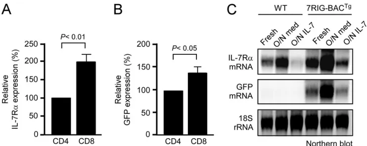
DP thymocytes (Fig. 2A). These results support the expectation that the BAC transgene retained all endogenous regulatory ele-ments for correct IL-7R␣ expression. Notably, IL-7R␣ surface levels on 7RIG-BACTgtransgenic cells were significantly higher compared with WT controls, presumably because Il7r gene transcription was active both from the endogenous and the transgenic Il7r locus. To demonstrate that the 7RIG-BACTg transgene faithfully reports Il7r gene transcription, we assessed GFP expression in thymocytes. GFP expression also followed the on-off-on pattern of IL-7R␣ expression in DN, DP, and SP thymocytes, respectively, indicating that transcription from the transgenic locus was correctly inhibited in DP thymocytes (Fig. 2B). We also assessed IL-7R␣ reporter expression in peripheral LN cells. Mature B cells do not express IL-7R␣. Accordingly, we found that 7RIG-BACTgDN lymph node cells, which include all mature B cells, were negative for GFP expression (Fig. 2C). CD4 and CD8 lymph node T cells, on the other hand, correctly expressed high levels of GFP. Interestingly, CD8 T cells reported higher levels of Il7r transcription than CD4 T cells based on their GFP levels (Fig. 2C). These data confirm that 7RIG-BACTg reporter mice faithfully represent endogenous IL-7R␣ expression in vivo and reveal a hitherto unappreciated difference in IL-7R␣ transcription levels in CD4 and CD8 T cells.
Cytokine-induced Regulation of 7RIG-BACTg Expression—
To further document distinct IL-7R␣ expression in CD4 and CD8 T cells, we quantified surface IL-7R␣ and intracellular GFP levels in freshly isolated 7RIG-BACTgLN T cells. We con-firmed statistically significant higher levels of both IL-7R␣
expression and transcription in CD8 T cells in multiple exper-iments (Fig. 3, A and B). Moreover, such lineage-specific IL-7R␣ expression was developmentally set as CD4 and CD8 T cells incubated overnight in medium in the absence of in vivo signals still displayed distinct levels of IL-7R␣ expression (data not shown).
The Il7r gene locus is exquisitely sensitive to cytokine signal-ing. For instance, in vivo IL-7 signaling down-regulates Il7r transcription and steady-state levels of IL-7R␣ mRNA in T cells (13). To determine whether the transgenic IL-7R␣ gene locus also responds to cytokine treatment, we incubated LN T cells from either WT or 7RIG-BACTgmice overnight in medium or in the presence of IL-7. The next day, total RNA was extracted and IL-7R␣ mRNA signals were assessed and compared with those from freshly isolated T cells. In both WT and 7RIG-BACTgT cells, overnight release from the cytokine-rich in vivo environment highly up-regulated IL-7R␣ mRNA expression (Fig. 3C). Furthermore, overnight IL-7 signaling potently sup-pressed IL-7R␣ mRNA expression in both WT and transgenic T cells. These results suggest that both the endogenous and the transgenic Il7r loci are regulated in a cytokine-dependent man-ner. More importantly, GFP mRNA levels also faithfully repli-cated IL-7R␣ expression, which confirms the validity of this transgenic model as an in vivo IL-7R␣ reporter (Fig. 3).
Restoring T Cell Development in IL-7R␣-deficient Mice by 7RIG-BACTg—IL-7R␣ deficiency results in severely impaired T
cell development and peripheral T cell homeostasis. Because 7RIG-BACTg replicates expression of endogenous Il7r, we wished to know if the BAC transgene could restore T cell
FIGURE 2. 7RIG-BACTgfaithfully reports IL-7R␣ expression in vivo. A, cell surface IL-7R␣ expression during thymocyte development. IL-7R␣ expression on
gated thymocyte subpopulations from WT mice (upper panel), 7RIG-BACTg(lower panel), and a human CD2 mini-cassette driven IL-7R␣Tgmice (middle panel) are
shown (solid line) over isotype control staining (dotted line). B, assessing IL-7R␣ transcription using GFP reporter activity in thymocyte subpopulations. Total
thymocytes from WT and 7RIG-BACTgmice were stained for CD4 and CD8 surface markers, and GFP expression was determined in individual subpopulations.
Data are representative of four independent experiments. C, lineage specific IL-7R␣ transcription in LNT cells. Total LN cells from WT and 7RIG-BACTgmice were
stained for CD4 and CD8 surface markers, and GFP expression was determined in CD8 and CD4 LN T cells. Mean fluorescence intensity of surface IL-7R␣ are
shown for CD8 and CD4 T cells, respectively. Data are representative of four independent experiments.
FIGURE 3. Transcriptional regulation of GFP reporter expression. A, relative surface IL-7R␣ expression on CD4 and CD8 LNT cells. Surface IL-7R␣ levels on
7RIG-BACTgT cells were quantified in mean fluorescence intensity and normalized to IL-7R␣ levels on CD4 cells. Bar graph shows mean ⫾ S.E. from three
independent experiments. B, relative GFP expression in CD4 and CD8 LN T cells. Intracellular GFP levels in 7RIG-BACTgT cells were quantified in the mean
fluorescence intensity and normalized to GFP levels in CD4 cells. Bar graph shows mean⫾ S.E. from three independent experiments. C, purified LN T cells from
WT or 7RIG-BACTgmice were assessed for IL-7R␣ and GFP mRNA expression by Northern blot analysis with probes indicated on the left. Total RNA was isolated
from fresh, overnight medium incubated, or overnight IL-7-treated LNT cells.
at BILKENT UNIVERSITY on November 9, 2017
http://www.jbc.org/
defects in IL-7R␣KOmice. To test this, we crossed 7RIG-BACTg into IL-7R␣KOmice to generate IL-7R␣KO7RIG-BACTgmice (supplemental Fig. S4A). In these mice, thymic␣ T cell devel-opment was largely restored as demonstrated by thymocyte CD4 versus CD8 profiles and TCR surface expression (Fig. 4A). Also, thymic NKT cell development and␥␦ T cell genera-tion was dramatically improved compared with IL-7R␣KOmice (supplemental Fig. S4, B and C). Notably, total thymocyte num-bers were also restored compared with IL-7R␣KOmice, but they did not fully recover to WT levels (Fig. 4B). To further
under-stand this, we analyzed surface IL-7R␣ levels on thymocytes, and we found that IL-7R␣KO7RIG-BACTgmice expressed sig-nificantly higher levels of IL-7R␣ than WT mice (Fig. 4C). This is presumably caused by insertion of multiple copies of the BAC transgene into the genome as usually observed in BAC trans-genesis. Elevated levels of IL-7R␣, however, have been shown to increase IL-7 consumption and competition for IL-7, which results in an overall decrease in thymocyte numbers (40, 41). Thus, decreased total thymocyte numbers might reflect quan-titative differences in surface IL-7R␣ expression between WT
FIGURE 4. 7RIG-BACTgrestores thymocyte development and T cell homeostasis in IL-7R␣KOmice. A, thymocyte development in IL-7R␣KO7RIG-BACTg.
Total thymocytes from WT and IL-7R␣KO7RIG-BACTgmice were assessed for CD4, CD8, and TCR surface marker expression (top and middle). Mature TCRhi
thymocytes were analyzed for CD4 and CD8 profiles (bottom). B, total thymocyte numbers in IL-7R␣KO7RIG-BACTgmice. Thymocyte numbers from WT,
IL-7R␣KO7RIG-BACTg, and IL-7R␣KOmice were determined. Bar graph shows mean⫾ S.E. from three independent experiments. C, surface IL-7R␣ on WT and
7RIG-BACTgthymocytes. IL-7R␣ expression was determined on WT and IL-7R␣KO7RIG-BACTgthymocyte subpopulations. Bar graph shows mean⫾ S.E. from
three independent experiments. D, peripheral T cell homeostasis in IL-7R␣KO7RIG-BACTgmice. LN cells were isolated, counted, and phenotyped for CD4 and
CD8 expression (top). Bar graph shows total LN numbers (bottom). Data show mean⫾ S.E. from four independent experiments. E, surface IL-7R␣ on WT and
7RIG-BACTgLNT cells. IL-7R␣ expression was assessed and quantified on WT and IL-7R␣KO7RIG-BACTgLN cell subpopulations. Data show representative
histograms from three independent experiments.
at BILKENT UNIVERSITY on November 9, 2017
http://www.jbc.org/
and IL-7R␣KO7RIG-BACTg thymocytes. Interestingly, how-ever, no major differences were observed in peripheral LN T cell numbers and CD4/CD8 profiles (Fig. 4D), despite surface IL-7R␣ levels on transgenic LN T cells being still higher than WT counterparts (Fig. 4E). Thus, thymocytes and LN T cells are differently affected by IL-7R␣ levels, likely because of the different mechanisms of proliferation and homeostasis in these two organs. Collectively, we find that BAC transgenic IL-7R␣ is expressed and regulated in a developmentally correct fashion, and consequently restores T cell development and mainte-nance in IL-7R␣-deficient mice.
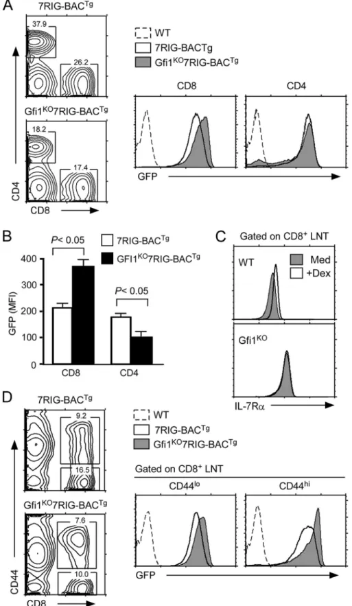
Gfi1 Overexpression Suppresses Il7r Gene Transcription in CD8 T Cells—Using these reporter mice, next we asked whether Gfi1 can suppress IL-7R␣ transcription and expression
in vivo. To this end, we introduced the 7RIG-BACTgonto mice expressing a T lineage-specific, human CD2 promoter driven Gfi1 transgene (25) to generate Gfi1Tg7RIG-BACTgmice and determined their IL-7R␣ transcription by analyzing GFP expression. Strikingly, in Gfi1Tg7RIG-BACTgthymocytes, we found that GFP levels were significantly down-regulated by Gfi1, but that this was specific and restricted to CD8SP thymo-cytes (Fig. 5A). These results suggest that transgenic Gfi1 expression fails to suppress IL-7R␣ transcription in any other thymocyte subpopulation than in CD8SP cells. Such a specific effect of Gfi1 on CD8 lineage cells was further confirmed in peripheral CD8 T cells. We found both surface IL-7R␣ and intracellular GFP levels selectively down-regulated in CD8, but not in CD4 T cells (Fig. 5B). Thus, these data document that Gfi1 only down-regulates IL-7R␣ gene transcription and sur-face protein expression in CD8 T lymphocytes, and that all other thymocyte subpopulations and CD4 T cells are not sus-ceptible to repression of this gene by Gfi1.
To further assess the specificity of Gfi1 transcriptional repression, we examined expression of other members of the␥c cytokine receptor family. The␥c cytokine family is composed of IL-2, IL-4, IL-7, IL-9, IL-15, and IL-21 (42), and antibodies are available for surface staining of each member of the␥c receptor family with the exception of IL-9. Consequently, we examined expression of ␥c cytokine receptors for IL-2R␣, -2R, ␥c, IL-4R␣, IL-7R␣, and IL-21R on WT and Gfi1TgCD8 T cells (Fig. 5C). Strikingly, IL-7R␣ was the only cytokine receptor that was significantly affected by Gfi1 overexpression (Fig. 5D), which revealed a highly selective effect of Gfi1 on IL-7R␣ and reaffirmed its specificity for CD8 T cells.
Gfi1 Deficiency Up-regulates Il7r Transcription in CD8 T Cells—Such a CD8 lineage-specific effect of Gfi1 was intrigu-ing. One potential explanation could be that Gfi1 is expressed in all thymocytes and T cells, but that its repressor activity is only limited to CD8 T cells. Consequently, we wished to test whether removal of Gfi1 would up-regulate IL-7R␣ expression in T cells other than in CD8 lineage cells. To this end, we generated Gfi1KO7RIG-BACTgmice and analyzed the GFP levels in thy-mocytes and mature LN T cells (supplemental Fig. S5Aand Fig. 6A) (24). Notably, Gfi1 deficiency failed to de-repress IL-7R␣ transcription in most thymocytes, with the exception of CD8SP thymocytes (supplemental Fig. S5A). DP thymocytes, which express high levels of Gfi1 and are completely silent for IL-7R␣ transcription, were still negative for GFP expression, when Gfi1
expression was ablated. These results indicate that Gfi1 is not required to suppress IL-7R␣ expression in this particular sub-set. Rather, the effect of Gfi1 was highly restricted to post-se-lection CD8 lineage cells as GFP expression was quantitatively increased in both CD8SP thymocytes and LN CD8 T cells only (supplemental Fig. S5Aand Fig. 6B).
Because absent Gfi1 was sufficient to up-regulate IL-7R␣ expression on CD8 T cells, next, we wished to know whether this would correlate with the Dex effect that we observed in 3B4.15 cells (Fig. 1, A and B). To this end, we stimulated WT and Gfi1TgT cells with Dex and assessed their surface IL-7R␣ expression after overnight culture (Fig. 6C). Although WT CD8⫹T cells significantly up-regulated IL-7R␣ as previously observed (17), IL-7R␣ levels on Gfi1KOCD8⫹T cells were unaf-fected by stimulation with Dex (Fig. 6C). Notably, IL-7R␣ expression on Gfi1KO CD4⫹T cells was still up-regulated by Dex indicating that Gfi1 effect is CD8 lineage specific ( supple-mental Fig. S5B). These data strongly suggest that a major role for Dex is to suppress Gfi1, and that Gfi1 directly controls IL-7R␣ expression in CD8 T cells.
Gfi1 deficiency has been proposed to promote CD8 memory phenotype cell generation (43). Because memory CD8 T cells express high levels of IL-7R (42), we wished to test whether increased IL-7R␣ is a consequence of memory cell differentia-tion or a direct effect of Gfi1 deficiency. GFP levels in CD8 T cell subsets demonstrated that IL-7R␣ transcription was signifi-cantly increased in both naive (CD44low) and activated/mem-ory (CD44high) phenotype CD8 T cells (Fig. 6D). Thus, we con-clude that absent Gfi1 expression de-represses IL-7R␣ transcription and expression in all CD8 T cells independently of their differentiation status.
In Vivo Analysis of IL-7R␣ Transcription Using 7RIG-BACTg on a Single Cell Basis—We wished to know if using the 7RIG-BACTgcould provide us with new insights on IL-7R␣ expres-sion that so far has not been experimentally feasible to assess. Adoptive transfer of CD8 T cells into chronic lymphopenic mice results in lymphopenia-induced homeostatic prolifera-tion (LIP). Slowly dividing homeostatic proliferaprolifera-tion is depend-ent on IL-7, whereas rapid proliferation is IL-7 independdepend-ent and driven by commensal antigens (44). To further understand the role of IL-7 in this process, it would be important to assess how IL-7R␣ expression is regulated during LIP. To do so, we assessed surface IL-7R␣ and intracellular GFP expression on day 5 adoptively transferred 7RIG-BACTgCD8 T cells. Analyz-ing IL-7R␣ transcription in adoptively transferred cells on a single cell basis has not been possible so far. IL-7 signaling sup-presses IL-7R␣ expression under steady-state conditions (13). Surprisingly, however, surface IL-7R␣ levels remained largely unchanged during IL-7-driven LIP as assessed on slowly divid-ing cells (Fig. 7, A, left, and B). Strikdivid-ingly, in the same cells, Il7r transcription was dramatically down-regulated upon cell divi-sion, as demonstrated by reduced GFP expression in cell trace-diluted cells (Fig. 7, A, right, and B). These data indicate that surface IL-7R␣ expression is not a reliable marker for Il7r tran-scription, at least during homeostatic proliferation. They fur-ther suggest the operation of a transcription-independent mechanism of surface IL-7R expression, which could be either increased recycling of endocytosed IL-7R␣, stabilization of
at BILKENT UNIVERSITY on November 9, 2017
http://www.jbc.org/
existing IL-7R␣ proteins or a yet unknown post-transcriptional mechanism. We are currently in the process of addressing these possibilities.
Because Gfi1 suppresses Il7r transcription, next we wished to assess the effect of Gfi1 on IL-7R␣ expression during LIP. Day 5 adoptively transferred Gfi1Tg7RIG-BACTg cells displayed a comparable pattern of surface IL-7R␣ and intracellular GFP expression to WT cells, in that GFP levels steadily decreased
upon proliferation (Fig. 7C, right) but surface IL-7R␣ levels remain largely unaffected (Fig. 7C, left). Thus, the dichotomoy of IL-7R␣ transcription and surface protein expression during LIP still remained distinct in Gfi1Tg7RIG-BACTgcells. More-over, LIP further down-regulated IL-7R␣ transcription in Gfi1Tgcells, which is presumably mediated by a mechanism independent of Gfi1. Importantly, Gfi1 overexpression signifi-cantly impaired CD8 T cell LIP, which correlated with lower
FIGURE 5. Gfi1 suppresses IL-7R␣ expression and transcription in vivo. A, suppression of GFP reporter activity by overexpression of Gfi1. IL-7R␣
transcrip-tional activities in individual thymocyte subpopulations were determined using the 7RIG-BACTgon WT or Gfi1Tgbackgrounds. Contour plots are representative
of four independent experiments with 4 WT and 5 Gfi1Tgmice transgenic for 7RIG-BACTg. Bar graph shows mean⫾ S.E. four independent experiments. B, IL-7R␣
surface expression and transcription in Gfi1Tgtransgenic CD4 and CD8 LNT cells. The effect of Gfi1 was assessed in WT or Gfi1Tgmice transgenic for 7RIG-BACTg.
Cell surface IL-7R␣ and GFP expression were determined by flow cytometry. Bar graph shows mean ⫾ S.E. from three independent experiments with 3 WT and
5 Gfi1Tg7RIG-BACTgmice. C, expression of␥c cytokine receptor families on Gfi1TgCD8⫹LNT cells. Surface expression of the indicated␥c cytokine receptors were
determined on gated CD8 T cells from WT and Gfi1Tgmice. Data are representative of three independent experiments. D, Gfi1Tgspecifically suppresses IL-7R␣
expression on CD8⫹T cells. Surface cytokine receptor levels on Gfi1TgCD8⫹T cells were quantified and normalized to levels on WT CD8⫹T cells. Bar graph
shows mean⫾ S.E. of three independent experiments.
at BILKENT UNIVERSITY on November 9, 2017
http://www.jbc.org/
FIGURE 6. Gfi1 deficiency de-represses IL-7R␣ transcription and expression in CD8 lineage cells. A, increased GFP reporter activity in Gfi1KO7RIG-BACTg
CD8 T cells. GFP expression was assessed in CD8 and CD4 T cells from WT or Gfi1KOmice expressing 7RIG-BACTg. Data are representative of four independent
experiments. B, quantification of GFP expression in Gfi1KO7RIG-BACTgT cells. Mean fluorescence intensity of intracellular GFP levels were determined from
7RIG-BACTgand Gfi1KO7RIG-BACTgLNT cells. Bar graph shows the mean⫾ S.E. of two independent experiments. C, Dex effect on CD8⫹LNT IL-7R␣ expression.
Surface IL-7R␣ expression was determined on WT and Gfi1KOCD8 T cells incubated overnight in medium or Dex. D, the effect of Gfi1 on IL-7R␣ transcription is
independent of activation/differentiation status in CD8 T cells. GFP reporter expression was assessed in freshly isolated CD44loand CD44hiCD8 T cells from
7RIG-BACTgand Gfi1KO7RIG-BACTgmice. Data are representative of four independent experiments.
at BILKENT UNIVERSITY on November 9, 2017
http://www.jbc.org/
IL-7R␣ levels on Gfi1TgCD8 T cells (Fig. 5B). Thus, Gfi1 sup-presses IL-7R␣ expression and also IL-7-dependent prolifera-tion. Taken together, these results uncover Gfi1 as a critical regulator of IL-7R␣ transcription and expression in vivo, but exclusively in CD8 lineage thymocytes and T cells.
DISCUSSION
To understand the molecular mechanisms of IL-7R␣ expres-sion during T cell development and activation, here we gener-ated a novel BAC GFP-reporter transgene and utilized this tool to assess IL-7R␣ transcription in vivo. The BAC transgene was constructed by inserting a GFP-reporter and an IRES element into the 3⬘ UTR of the murine Il7r gene. Consequently, BAC reporter mice overexpressed full-length IL-7R␣ proteins in addition to GFP. We affirmed the developmentally correct expression of reporter transgenes by assessing lymphocyte dif-ferentiation in IL-7R␣-deficient mice reconstituted with the BAC construct (IL-7R␣KO7RIG-BACTg). In these mice, devel-opment of all IL-7R␣-dependent lymphoid cell populations, including␣-, ␥␦-T cells, B-cells, and NKT cells, were restored. Thus, the transgenic Il7r gene locus in 7RIG-BACTgmice is correctly and lineage specifically regulated, and it equipped us with a new tool to assess IL-7R␣ transcription and expression in
vivo.
IL-7R␣ is the ligand-specific subunit of the functional IL-7 receptor, which is composed of the IL-7R␣ chain and the ␥c-chain (4). In contrast to the ␥c-chain, IL-7R␣ expression is
dynamic and is actively regulated during T cell development and differentiation. All developing thymocytes and all mature T cells express IL-7R␣, albeit at varying degrees. However, imma-ture DP thymocytes are unique in that they have completely terminated IL-7R␣ expression. Such peculiar absence of IL-7R␣ on pre-selection DP cells was proposed to reflect a crit-ical thymic selection mechanism that ensures a random but self-MHC-specific TCR repertoire (39, 45, 46). Accordingly, absent IL-7R␣ expression renders DP thymocytes dependent on selecting TCR signals and not on nondiscriminatory IL-7 signaling for survival. However, the molecular mechanisms that terminate IL-7R␣ expression in DP cells remain unclear. Along this line, the molecular circuitry that re-induces IL-7R␣ expres-sion upon positive selection also remains unmapped. This is even more intriguing as TCR signaling in mature T cells down-regulates IL-7R␣ expression, but in immature DP thymocytes, TCR signaling induces IL-7R␣ expression. Recent studies have suggested that re-expression of IL-7R␣ on DP cells is depend-ent on positive selecting TCR signals in a NFAT- and MAPK-dependent fashion and to a lesser degree on Akt (47). However, the detailed mechanism and molecular events remain unknown. Using 7RIG-BACTg mice, here we establish that absent IL-7R␣ expression in DP cells is a transcriptionally reg-ulated event. Pre-selected DP cells failed to express GFP, while positive selection and maturation resulted in re-expression of GFP. With the 7RIG-BACTgreporter mouse, it is now feasible
FIGURE 7. In vivo effects of Gfi1 on IL-7R␣ expression. A, IL-7R␣ expression during lymphopenia-induced homeostatic proliferation. Surface IL-7R␣
expres-sion and intracellular GFP expresexpres-sion were assessed on day 5 adoptively transferred 7RIG-BACTgCD8⫹T cells in RAG-2-deficient host mice. Cell division was
monitored by CellTrace Violet dye dilution. Dot plots are representative of three independent experiments. B, IL-7R␣ expression and transcription in
prolifer-ating donor cells. Mean fluorescence intensity of surface IL-7R␣ (open circle) and intracellular GFP (closed circle) were assessed for each cell division (D1 to D6),
and normalized to nondividing cells (D1), which was set to 100 (%). Graph shows the mean⫾ S.E. from three independent experiments. C, IL-7R␣ expression
on proliferating Gfi1Tg7RIG-BACTgCD8⫹T cells. Surface IL-7R␣ expression and intracellular GFP expression was assessed on day 5 adoptively transferred
Gfi1Tg7RIG-BACTgCD8⫹T cells in RAG-2-deficient host mice. Cell division was monitored by CellTrace Violet dye dilution. Dot plots are representative of three
independent experiments. D, Gfi1 impairs lymphopenia-induced homeostatic proliferation. CellTrace Violet dilutions on slow dividing donor 7RIG-BACTgor
Gfi1Tg7RIG-BACTgwere quantitated. Numbers indicate the percentage of cells that have undergone one or more divisions during homeostatic proliferation.
Data are representative of three independent experiments.
at BILKENT UNIVERSITY on November 9, 2017
http://www.jbc.org/
to test nuclear factors that regulate IL-7R␣ transcription in
vivo. We anticipate further applications of these reporter mice in dissecting the thymic signals that lead to termination as well as re-induction of Il7r transcription under various in vitro and
in vivosettings.
In this regard, we were able to assess the effect of a transcrip-tional repressor on Il7r gene expression in vivo. Gfi1 has been considered as a potential Il7r regulator in immature thymo-cytes because of its high level of expression in pre-selection DP thymocytes and its potent suppression of IL-7R␣ in pre-B cells. As such, Gfi1KO7RIG-BACTgmice offered a unique opportu-nity to assess the effect of Gfi1 on IL-7R␣ transcription in DP thymocytes in vivo. The complete absence of GFP signals in Gfi1KO7RIG-BACTg DP thymocytes strongly suggested that transcriptional silencing of Il7r gene expression does not require Gfi1, at least in pre-selection DP thymocytes. In mature T cells, Gfi1 clearly plays a more critical role. In fact, Gfi1 was first reported to suppress IL-7R␣ expression by analyzing peripheral mature T cells (13). Nevertheless, a definitive and direct role of Gfi1 in IL-7R␣ transcription has remained highly controversial. In some cases, surface IL-7R␣ expression on Gfi1KOT cells was found to be not, or insignificantly different (38), and also increased IL-7R␣ levels in Gfi1KOmice had been suggested to be indirect, i.e. due to high proportions of memory cells under Gfi1 deficiency (43). Moreover, increased Gfi1 mRNA levels in human IL-7R␣higheffector memory CD8 T cells contradicted a repressor role for Gfi1 in IL-7R␣ expression (48). Our current data showing significantly increased GFP expression in GfiKOCD8 T cells, however, clearly documents Gfi1 as an in vivo repressor for IL-7R␣ transcription.
Nevertheless, contradictory observations still keep open the possibility of a more complex regulatory network, with several Gfi1 co-factors being involved. As such, the cis-elements par-ticipating in the Gfi1 control of Il7r gene transcription are still not well defined. In B lymphocytes, Gfi1 can bind to the second intron of the Il7r gene to suppress transcription (14). In myeloid cells, Gfi1 can also associate with PU.1, an ETS family transcrip-tion factor that binds to the promoter of IL-7R␣ and activates its transcription (49). However, PU.1 is not expressed in T lym-phocytes. Rather another ETS family transcription factor, GABP, is thought to occupy the ETS site in the promoter of the
Il7rgene (50). Whether Gfi1 can interact with the GABP pro-tein is not known. But in T cells, GABP and Gfi1 were shown to have opposing roles so that a direct interaction and mutual suppression cannot be excluded (51). Thus, in addition to its role in binding the Il7r gene promoter, GABP may control Gfi1 expression as GABP-deficient splenocytes lack Gfi1 expression, presumably due to the positively acting GABP binding sites in the Gfi1 promoter (52).
With Gfi1 emerging as a key control factor in IL-7R␣ expres-sion and also in many aspects of T and B cell immunology, it is critical to know what controls Gfi1 expression. So far, an auto-regulatory role for Gfi1 as well as a transauto-regulatory role for Gfi1b has been documented (53). Also, downstream signaling of the GTPase Cdc42 has been proposed to suppress Gfi1 expression in resting T cells (54). The current study now pro-poses that glucocorticoids are a new family of molecules that can control Gfi1 expression. Glucocorticoids have been known
to take part in thymocyte development and selection (55–58). Recently, conditional deletion of GR in pre-selection thymo-cytes documented significantly reduced thymus cellularity with an altered TCR repertoire and increased negative selection (59). Mechanistically, glucocorticoids have been shown to intersect with TCR signaling and increase TCR signaling thresholds to promote thymic selection (60). Our current data now also pro-poses a role for Gfi1 downstream of GR signaling, and it would be informative to assess the contribution of Gfi1 to the GR-de-ficient phenotype.
In peripheral T cells, glucocorticoids are immunosuppres-sive, but they also up-regulate IL-7R␣ transcription and expres-sion. The identification of Gfi1 as a novel target of Dex sheds new light onto pre-existing observations on the effects of glu-cocorticoids and in this regard, the discovery of Gfi1 as an inter-mediary of Dex in IL-7R␣ expression suggests that other Dex-induced events might also employ such a mechanism. Although the up-regulation of IL-7R␣ expression in Dex-treated B cells, which normally do not express IL-7R␣, could be another case of an Gfi1-mediated Dex effect (34), inhibition of cytokine pro-duction in Dex-treated cells also could be explained along this line. Further studies are planned to address these possibilities and to delineate a direct Dex effect versus Gfi1-mediated Dex effects.
Although Gfi1 expression is down-regulated by Dex signal-ing, and overexpression of Gfi1 can overcome Dex-mediated up-regulation of IL-7R␣, we found, as expected, that GR is crit-ical for Dex-mediated IL-7R␣ up-regulation. GR binds to a putative Il7r enhancer in an evolutionarily conserved sequence 3.5 kb upstream of the transcriptional start site (16). The GR site in this evolutionarily conserved sequence is 50 base pairs away from a FoxO1 transcription factor binding site, which positively regulates gene expression in both naive CD4⫹and CD8⫹T cells (20, 21). Whether this enhancer, the PU.1/GABP binding promoter, and the putative Gfi1-binding intronic silencer are the only functional control elements in the Il7r gene locus is not known. In fact, a series of nuclear factors are known to regulate IL-7R␣ expression. In addition to Gfi1, two other transcriptional repressors have been identified that down-reg-ulate Il7r gene expression in T cells. The forkhead family tran-scription factor FoxP1 suppresses IL-7R␣ expression by com-peting with the Il7r transactivator FoxO1 for enhancer occupancy (19). On the other hand, FoxP3, which is specifically expressed in regulatory T cells, directly suppress IL-7R␣ expression (22). Overexpression of a Gfi1 family member, Gfi1b, can also repress IL-7R␣ expression in T lymphocytes, but presumably this effect is through the same intronic Gfi1 binding site, because the DNA binding domains of these two factors are very similar (61). Notably, in our expression profil-ing experiments, Gfi1 was the only nuclear factor that showed a significant difference in expression upon Dex treatment.
Collectively, the present study identified and tested a novel
Il7rtranscriptional control mechanism in vivo using a newly established IL-7R␣ reporter mouse. We validated these reporter mice by complementing IL-7R␣ deficiency, and we utilized this tool to assess Il7r transcription downstream of Gfi1. Using a lymphopenia-induced homeostatic proliferation model, we further documented the superiority of these reporter
at BILKENT UNIVERSITY on November 9, 2017
http://www.jbc.org/
mice in assessing IL-7R␣ gene transcription in T cells to tradi-tional methods. Finally, the current study not only resolves the controversies surrounding the role of Gfi1 as a transcriptional repressor of IL-7R␣ in CD8 T cells but also demonstrates that IL-7R␣ expression in any other T cell population is indepen-dent of Gfi1. The molecular basis for such lineage-specific reg-ulation of cytokine receptor expression is intriguing and impor-tant, and we think that our findings will provide a new venue to identify critical players for controlling IL-7R␣ expression. Acknowledgments—We are grateful to A. Singer for critical reading of the manuscript, helpful discussions, and support throughout the course of this study; S. Kaech for critical reading of the manuscript; O. Leo for providing 3B4.15 T cell hybridoma; S. Sharrow, A. Adams, and L. Granger for expert flow cytometry; and A. Alag and T. Guinter for screening mice.
REFERENCES
1. Peschon, J. J., Morrissey, P. J., Grabstein, K. H., Ramsdell, F. J., Marask-ovsky, E., Gliniak, B. C., Park, L. S., Ziegler, S. F., Williams, D. E., Ware, C. B., Meyer, J. D., and Davison, B. L. (1994) Early lymphocyte expansion is severely impaired in interleukin 7 receptor-deficient mice. J. Exp. Med.
180,1955–1960
2. Sudo, T., Nishikawa, S., Ohno, N., Akiyama, N., Tamakoshi, M., and Yo-shida, H. (1993) Expression and function of the interleukin 7 receptor in murine lymphocytes. Proc. Natl. Acad. Sci. U.S.A. 90, 9125–9129 3. Yu, Q., Erman, B., Park, J. H., Feigenbaum, L., and Singer, A. (2004) IL-7
receptor signals inhibit expression of transcription factors TCF-1, LEF-1, and ROR␥t: Impact on thymocyte development. J. Exp. Med. 200, 797– 803
4. Mazzucchelli, R., and Durum, S. K. (2007) Interleukin-7 receptor expres-sion: Intelligent design. Nat. Rev. Immunol. 7, 144 –154
5. Kaech, S. M., Tan, J. T., Wherry, E. J., Konieczny, B. T., Surh, C. D., and Ahmed, R. (2003) Selective expression of the interleukin 7 receptor iden-tifies effector CD8 T cells that give rise to long-lived memory cells. Nat. Immunol. 4,1191–1198
6. Hand, T. W., Morre, M., and Kaech, S. M. (2007) Expression of IL-7
receptor␣ is necessary but not sufficient for the formation of memory
CD8 T cells during viral infection. Proc. Natl. Acad. Sci. U.S.A. 104, 11730 –11735
7. Klonowski, K. D., Williams, K. J., Marzo, A. L., and Lefrançois, L. (2006)
Cutting edge. IL-7-independent regulation of IL-7 receptor␣ expression
and memory CD8 T cell development. J. Immunol. 177, 4247– 4251 8. DeKoter, R. P., Lee, H. J., and Singh, H. (2002) PU.1 regulates expression of
the interleukin-7 receptor in lymphoid progenitors. Immunity 16, 297–309
9. DeKoter, R. P., Schweitzer, B. L., Kamath, M. B., Jones, D., Tagoh, H., Bonifer, C., Hildeman, D. A., and Huang, K. J. (2007) Regulation of the
interleukin-7 receptor␣ promoter by the Ets transcription factors PU.1
and GA-binding protein in developing B cells. J. Biol. Chem. 282, 14194 –14204
10. Xue, H. H., Bollenbacher, J., Rovella, V., Tripuraneni, R., Du, Y. B., Liu, C. Y., Williams, A., McCoy, J. P., and Leonard, W. J. (2004) GA-binding
protein regulates interleukin 7 receptor␣-chain gene expression in T cells.
Nat. Immunol. 5,1036 –1044
11. Kim, H. R., Hwang, K. A., Kim, K. C., and Kang, I. (2007) Down-regulation of IL-7R␣ expression in human T cells via DNA methylation. J. Immunol.
178,5473–5479
12. González-García, S., García-Peydró, M., Martín-Gayo, E., Ballestar, E., Esteller, M., Bornstein, R., de la Pompa, J. L., Ferrando, A. A., and Toribio, M. L. (2009) CSL-MAML-dependent Notch1 signaling controls T lin-eage-specific IL-7R␣ gene expression in early human thymopoiesis and leukemia. J. Exp. Med. 206, 779 –791
13. Park, J. H., Yu, Q., Erman, B., Appelbaum, J. S., Montoya-Durango, D.,
Grimes, H. L., and Singer, A. (2004) Suppression of IL-7R␣ transcription
by IL-7 and other prosurvival cytokines. A novel mechanism for maximiz-ing IL-7-dependent T cell survival. Immunity 21, 289 –302
14. Deleted in proof
15. Zweidler-Mckay, P. A., Grimes, H. L., Flubacher, M. M., and Tsichlis, P. N. (1996) Gfi1 encodes a nuclear zinc finger protein that binds DNA and functions as a transcriptional repressor. Mol. Cell. Biol. 16, 4024 – 4034 16. Lee, H. C., Shibata, H., Ogawa, S., Maki, K., and Ikuta, K. (2005)
Transcrip-tional regulation of the mouse IL-7 receptor␣ promoter by glucocorticoid
receptor. J. Immunol. 174, 7800 –7806
17. Franchimont, D., Galon, J., Vacchio, M. S., Fan, S., Visconti, R., Frucht, D. M., Geenen, V., Chrousos, G. P., Ashwell, J. D., and O’Shea, J. J. (2002) Positive effects of glucocorticoids on T cell function by up-regulation of
IL-7 receptor␣. J. Immunol. 168, 2212–2218
18. Egawa, T., Tillman, R. E., Naoe, Y., Taniuchi, I., and Littman, D. R. (2007) The role of the Runx transcription factors in thymocyte differentiation and in homeostasis of naive T cells. J. Exp. Med. 204, 1945–1957 19. Feng, X., Wang, H., Takata, H., Day, T. J., Willen, J., and Hu, H. (2011)
Transcription factor Foxp1 exerts essential cell-intrinsic regulation of the quiescence of naive T cells. Nat. Immunol. 12, 544 –550
20. Kerdiles, Y. M., Beisner, D. R., Tinoco, R., Dejean, A. S., Castrillon, D. H., DePinho, R. A., and Hedrick, S. M. (2009) Foxo1 links homing and survival of naive T cells by regulating L-selectin, CCR7, and interleukin 7 receptor. Nat. Immunol. 10,176 –184
21. Ouyang, W., Beckett, O., Flavell, R. A., and Li, M. O. (2009) An essential role of the Forkhead-box transcription factor Foxo1 in control of T cell homeostasis and tolerance. Immunity 30, 358 –371
22. Liu, W., Putnam, A. L., Xu-Yu, Z., Szot, G. L., Lee, M. R., Zhu, S., Gottlieb, P. A., Kapranov, P., Gingeras, T. R., Fazekas de St Groth, B., Clayberger, C., Soper, D. M., Ziegler, S. F., and Bluestone, J. A. (2006) CD127 expression
inversely correlates with FoxP3 and suppressive function of human CD4⫹
T reg cells. J. Exp. Med. 203, 1701–1711
23. Van Laethem, F., Baus, E., Smyth, L. A., Andris, F., Bex, F., Urbain, J., Kioussis, D., and Leo, O. (2001) Glucocorticoids attenuate T cell receptor signaling. J. Exp. Med. 193, 803– 814
24. Hock, H., Hamblen, M. J., Rooke, H. M., Traver, D., Bronson, R. T., Cam-eron, S., and Orkin, S. H. (2003) Intrinsic requirement for zinc finger transcription factor Gfi1 in neutrophil differentiation. Immunity 18, 109 –120
25. Rödel, B., Tavassoli, K., Karsunky, H., Schmidt, T., Bachmann, M., Schaper, F., Heinrich, P., Shuai, K., Elsässer, H. P., and Möröy, T. (2000) The zinc finger protein Gfi1 can enhance STAT3 signaling by interacting with the STAT3 inhibitor PIAS3. EMBO J. 19, 5845–5855
26. Liu, P., Jenkins, N. A., and Copeland, N. G. (2003) A highly efficient re-combineering-based method for generating conditional knockout muta-tions. Genome Res. 13, 476 – 484
27. Brinster, R. L., Chen, H. Y., Trumbauer, M. E., Yagle, M. K., and Palmiter, R. D. (1985) Factors affecting the efficiency of introducing foreign DNA into mice by microinjecting eggs. Proc. Natl. Acad. Sci. U.S.A. 82, 4438 – 4442
28. Yu, S., Zhou, X., Hsiao, J. J., Yu, D., Saunders, T. L., and Xue, H. H. (2012) Transgenic Res. 21,201–215
29. Thomas, K. R., and Capecchi, M. R. (1987) Site-directed mutagenesis by gene targeting in mouse embryo-derived stem cells. Cell 51, 503–512 30. Naviaux, R. K., Costanzi, E., Haas, M., and Verma, I. M. (1996) The pCL
vector system. Rapid production of helper-free, high-titer, recombinant retroviruses. J. Virol. 70, 5701–5705
31. Simon, A., and Biot, E. (2010) ANAIS, analysis of NimbleGen arrays inter-face. Bioinformatics 26, 2468 –2469
32. Sturn, A., Quackenbush, J., and Trajanoski, Z. (2002) Genesis. Cluster analysis of microarray data. Bioinformatics 18, 207–208
33. Edgar, R., Domrachev, M., and Lash, A. E. (2002) Gene Expression Omni-bus. NCBI gene expression and hybridization array data repository. Nu-cleic Acids Res. 30,207–210
34. Shibata, H., Tani-ichi, S., Lee, H. C., Maki, K., and Ikuta, K. (2007)
Induc-tion of the IL-7 receptor␣ chain in mouse peripheral B cells by
glucocor-ticoids. Immunol. Lett. 111, 45–50
35. D’Adamio, F., Zollo, O., Moraca, R., Ayroldi, E., Bruscoli, S., Bartoli, A.,
at BILKENT UNIVERSITY on November 9, 2017
http://www.jbc.org/
Cannarile, L., Migliorati, G., and Riccardi, C. (1997) A new dexametha-sone-induced gene of the leucine zipper family protects T lymphocytes from TCR/CD3-activated cell death. Immunity 7, 803– 812
36. Riccardi, C., Cifone, M. G., and Migliorati, G. (1999) Glucocorticoid hor-mone-induced modulation of gene expression and regulation of T-cell death. Role of GITR and GILZ, two dexamethasone-induced genes. Cell Death Differ 6,1182–1189
37. Zhan, Y., Funda, D. P., Every, A. L., Fundova, P., Purton, J. F., Liddicoat, D. R., Cole, T. J., Godfrey, D. I., Brady, J. L., Mannering, S. I., Harrison, L. C., and Lew, A. M. (2004) TCR-mediated activation promotes GITR up-regulation in T cells and resistance to glucocorticoid-induced death. Int. Immunol. 16,1315–1321
38. Pargmann, D., Yücel, R., Kosan, C., Saba, I., Klein-Hitpass, L., Schimmer, S., Heyd, F., Dittmer, U., and Möröy, T. (2007) Differential impact of the
transcriptional repressor Gfi1 on mature CD4⫹and CD8⫹T lymphocyte
function. Eur. J. Immunol. 37, 3551–3563
39. Yu, Q., Park, J. H., Doan, L. L., Erman, B., Feigenbaum, L., and Singer, A. (2006) Cytokine signal transduction is suppressed in preselection double-positive thymocytes and restored by double-positive selection. J. Exp. Med. 203, 165–175
40. Laouar, Y., Crispe, I. N., and Flavell, R. A. (2004) Overexpression of IL-7R␣
provides a competitive advantage during early T-cell development. Blood
103,1985–1994
41. Munitic, I., Williams, J. A., Yang, Y., Dong, B., Lucas, P. J., El Kassar, N., Gress, R. E., and Ashwell, J. D. (2004) Dynamic regulation of IL-7 receptor expression is required for normal thymopoiesis. Blood 104, 4165– 4172 42. Rochman, Y., Spolski, R., and Leonard, W. J. (2009) New insights into the
regulation of T cells by ␥c family cytokines. Nat. Rev. Immunol. 9,
480 – 490
43. Zhu, J., Jankovic, D., Grinberg, A., Guo, L., and Paul, W. E. (2006) Gfi1 plays an important role in IL-2-mediated Th2 cell expansion. Proc. Natl. Acad. Sci. U.S.A. 103,18214 –18219
44. Kieper, W. C., Troy, A., Burghardt, J. T., Ramsey, C., Lee, J. Y., Jiang, H. Q., Dummer, W., Shen, H., Cebra, J. J., and Surh, C. D. (2005) Recent immune status determines the source of antigens that drive homeostatic T cell expansion. J. Immunol. 174, 3158 –3163
45. Park, J. H., Adoro, S., Guinter, T., Erman, B., Alag, A. S., Catalfamo, M., Kimura, M. Y., Cui, Y., Lucas, P. J., Gress, R. E., Kubo, M., Hennighausen, L., Feigenbaum, L., and Singer, A. (2010) Signaling by intrathymic cyto-kines, not T cell antigen receptors, specifies CD8 lineage choice and pro-motes the differentiation of cytotoxic-lineage T cells. Nat. Immunol. 11, 257–264
46. Kim, G. Y., Hong, C., and Park, J. H. (2011) Seeing is believing. Illuminating the source of in vivo interleukin-7. Immune Netw. 11, 1–10
47. Sinclair, C., Saini, M., van der Loeff, I. S., Sakaguchi, S., and Seddon, B. (2011) The long-term survival potential of mature T lymphocytes is pro-grammed during development in the thymus. Sci. Signal 4, ra77
48. Kim, H. R., Hong, M. S., Dan, J. M., and Kang, I. (2006) Altered IL-7R␣
expression with aging and the potential implications of IL-7 therapy on
CD8⫹T-cell immune responses. Blood 107, 2855–2862
49. Spooner, C. J., Cheng, J. X., Pujadas, E., Laslo, P., and Singh, H. (2009) A recurrent network involving the transcription factors PU.1 and Gfi1 or-chestrates innate and adaptive immune cell fates. Immunity 31, 576 –586 50. Anderson, M. K. (2006) At the crossroads. Diverse roles of early
thymo-cyte transcriptional regulators. Immunol. Rev. 209, 191–211
51. Chandele, A., Joshi, N. S., Zhu, J., Paul, W. E., Leonard, W. J., and Kaech,
S. M. (2008) Formation of IL-7R␣ high and IL-7R␣ low CD8 T cells during
infection is regulated by the opposing functions of GABP␣ and Gfi1. J. Im-munol. 180,5309 –5319
52. Yang, Z. F., Drumea, K., Cormier, J., Wang, J., Zhu, X., and Rosmarin, A. G. (2011) GABP transcription factor is required for myeloid differentiation, in part, through its control of Gfi1 expression. Blood 118, 2243–2253 53. Doan, L. L., Porter, S. D., Duan, Z., Flubacher, M. M., Montoya, D.,
Tsich-lis, P. N., Horwitz, M., Gilks, C. B., and Grimes, H. L. (2004) Targeted transcriptional repression of Gfi1 by GFI1 and GFI1B in lymphoid cells. Nucleic Acids Res. 32,2508 –2519
54. Guo, F., Hildeman, D., Tripathi, P., Velu, C. S., Grimes, H. L., and Zheng, Y. (2010) Coordination of IL-7 receptor and T-cell receptor signaling by cell-division cycle 42 in T-cell homeostasis. Proc. Natl. Acad. Sci. U.S.A.
107,18505–18510
55. Ashwell, J. D., Lu, F. W., and Vacchio, M. S. (2000) Glucocorticoids in T cell development and function. Annu. Rev. Immunol. 18, 309 –345 56. Brewer, J. A., Sleckman, B. P., Swat, W., and Muglia, L. J. (2002) Green
fluorescent protein-glucocorticoid receptor knockin mice reveal dynamic receptor modulation during thymocyte development. J. Immunol. 169, 1309 –1318
57. Brewer, J. A., Kanagawa, O., Sleckman, B. P., and Muglia, L. J. (2002) Thymocyte apoptosis induced by T cell activation is mediated by gluco-corticoids in vivo. J. Immunol. 169, 1837–1843
58. Purton, J. F., Boyd, R. L., Cole, T. J., and Godfrey, D. I. (2000) Intrathymic T cell development and selection proceeds normally in the absence of glucocorticoid receptor signaling. Immunity 13, 179 –186
59. Mittelstadt, P. R., Monteiro, J. P., and Ashwell, J. D. (2012) Thymocyte responsiveness to endogenous glucocorticoids is required for immuno-logical fitness. J. Clin. Invest. 122, 2384 –2394
60. Vacchio, M. S., Lee, J. Y., and Ashwell, J. D. (1999) Thymus-derived glu-cocorticoids set the thresholds for thymocyte selection by inhibiting TCR-mediated thymocyte activation. J. Immunol. 163, 1327–1333
61. Doan, L. L., Kitay, M. K., Yu, Q., Singer, A., Herblot, S., Hoang, T., Bear, S. E., Morse, H. C., 3rd, Tsichlis, P. N., and Grimes, H. L. (2003) Growth factor independence-1B expression leads to defects in T cell activation,
IL-7 receptor␣ expression, and T cell lineage commitment. J. Immunol.
170,2356 –2366
at BILKENT UNIVERSITY on November 9, 2017
http://www.jbc.org/