ORIGINAL ARTICLE
A biomechanically based concept for a stronger obstetric anal
sphincter repair
Peter Petros1 &Akin Sivaslioglu2&Elvira Bratila3&Petre Bratila3&Burghard Abendstein4 Received: 28 February 2020 / Accepted: 14 May 2020
# The International Urogynecological Association 2020 Abstract
Introduction and hypothesis This study emanates from the ISPP OASIS and fecal incontinence study group at the 2018 annual meeting of the International Society for Pelviperineology (ISPP) in Bucharest, Romania. The aim was to analyze the biome-chanical factors leading to the breakdown of anal sphincter repair and to suggest a more robust technique for external anal sphincter (EAS) repair.
Methods Our starting point was what happens to the EAS wound repair site during defecation following EAS repair, with special reference to the process of wound healing.
Results We concluded that a graft no more than 1 × 1.5 cm sutured across the EAS tear line would mechanically support the tear line, vastly reduce the internal centrifugal forces acting on it during defecation, thereby giving the wound time to heal. Three different grafts were discussed, autologous, biological, and mesh. Also analyzed were the effects on EAS muscle contractility of overly tight repair and overly loose sphincter repair, the latter occasioned by the tearing out of sutures and repair by secondary intention.
Conclusions We have analyzed causes of sphincter repair failure, introduced a graft method, preferably autologous, for the prevention thereof and supported ultrasound assessment, rather than the absence of fecal incontinence as the criterion for success of EAS repair. Although based on well-established biomechanical principles, our proposal at this stage remains unproven. Our hope is that these concepts will be challenged, clarified, and tested, preferably in a randomized controlled trial.
Keywords OASIS . Anal sphincter tears . Biomechanics . Autologous . Graft
Introduction
The concepts in this paper evolved from discussions by the ISPP OASIS and fecal incontinence (FI) study group at the 2018 annual meeting of the International Society for Pelviperineology (ISPP) in Bucharest, Romania. Our aim
was to analyze the biomechanical factors leading to the break-down of anal sphincter repair and to suggest a more robust technique for external anal sphincter (EAS) repair.
Anal sphincter damage for primiparous women is becom-ing a serious problem in some places in the world. In Sydney, Australia, the incidence of third-/fourth-degree tears reached 11.49% for primiparas in one teaching hospital [1].
Epidemic or not, anal sphincter rupture at birth is a serious condition. It has long-term consequences for the patient’s quality of life. This has been recognized by an increasing number of articles in specialist journals on the subject. Sphincter repair is often performed by junior staff in the de-livery room. The results are often poor. Pinta et al. found that 75% of women who had been repaired at delivery after an obstetric tear had a persistent defect in the anal sphincter mus-culature 15 months (median) after a repair, and 60% were incontinent. [2].
A major step forward in helping to solve this problem has been the establishment of workshops to upskill obstetricians and pelvic floor surgeons [3].
From the International Society for Pelviperineology (ISPP) OASIS and fecal incontinence study group
* Peter Petros [email protected]
1 School of Mechanical and Biomechanical Engineering, University of Western Australia, Perth, Australia
2 Department of Obstetrics and Gynecology, Muğla Sitki Kocman University, Mugla, Turkey
3
Department of Obstetrics and Gynecology, University of Bucharest, Bucharest, Romania
4 Department of Obstetrics and Gynecology, Landeskrankenhaus Feldkirch, Feldkirch, Austria
https://doi.org/10.1007/s00192-020-04350-0
Materials and methods
Biomechanics
In addressing the question,“What is it that causes surgical repairs of EAS to fail?,” we examined the biomechanics of the tissues, the post-repair defecation forces acting on the EAS, the role of normal and defective wound healing, how sutures would work in edematous tissues, whether it was only EAS/internal anal sphincter (IAS) damage that caused FI, all of which begged the question,“how to mitigate these factors when performing EAS repair?”
The surgical difficulties repairing muscle tears are not con-fined to EAS sphincter tears. In a general surgical sense, repair of any muscle belly lacerations is difficult, technically de-manding, and the likelihood of clinical failure is high [4]. A major reason for this failure is that the regenerative capacity of injured skeletal muscle is limited. Fibrotic tissue forms at the injury site, thus delaying the muscle’s functional recovery [4]. The limited ability of the skeletal muscle to self-regenerate may justify the need for biological or synthetic augmentations for repairing large damage [4].
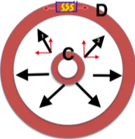
We see expansion by the fecal bolus as a key factor causing the breakdown of post-delivery EAS repair. In order to ex-trude the fecal bolus, the anal orifice must expand from“C″ (closed) to“D” (dilated; Fig. 1). The expansionary forces (large arrows) have transverse and vertical vector components (small arrows) that would pull and push against the suture lines“SSS.” Whether or not the repair will be successful de-pends largely on the ability of the EAS sutures and the tissues into which they are inserted to resist the tearing effect of the
centrifugal forces generated by passage of the fecal bolus for sufficient time for the scar to heal and restore normal function. The role of wound healing is, in our view, a critical factor in EAS surgical failure. It takes 6 weeks to achieve 40% strength in the wound (Fig.2) [5]. If the sutures move or tear through, the wound must heal by secondary intention. This will effec-tively lengthen EAS sarcomeres and decrease their contractile strength (Fig.3) [6].
The biomechanics of an EAS graft
We considered that an implanted graft over the approximated sutured limbs of the EAS would mechanically support the suture line, divert the centrifugal forces away from the graft, and protect the wound for 6 weeks while it healed.
The internal centrifugal pressure in a standard EAS repair is exerted over a 1-mm wide suture line and sutures that easily cut through: Pressure = Force/Area. At least theoretically, a firmly attached graft measuring 15 mm lessens the pressure exerted by a factor of 15.
As we see it, one advantage of a graft support method is that it is eminently feasible with a partial third-degree tear. We were all in general agreement with the biomechanical princi-ples behind this method and the surgical technique, which concerns only EAS repair. The repair suggested is in accord with aspects of a well-established technique, for example, identification of the full muscle bellies, IAS repair, post-operative care, as generally advised [3].
Kragh et al. [7] experimentally tested the differential strength of a muscle wound repair only the epimysium against the perimysium (Fig.4). They found that the preservation and suturing of the epimysium increased the resistance of the su-ture to tensile force, by 50% more. The key factor appeared to be that the fibrous tissue (collagen) component of the muscle provided far stronger anchoring points for the sutures than the muscle fibrils.
Fig. 1 Expansionary forces (black arrows) are generated by the fecal bolus so as to progress from the closed stage (C) to the dilated stage (D). Near the suture line (SSS), the vectors (small arrows) have transverse and vertical components, both of which would work against the healing wound (SSS)
Fig. 2 Tensile strength of the healing fascia according to time. EAS external anal sphincter. (After Douglas [6])
Suggested technique
Having fully identified the tear and the EAS muscle, a preliminary suture (blue; Fig.3) brings the torn ends together. The graft su-tures pierce the full thickness of the muscle, pass into the graft, and back through the EAS, as in Fig.3, and are gently sutured with minimal tension. This technique passes twice through the endomysium and perimysium of each limb of the torn sphincter, adding to the strength of the operation [7]. Furthermore, the graft sutures are in tough fascial tissue, which is far stronger than mus-cle tissue. The estimated nonpregnant breaking strain of the vagi-na is approximately 60 mg/mm2, whereas striated muscle has a breaking strain of only 5 mg/mm2[8].
The graft method does not alter the length of the muscle and therefore its contractility. It will divert the vector forces away from the healing wound to the healthy muscle. Other than being dis-solvable, we did not think that the type of suture used to repair the damaged sphincter was in itself a critical factor.
Edematous tissues
Edematous tissue will diminish the blood flow and inhibit the first rush of blood factors required by the healing wound. Edema in-creases directly with a delay in suturing; hence, the well-known directive, to repair as soon as possible. Undoubtedly, suturing edematous and less vascularized tissues will increase the risk for the tearing out of sutures, malpositioning of the structures, surgical failure, and infection. Muscle is not a strong structure. Edematous muscle less so. It was our view that the graft would provide a firmer anchoring point for the sutures than the current practice, where the sutures would be placed in the edematous tissues.
Discussion
In our discussions, we compared direct EAS suturing methods against these fundamental physiological qualities of muscle and their effect on the wound healing that is so necessary to restore function. We had no comment as regards the suturing of anal mucosa and the IAS beyond the recommendations of Sultan and Thakar [3]. Our comments concern only restora-tion of the EAS itself.
We considered that direct suturing of the tear does not alter muscle length; thus, if the suture holds, there are no issues as regards loss of contractile force according to Gordon et al. [5]. As we see it, direct sutures in the muscle are vulnerable to the inter-nal expansionary forces exerted by the distending fecal bolus. Loosening or tearing through of the sutures by these forces may lead to separation of the edges of the wound to result in healing by secondary intention. This effectively lengthens the EAS. Lengthening diminishes the contractile force of muscle (Fig.5) [5]. If the sutures tear out completely, there will be total wound failure and repair by secondary intention, which will lengthen the EAS by“L” (Fig.5), resulting in loss of contractile force for the EAS. We believe that tight figure-of-eight sutures are contraindicated, as they may devascularize the tissues.
As we see it, overlapping of the torn ends of the sphincter tear potentially provides a double layer of perimysium to hold the sutures, so that theoretically, the wound is stronger. With reference to Fig.5, however, excess overlapping may lead to shortening of the striated muscle of the EAS and weakening of the EAS muscle force, at least potentially. Furthermore, as part of this technique, if there is only a partial third-degree tear, the untorn healthy part of the sphincter needs to be cut, so that the overlay can take place. Such a practice, as we see it, goes against a basic surgical principle, to avoid destruction of Fig. 4 Perimysium and epimysium—schematic view. The graft is
su-tured to the perimysium
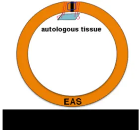
Fig. 3 Proposed graft technique for external anal sphincter (EAS) repair. The two limbs of the EAS are brought together with one standard 00 Vicryl suture (blue). A small 1.5 × 1 cm autologous (preferably) or other graft is sutured below the two joined limbs of the EAS (red). This protects the healing wound and spreads the expansionary force created by the fecal bolus during defecation
healthy tissue. Also, in our view, excessive tension on the EAS from the overlap may create a contractile force pulling in opposite directions, which may disrupt the wound sutures.
What type of graft?
Each type of graft has risks and benefits. In our view, an autolo-gous graft is the cheapest and best option: excision of a full-thickness elliptical graft from the vagina or abdominal skin; re-move the epithelial layer with a sharp scalpel and trim accordingly to a 1.5 × 1 cm rectangle. The downside is that an autologous graft is more invasive, needs more surgical skill and experience, and is not easily applicable in a birth suite. We believe that a graft repair is best done in the operating room. What type of autologous graft? Those of us who do grafts anecdotally agreed that an abdominal graft would be stronger, but less accessible. A vaginal graft is weaker but easily accessible if the decision is primary repair.
A small-intestinal submucosa (SIS) graft seemed ideal in that it can be applied directly and dissolves in 6–8 weeks. However, it is expensive, not universally available, and not so easy to use. It needs to be soaked and stretched, which is not an easy task for a small postage stamp-sized graft, especially in a labor ward en-vironment. Polypropylene mesh is cheap, simpler, and more pre-dictable technically to use than SIS, especially for surgeons who have experience in using mesh. It will create a firm structure on the inferior margin of the EAS (Fig.3). However, like all mesh grafts, there is a possibility of tissue reaction, surfacing, and erosion.
The new method awaits a systematic assessment, prefera-bly by RCT, and compared with standard methods.
Should fecal incontinence be the criterion for EAS
cure?
Finally, we discuss using cure of FI as the criterion for the success of anal sphincter repair. This would work if FI was exclusively caused by an EAS defect. It is not. In a study involving 1,420 patients who had a posterior polypropylene sling to cure apical
prolapse [9], 162 had FI and approximately 70% were cured of their FI by a posterior sling. None had prior EAS damage. The posterior sling shortened and reinforced the uterosacral liga-ments. This study demonstrated that FI could occur without EAS damage and could be cured by a posterior sling. Put another way, persistence of FI could occur in patients with a perfectly restored EAS, but who developed FI with another cause, such as in the case of Wagenlehner et al. [9], because of loose or dam-aged uterosacral ligaments [9]. On this basis alone, as suggested previously [10], ultrasound evidence of anatomical restoration would seem a more reasonable criterion for EAS cure than ab-sence of FI. For further information on connective tissue patho-genesis of FI, interested readers are referred to Petros and Swash [11], which is free online.
Conclusions
We have analyzed causes of sphincter repair failure, intro-duced a graft method for the prevention thereof and supported ultrasound assessment, rather than the absence of FI as the criterion for success of EAS repair. Although based on well-established biomechanical principles, our proposal at this stage remains unproven. Our hope is that these concepts will be challenged, clarified, and tested, preferably in an RCT. Authors’ contributions All authors: conceptualization, planning, writing; P.P.: figures.
Compliance with ethical standards
Conflicts of interest None.
References
1. Beale RM, Petros PE. Passive management of labour may predis-pose to anal sphincter injury. Int Urogynecol J.https://doi.org/10. 1007/s00192-019-04183-6.
Fig. 5 Striated muscle contracts efficiently only over a finite length [5]. Maximal contractile force is at 2. 1 represents shortening of the sarcomere. 3 represents lengthening of the sarcomere. Shortening beyond 1 results in an exponential fall in contractile strength
2. Pinta TM, Kylanpaa ML, Salmi TK, Teramo KA, Luukkonen PS. Primary sphincter repair: are the results of the operation good enough? Dis Colon Rectum. 47:18–23.
3. Sultan AH, Thakar R. Posterior compartment disorders and man-agement of acute anal sphincter trauma. In: Santoro GA, Wieczorek AP, Bartram CI, editors. Pelvic floor disorders. Milan: Springer; 2010.
4. Oliva F, Via AG, Kiritsi O, Foti C, Maffulli N. Surgical repair of muscle laceration: biomechanical properties at 6 years follow-up. Muscles Ligaments Tendons J. 2013;3(4):313–7.
5. Gordon AM, Huxley AF, Julian FJ. The variation in isometric ten-sion with sarcomere length in vertebrate muscle fibres. J Physiol. 1966 May;184(1):170–92.
6. Douglas DM. The healing of aponeurotic incisions. Br J Surg. 1952;40:79–84.
7. Kragh JF Jr, Svoboda SJ, Wenke JC, Ward JA, Walters TJ. Epimysium and perimysium in suturing in skeletal muscle lacera-tions. J Trauma. 2005;59:209–12.
8. Yamada H. Aging rate for the strength of human organs and tissues. In: Evans FG, editors. Strength of biological materials. Baltimore: Williams & Wilkins;1970. p. 272–280.
9. Wagenlehner F, Andrei Muller-Funogea I-A, Perletti G, Abendstein B, Goeschen K, Inoue H, et al. Vaginal apical prolapse repair using two different sling techniques improves chronic pelvic pain, urgency and nocturia—a multicentre study of 1420 patients. Pelviperineology. 2016;35:99–104.
10. Abdool Z, Sultan AH, Thakar R. Ultrasound imaging of the anal sphincter complex: a review. Br J Radiol. 2012;85(1015):865–75.
https://doi.org/10.1259/bjr/27314678.
11. Petros PE, Swash MA. The musculoelastic theory of anorectal func-tion and dysfuncfunc-tion in the female. J Pelviperineol. 2008;27:89–93. Publisher’s note Springer Nature remains neutral with regard to jurisdic-tional claims in published maps and institujurisdic-tional affiliations.
![Fig. 5 Striated muscle contracts efficiently only over a finite length [ 5 ]. Maximal contractile force is at 2](https://thumb-eu.123doks.com/thumbv2/9libnet/3819612.33793/4.892.346.816.85.324/striated-muscle-contracts-efficiently-finite-length-maximal-contractile.webp)
