1 Altıparmak B, et al. BMJ Case Rep 2019;12:e227495. doi:10.1136/bcr-2018-227495
Case report
Double axillary vein variation diagnosed with
ultrasound guidance during infraclavicular nerve
block intervention
Başak altıparmak,
1Melike Korkmaz toker,
2ali İhsan Uysal,
2semra Gümüş Demirbilek
1Reminder of important clinical lesson
To cite: altıparmak B, Korkmaz toker M, Uysal aİ, et al. BMJ Case Rep 2019;12:e227495. doi:10.1136/bcr-2018-227495
1anesthesiology and reanimation, Muğla sıtkı Koçman University, Muğla, turkey
2anesthesiology and reanimation, Muğla sıtkı Koçman University training and research Hospital, Muğla, turkey Correspondence to Dr Başak altıparmak, basak_ ugurlu@ yahoo. com accepted 9 December 2018
© BMJ publishing Group Limited 2019. No commercial re-use. see rights and permissions. published by BMJ.
Summary
the use of ultrasound guidance increases the safety of peripheral block interventions by allowing anaesthesiologists to simultaneously see the position of block needle, the targeted nerves and surrounding vessels. In this report, we represented three patients diagnosed with double axillary vein variation with ultrasound guidance during infraclavicular nerve block intervention. the patients were scheduled for different types of upper limb surgeries. all patients received infraclavicular nerve block for anaesthetic management. a double axillary vein variation was diagnosed with ultrasound during block interventions. Hydro-location technique was used in all cases and the procedures were completed uneventfully. In the current literature, there is limited number of reports concerning double axillary vein variation. Detailed knowledge of the axillary anatomy is important to avoid complications such as intravascular injection during peripheral nerve block interventions. the use of ultrasound guidance and hydro-location technique should be considered for nerve blocks, especially in the axillary area.
BaCkground
The use of ultrasound guidance during periph-eral block interventions has gained popularity in the recent years. As it allows anaesthesiologists to simultaneously see the position of block needle, the targeted nerves and surrounding vessels, the use of ultrasound increases the success rate and safety of block interventions.1 2
The infraclavicular nerve block is a frequently preferred anaesthetic technique for elbow and upper limb surgery. It has several advantages such as lower likelihood of tourniquet pain, a reliable blockade of the musculocutaneous nerve and a simpler and safer approach when compared with multi-injec-tion axillary nerve block and mid-humeral blocks.3
However, the risk of complications increases in the presence of anatomical variations. In this report, we aimed to represent the management of three patients diagnosed with double axillary vein varia-tion during infraclavicular nerve block intervenvaria-tion under ultrasound guidance.
CaSe preSenTaTion
Written informed consents for block procedures and sharing of the data were obtained from each patient. The first patient was a 27-year-old man with
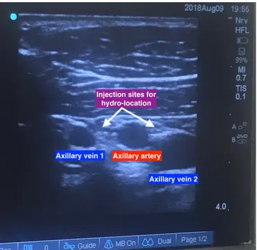
American Society of Anaesthesiologists status I. He was scheduled for an elbow surgery due to distal radius fracture. The patient was planned to receive infraclavicular nerve block for anaesthetic manage-ment. The arm of the patient was adducted to the side of the operation and the coracoid process was palpated. Under aseptic conditions, the ultrasound probe was located to the intersection point between the coracoid process and the clavicula. Two axillary veins, a single axillary artery and cords were identi-fied (figure 1). The skin was infiltrated with 3–4 mL of lidocaine under ultrasound guidance. Then 8 cm, 22-gauge Stimuplex needle was inserted along the same path to the axillary vein medial to the axillary artery with inplane technique. The targeted vein was moved away from the axillary artery by using hydro-location technique with 2–3 mL of saline solution. We aspirated the syringe before each local anaesthetic application in order to avoid intravas-cular injection. A total of 30 mL 0.25% bupiva-caine was then infiltrated around the artery until each cord was surrounded by a ring of local anaes-thetic solution (figure 2). The second patient was a 46-year-old man with American Society of Anaes-thesiologists status II and he was scheduled for an elective hand surgery. Using the same technique, we identified two axillary veins and a single axillary artery. The nerve block was performed unevent-fully with the same technique. And the third patient was a 19-year-old man with American Society of Anaesthesiologists status I. He was planned to have an operation for distal humerus fracture. We again identified two axillary veins and a single axillary artery during infraclavicular nerve block with ultrasound guidance. As the vessels were very close to each other, we confirmed the positions of veins and artery by using Doppler ultrasonography. Then again, we used hydro-location technique with 3–4 mL of saline solution to avoid intravascular injection and approach near the artery. In all cases, an adequate anaesthesia was provided without any complications.
ouTCome and follow-up
None of the patients had any complications related to infraclavicular nerve block intervention. The operations were completed uneventfully. The patients were informed about their axillary vein variation.
2 altıparmak B, et al. BMJ Case Rep 2019;12:e227495. doi:10.1136/bcr-2018-227495
reminder of important clinical lesson
diSCuSSion
The connection of the basilic and the brachial veins forms the axillary vein which is located between the lateral edge of the teres major muscle and the lateral edge of the first rib. The axillary artery runs slightly above and posterior to the axillary vein.4 In the
liter-ature, there is limited information about the variations of axillary vessels. Kutiyanawala et al studied on 100 patients and reported the incidence of double axillary veins as 10%.5 Khan et al
docu-mented the variants of axillary region in 73 patients. They reported the incidence of double axillary veins as 14%.6 Both studies were
performed during axillary dissection as part of the breast cancer surgery. Berthier et al conducted a study to show the correlations
between the anatomical structures seen in the cadaveric dissection and ultrasound images during axillary nerve block.7 They reported
to detect several cases with multiple axillary veins during ultrasound guided axillary nerve block procedure. However, the number of patients or the cases with double axillary veins were not mentioned in this study. Other than these, the current literature contains a single case report to represent detection of double axillary veins during peripheral nerve block intervention under ultrasound guid-ance, and another report describing a double axillary vein variation preoperatively diagnosed by CT.8 9 In these reports, the authors
highlighted the importance of detailed knowledge of the axillary anatomy to avoid complications such as intravascular injection of local anaesthetics during peripheral nerve block interventions or injury to the vessels and nerves during surgical axillary dissec-tion.5–8 Uysal et al reported that the use of hydro-location technique
provided a safer area during infraclavicular nerve block intervention of a patient with double axillary vein.8 Hydro-location technique
is a principle to locate needle-tip by using ultrasound liquid detec-tion. The technique is performed with the injection of 0.5–1 mL liquid under ultrasound beam to identify the anatomical position of needle tip.10 We used hydro-location technique in all patients with
double axillary vein variation and the interventions were completed without any complications. Particularly, an anatomical variation of the axillary vessels have an increased risk of vascular puncture during peripheral nerve block interventions performed blinded or guided only by electrical nerve stimulation.7 In the light of these
information, the use of ultrasound guidance should be considered during nerve blocks, especially in the axillary area.
As a conclusion; variations in the anatomy of axillary area are common but descriptions remain rare in the literature. In the presence of multiple vascular structures, the anaesthesiologists should perform real time observation of anatomical structures under ultrasound guidance. Using hydro-location technique and intermittent aspiration of the syringe during local anaesthetic injection may provide a safer condition for peripheral block interventions.
learning points
► The use of ultrasound guidance increases the safety of peripheral block interventions by allowing anaesthesiologists to simultaneously see the position of block needle.
► Hydro-location technique is a principle to locate needle-tip by using ultrasound liquid detection.
► Double axillary vein is a common but poorly described variation of the axillary area.
► An anatomical variation of the axillary vessels have an increased risk of vascular puncture during peripheral nerve block interventions performed blinded or guided only by electrical nerve stimulation.
Contributors Ba (Muğla sıtkı Koçman University) has contributions in; 1. Conception and design, acquisition of data or analysis and interpretation of data. 2. Drafting the article or revising it critically for important intellectual content. 3. Final approval of the version published. 4. agreement to be accountable for the article and to ensure that all questions regarding the accuracy or integrity of the article are investigated and resolved. MKt (Muğla sıtkı Koçman University training and research Hospital) has contributions in; 1. Conception and design, acquisition of data or analysis and interpretation of data. 2. Drafting the article or revising it critically for important intellectual content. 3. Final approval of the version published. 4. agreement to be accountable for the article and to ensure that all questions regarding the accuracy or integrity of the article are investigated and resolved. aİU (Muğla sıtkı Koçman University training and research Hospital) has contributions in; 1. Conception and design, acquisition of data or analysis and interpretation of data. 2. Drafting the article or revising it critically for important intellectual content. 3. Final approval of the version published. 4. agreement to be accountable for
figure 1 Double axillary veins and single axillary artery diagnosed with ultrasonography.
3 Altıparmak B, et al. BMJ Case Rep 2019;12:e227495. doi:10.1136/bcr-2018-227495
reminder of important clinical lesson
the article and to ensure that all questions regarding the accuracy or integrityof the article are investigated and resolved.semra GÜMÜŞ DeMİrBİLeK (Muğla sıtkı Koçman University training and research Hospital) has contributions in; 1. Conception and design, acquisition of data or analysis and interpretation of data. 2. Drafting the article or revising it critically for important intellectual content.3. Final approval of the version published. 4. agreement to be accountable for the article and to ensure that all questions regarding the accuracy or integrity of the article are investigated and resolved.
funding the authors have not declared a specific grant for this research from any funding agency in the public, commercial or not-for-profit sector.
Competing interests None declared. patient consent obtained.
provenance and peer review Not commissioned; externally peer reviewed.
RefeRences
1 perlas a, Brull r, Chan VW, et al. Ultrasound guidance improves the success of sciatic nerve block at the popliteal fossa. Reg Anesth Pain Med 2008;33:259–65.
2 ootaki C, Hayashi H, amano M. Ultrasound-guided infraclavicular brachial plexus block: an alternative technique to anatomical landmark-guided approaches. Reg Anesth Pain Med 2000;25:600–4.
3 Chin KJ, singh M, Velayutham V, et al. Infraclavicular brachial plexus block for regional anaesthesia of the lower arm. Anesth Analg 2010;111:1072.
4 Galloway s, Bodenham a. Ultrasound imaging of the axillary vein-anatomical basis for central venous access. Br J Anaesth 2003;90:589–95.
5 Kutiyanawala Ma, stotter a, Windle r. anatomical variants during axillary dissection.
Br J Surg 1998;85:393–4.
6 Khan a, Chakravorty a, Gui Gp. In vivo study of the surgical anatomy of the axilla. Br J Surg 2012;99:871–7.
7 Berthier F, Lepage D, Henry Y, et al. anatomical basis for ultrasound-guided regional anaesthesia at the junction of the axilla and the upper arm. Surg Radiol Anat
2010;32:299–304.
8 Uysal aİ, tarakçı e, altıparmak B, et al. [the anatomical variation to be considered in upper extremity blocks: double axillary vein]. Agri 2017;29:147–8.
9 Fujii t, Yajima r, Yamaguchi s, et al. Double axillary vein: a case detected by Ct. Int J Case Reports Images 2013;3:58–9.
10 Bloc s, ecoffey C, Dhonneur G. Controlling needle tip progression during ultrasound-guided regional anesthesia using the hydrolocalization technique. Reg Anesth Pain Med 2008;33:382–3.
Copyright 2019 BMJ publishing Group. all rights reserved. For permission to reuse any of this content visit https://www.bmj.com/company/products-services/rights-and-licensing/permissions/
BMJ Case report Fellows may re-use this article for personal use and teaching without any further permission. Become a Fellow of BMJ Case reports today and you can:
► submit as many cases as you like
► enjoy fast sympathetic peer review and rapid publication of accepted articles ► access all the published articles
► re-use any of the published material for personal use and teaching without further permission For information on Institutional Fellowships contact [email protected]