Endovascular Treatment of Acute Stroke due to the
Floating Carotid Thrombus with Intra-Arterial Tissue
Plasminogen Activator and Stenting
Hareketli
Karotis Trombüsüne Bağlı Akut Strokun Stentleme ve İntra-arteryel
Doku Plazminojen Aktivatörü ile Endovasküler Tedavisi
Enes Duman
Başkent University, Konya Research Center, Department of Interventional Radiology, KONYA Cukurova Medical Journal 2015;40 (Ek Sayı 1):142-146.
ABSTRACT
54-year-old male patient was admitted to emergency service with bilateral loss of sight, consciousness and a Glasgow Coma Scale score of 15. His neurologic examination revealed no lateralized deficit but left homonymous hemianopia. Brain CT and diffusion MRI showed acute infarction in the right parietal and occipital lobes. There was a wide penumbra region on brain perfusion CT. The selective angiography showed preocclusive stenosis at the origin of right internal carotid artery (ICA) and massive floating thrombus (FT) in the cervical ICA segment.We report successful endovascular treatment of FT in the ICA using intra-arterial tissue plasminogen activator and stenting.
Key words: Floating thrombus, tPA, endovascular stent placement, carotid stenosis.
ÖZET
54 yaşında Glasgow koma skala skoru 15 olan erkek hasta acil servise bilateral görme kaybı, bilinç kaybı şikayeti ile başvurdu. Nörolojik muayenesinde lateralizasyon bulgusu izlenmeyen hastanın sol homonymous hemianopia’ sı mevcuttu. Beyin tomografisi ve beyin difüzyon MRG’ sinde sağ parietal ve oksipital lobda akut enfarkt saptandı. Perfüzyon BT tetkikinde geniş penumbra (kurtarılabilir alan- mismatch) alanı izlendi. Selektif karotit anjiyografi tetkikinde sağ internal karotit arter orjininde preokluziv darlık ve masif floating thrombus izlendi. Bu vakada intraarteriyel tPA ve stent kullanarak floating trombüsün başarılı endovasküler tedavisini paylaştık.
Anahtar kelimeler: Floating trombüs, tPA, endovasküler stent yerleştirilmesi, karotit darlığı
INTRODUCTION
Although the exact etiology of floating thrombus (FT) in the carotid artery is unknown, most cases are due to embolic thrombus or plaque rupture. FFT affects men more than women, with a ratio of nearly 2:1 at a younger age than most patients with carotid disease1. FT is a particularly difficult problem for the physician because most patients present with acute neurologic symptoms,
and it is unclear whether surgical or medical treatment is the best option1. Most of the reports in the literature to date describe surgical intervention and medical treatment with similar success rates
2-4
. Medical management for stabilizing neurologic deficits has less risk and less benefit than surgical intervention1.
Here, we report the successful endovascular treatment of acute FT in the carotid artery using
intra-arterial tissue plasminogen activator (tPA) and stenting.
CASE
A 54-year-old male patient was admitted to emergency service with bilateral loss of sight, consciousness and a Glasgow Coma Scale score of 15. His neurologic examination revealed no lateralized deficit but left homonymous hemianopia. Brain CT and diffusion MRI showed acute infarction in the right parietal and occipital lobes. Because his symptoms began one hour earlier and a wide penumbra region was apparent on brain perfusion CT (Aqullion, Toshiba, Japan) , the patient was taken to the interventional radiology unit for endovascular revascularization. There was a miss-match between mean transmitting time and cerebral blood volume and it showed that patient had a wide penumbra region on brain perfusion CT. Perfusion CT levels are below.
CBV MTT 1-Right 0.49 14.71 2-Left 3.15 9.78 % -74 +50
An informed consent was obtained from the patient and family members. The right carotid angiograph showed massive floating thrombus reaching from the origin of the ICA to the petrous segment of the ICA (Fig.1). There was reduced blood flow in the branches of the middle cerebral artery and no flow in the anterior cerebral artery (Fig.2). Because of high risk of distal embolism with massive FT, endarterectomy and endovascular mechanical thrombectomy were not performed. We decided to perform intra-arterial thrombolytic therapy. We placed 5F vertebral catheter into the right common carotid artery and the 4 mg tirofiban HCl (Aggrastat®, DSM Pharmaceuticals, North Carolina, ABD), 10 mg tPA
(Actilyse®, Boehringer Ingelheim Pharma, Riss, Germany) were given in fifteen minutes over the catheter with bolus-style. For sixteen hours, we continued to tPA infusion 1 mg per hour with controlling blood fibrinogen level every two hours. We stopped the tPA infusion because fibrinogen level dropped 425 mg/dL to 110 mg/dL at end of 16 hour of tPA infusion. Then we continued to the anticoagulant treatment with 500 IU/hour heparin (Vasparin®, Mefar, Istanbul, Turkey) infusion for 8 hours. The 24-hour control angiography showed that floating thrombus was resolved almost completely (Fig.3) and there was a critic stenosis at the origin of ICA, and quantity of residual thrombus in the distal segment of the stenosis and small thrombus particles were on the wall of the main carotid artery. So we decided to apply endovascular stent placement and angioplasty to the stenotic segment of ICA. 600 mg clopidogrel and 300 mg corasprin was given to the patient eight hour before the carotid artery stenting (CAS). In CAS procedure firstly, we reached to the distal branch of the external carotid artery with 0,035-inch hydrophilic glide wire, and 5F vertebral catheter. Then, we exchanged available 5 Fr sheath on femoral artery with 6F longer sheath (Epsylar, Optimed, Ettlingen, Germany).We placed the filter device (Angioguard RX, Cordis Corporation, Miami, USA) to the petrous segment of ICA to avoid the distal embolism. Then 7mm x 40 mm self-expandableclosed cell carotid stent (Carotid wall stent,Boston scientific,Galway, Ireland) were placed, and angioplasty with 5 mm balloon (Ultra-thin Diamond, Boston scientific, Galway, Ireland) was done. The control angiography, after CAS procedure, showed complete patency of ICA and distal flow (Fig. 4a, b). 75 mg clopidogrel and 300 mg corasprin were given daily after the procedure, and the patient was discharged on post-operative 7th day. The patient was followed-up with carotid artery Doppler US and brain CT at sixth month. He has no problem and followed-up uneventfully.
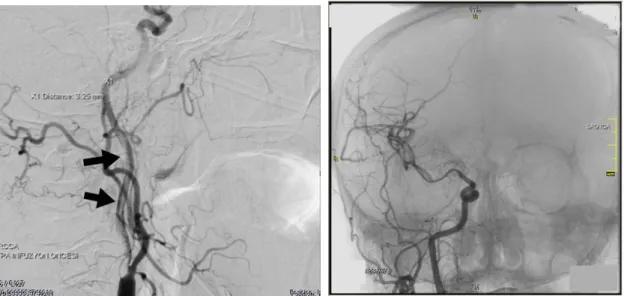
Figure 1. Lateral selective right carotid DSA image shows Figure 2. AP cerebral angiography shows; partial blood
preocclusive stenosis at the origin of ICA and massive flow in the branches of middle cerebral artery, and no floating thrombus (black arrows) in the cervical ICA flow in the anterior cerebral artery
segment.
Figure 3. Lateral selective right carotid DSA image shows almost complete resolution of thrombus and pre-occlusive
stenosis at the origin of the right ICA.
Figure 4a, b. Lateral right carotid (a), and AP cerebral (b) DSA images shows; 100% patency of right ICA after
endovascular stent placement, and normal blood flow of both anterior and middle cerebral arteries and their branches.
DISCUSSION
The suggested incidence of FT, because of the lack of a unified definition of FT, ranges between 0.4% and 0.7%. Additionally, FT may be misidentified intraoperatively as an atherosclerotic plaque instead of thrombus, thereby contributing to the underestimation of the incidence of this entity1.
Although the exact etiology of FT in the carotid artery is unknown, most cases are due to embolic thrombus or plaque rupture. FT has been reported as a rare cause of ischemic stroke. FTs usually show significant mobility with intact antegrade flow because portions are stuck to the injured arterial wall. Their instability can make them sources of recurrent artery-to-artery embolism. The most serious complication is detachment resulting in complete occlusion of the distal cerebral arteries. FT is a particularly difficult problem for the physician because most patients present with acute neurologic symptoms, and it is unclear whether surgical or medical treatment is superior. Medical management for stabilizing neurologic deficits carries less risk but also less potential benefit than surgical intervention1. Several treatment options for carotid stenosis with
literature, such as endarterectomy, medical therapy and endovascular stent placement2-4.
The selective intra-arterial thrombolytic therapy with tPA has been used for acute intracranial occlusions, and lower extremity thromboembolism, but the use of tPA for the carotid thrombosis is not well established. Randomized studies of unfractionated heparin, low-molecular-weight heparins, or heparinoids for the treatment of acute ischemic strokes showed no proven benefit in reducing stroke-related mortality and morbidity, stroke recurrence, and prognosis5-8. The major concerns about the use of tPA are bleeding complication and dosage. Although it is not proven, the blood fibrinogen level monitoring may be prevent bleeding complication, and can be used as an independent indicator if it drops rapidly or below 150mg/dL9. We need clinical investigations that show relationships between blood fibrinogen level and bleeding complication when tPA used for treatment.
Emergent CEA is one of treatment options for these patients, but it has higher risk of distal embolism. The CEA may be the treatment of
symptom onset or absence of penumbra region on perfusion CT or MRI examinations.
In our case we performed intra-arterial thrombolytic therapy. Because there was a high risk of distal embolism with massive FT, endarterectomy and endovascular mechanical thrombectomy were not performed. After intra-arterial thrombolytic treatment, endovascular treatment or CEA is suitable for patients.
In conclusion, we must evaluate each patient individually to determine the best treatment approach. For patients who have acute carotid FT and large penumbra region on brain perfusion examinations using of intra-arterial tPA is relatively safe and feasible and should be kept in mind.
Statement of Human and Animal Rights: All procedures involving human participants were in accordance with the ethical standards of the institutional and/or national research committee and with the 1964 Helsinki declaration and its later amendments or comparable ethical standards.
REFERENCES
1. Ahmad F, Bhatti, Luis R. Leon, Nicos Labropoulos, et.al. Free-floating thrombus of the carotid artery: literature review and case reports. J Vasc Surg. 2007;45:199-205.
2. Sallustio F, Di Legge S, Marziali S, Ippoliti A, Stanzione P. Floating carotid thrombus treated by intravenous heparin and endarterectomy. J Vasc Surg. 2011;53-2:489-91.
3. Tsumoto T, Terada T, Tsuura M, et.al. Carotid artery stenting for stenosis with intraluminal thrombus. Neuroradiology. 2006; 48-1:54-9.
4. Sugg RM, Malkoff MD, Noser EA, et al. Endovascular recanalization of internal carotid artery occlusion in acute ischemic stroke. AJNR Am J Neuroradiol. 2005; 26-10:2591-4.
5. Sandercock P. Intravenous unfractionated heparin in patients with acute ischemic stroke: a treatment to be used in the context of randomized trials only. Stroke. 2001;32:579.
6. Gubitz G, Counsell C, Sandercock P, Signorini D. Anticoagulants for acute ischemic stroke. Cochrane Database Syst Rev. 2000;2:CD000024.
7. Swanson RA. Intravenous heparin for acute stroke: what can we learn from megatrials? Neurology 1999;52:1746-50.
8. Coull BM, Williams LS, Goldstein LB, et al. Anticoagulants and antiplatelet agents in acute ischemic stroke: report of the Joint Stroke Guideline Development Committee of the American Academy of Neurology and the American Stroke Association (a Division of the American Heart Association). Stroke, 2002;33:1934-42.
9. Skeik N, Gits CC, Ehrenwald E, Cragg AH. Fibrinogen level as a surrogate for the outcome of thrombolytic therapy using tissue plasminogen activator for acute lower extremity intravascular thrombosis. Vasc Endovascular Surg. 2013;47-7:519-23.
Yazışma Adresi / Address for Correspondence:
Dr. Enes Duman
Başkent Üniversitesi Tıp Fakültesi Konya Uygulama ve Araştırma Merkezi KONYA
e-mail:[email protected] Geliş tarihi/Received on : 26.05.2015 Kabul tarihi/Accepted on: 14.07.2015