245 © 2018 Journal of Pediatric Neurosciences | Published by Wolters Kluwer - Medknow
Access this article online Quick Response Code:
Website:
www.pediatricneurosciences.com
DOI: 10.4103/jpn.JPN_96_17
Seckel syndrome and Moyamoya diseases are different entities that rarely associated with each other. Several cases presenting with both these diseases were reported. Intracerebral artery aneurysms and collateral vessels can be seen with Moyamoya. They are commonly treated with medical treatment. We present a 12-years old patient with both Seckel syndrome and Moyamoya disease presented with middle cerebral artery aneurysm which was treated with endovascular modalities.
Keywords: Endovascular treatment, intracranial aneurysm, moyamoya disease,
Seckel syndrome
I
ntroductIonS
eckel syndrome (SS), the so-called bird-headed dwarfism, is a rare autosomal recessive disorder characterized by prenatal and postnatal growth, mental retardation, microcephaly, and also facial abnormalities.[1] Association of different vascular anomalies such as multiple aneurysms, polyarteritis nodosa, and moyamoya (MM) disease has been reported with this syndrome, and the patients could show a wide range of symptoms, including malignant hypertension, intracranial hemorrhage, and cerebrovascular accident.[1,2] Here, we describe a case report of a 12-year-old child with SS associated with MM disease and multiple aneurysms, and who presented with cranial nerve palsy (CN III), resulting from a mass effect of the left dissecting middle cerebral artery (MCA) aneurysm, and also with left subdural hematoma surrounding the whole hemisphere. Furthermore, the treatment of subdural hematoma by surgical approach and endovascular treatment of the symptomatic MCA aneurysm are described.c
aser
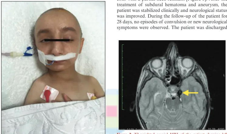
eportA 12-year-old boy, who had been diagnosed with SS and followed up by another institution, was referred to our hospital with convulsion. Patient’s physical examination revealed microcephalia, dwarfism, large-beaked nose,
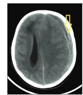
receding forehead, and micrognathia [Figure 1]. Furthermore, left ptosis and pupillary dilatation pointing to third nerve oculomotor palsy were present. The patient was hospitalized. Magnetic resonance imaging (MRI) and magnetic resonance angiography (MRA) showed hyperintense areas of periventricular deep white matter, left subdural hematoma, left MCA aneurysm, and abnormal collateralized signal void areas in basal cisterna with signs of MM [Figure 2]. Digital subtraction angiography (DSA) showed MM-like vasculopathic changes and pial and dural collateral vessels in the basal ganglia and basal cisterna. Moreover, two saccular aneurysms of right internal carotid artery (ICA), MCA not greater than 2 mm, and a dissecting wide-necked 10-mm left MCA aneurysm were seen. Vertebrobasilar angiograms showed a wide-necked complex basilar tip aneurysm [Figure 3]. The following day, neurological status of the patient worsened and consciousness deteriorated. Cranial computerized tomography (CT) showed an increase in the size of subdural hematoma by 1 cm and midline shift of the brain to the right side and parenchymal edema
a
bstra
ct
jopn
MEDKNOW
Journal of Pediatric Neurosciences JOPN
1817-1745 1998-3948
Medknow Publications & Media Pvt LtdIndia
10.4103/jpn.JPN_96_17 9617 000000 000000 00 00 00 00 20171127
© 2017 Journal of Pediatric Neurosciences | Published by Wolters Kluwer - Medknow 2017
Address for correspondence: Dr. Aylin Gunesli MD. Baskent University, Adana Teaching and Research Center, Department of Radiology, Dadaloglu Mh. 2591 Sk. No:4/A PK:01250 Yuregir/ADANA, Turkey. E-mail: [email protected] This is an open access journal, and articles are distributed under the terms of the Creative Commons Attribution-NonCommercial-ShareAlike 4.0 License, which allows others to remix, tweak, and build upon the work non-commercially, as long as appropriate credit is given and the new creations are licensed under the identical terms. For reprints contact: [email protected]
How to cite this article: Gunesli A, Andic C, Alkan O, Erol I, Suner HI. Endovascular treatment of a patient with Moyamoya disease and Seckel syndrome: A case report. J Pediatr Neurosci 2018;13:245-8.
Case Report
Endovascular Treatment of a Patient with Moyamoya Disease and Seckel
Syndrome: A Case Report
Aylin Gunesli, Cagatay Andic, Ozlem Alkan, Ilknur Erol,1 Halil Ibrahim Suner2
Department of Radiology,
1Department of Pediatric
Neurology, 2Department
of Neurosurgery, Baskent University, Teaching and Research Center, Adana/ TURKEY
Gunesli, et al.: Endovascular treatment of a patient with MM and SS
246 Journal of Pediatric Neurosciences ¦ Volume 13 ¦ Issue 2 ¦ April-June 2018
[Figure 4]. Urgent subdural evacuation was performed by neurosurgery. After the operation, the patient was transferred to the interventional radiology department and for the treatment of the left MCA aneurysm because of its dissecting nature and the presence of mass effect symptoms. Endovascular treatment was
performed under general anesthesia by standard transfemoral approach with a guiding sheath and guiding catheter combination. The left MCA aneurysm was selectively catheterized under road mapping with a microcatheter (Headway Duo; Microvention, Tustin, CA) and a 0.008-in microguidewire (Mirage; Covidien, Irvine, CA). The aneurysm sac had bare platinum coils with a partial neck remnant [Figure 5]. After the treatment of subdural hematoma and aneurysm, the patient was stabilized clinically and neurological status was improved. During the follow-up of the patient for 28 days, no episodes of convulsion or new neurological symptoms were observed. The patient was discharged
Figure 1: Characteristic appearance of the patient
Figure 2: T2-weighted cranial MRI of the patient showing left middle cerebral artery aneurysm pointed by yellow arrow
Figure 3: Diagnostic cerebral angiogram shows moyamoya-like vasculopathic changes in both hemispheres involving MCA and Anterior Cerebral Artery (ACA) territories and also a dissecting wide-necked left MCA aneurysm and wide-necked complex basilar tip aneurysm
Gunesli, et al.: Endovascular treatment of a patient with MM and SS
247 Journal of Pediatric Neurosciences ¦ Volume 13 ¦ Issue 2 ¦ April-June 2018
with only partial improvement on ophthalmoplegia. No complication other than ophthalmoplegia was observed during the 9-month follow-up of the patient.
d
IscussIonMoyamoya was first described in Japan; the term means puff of smoke, which has taken the name from the angiographic appearance of the disease.[3] Patients mostly presented with hemiparesis, ischemic attack or involuntary movement, and speech and visual disturbance. Gold standard for the diagnosis is DSA.[4] However, MRI and MRA can also be used
for the diagnosis to establish vascular abnormalities and acute stroke. The role of CT examination in these patients is to evaluate hemorrhage or large infarction.[3] Angiographic findings of MM are intracranial ICA stenosis and dilated collateral vessels in the area of basal ganglia and skull base.[3] MM may be associated with other congenital malformations and genetic syndromes. Rahme et al.[1] were the first to describe the association of SS and MM disease; however, they did not perform endovascular or surgical treatment. The presence of multiple intracranial aneurysms was also reported earlier with SS.[5,6] However, knowledge about the management of the patients with MM and SS was limited. As mentioned in the previous reports, the patients were managed conservatively. Our study differs from the previous cases in terms of both endovascular treatment of symptomatic aneurysms and surgical treatment of subdural hemorrhage. Endovascular treatment of intracranial aneurysms in MM and SS is more challenging because of the presence of vasculopathic changes, physical characteristics of the patients such as growth retardation, and the relatively small vessel diameters that make endovascular procedures difficult.[5] Patients with SS should be treated akin to infantile patients because the autoregulation of cerebrum is not appropriate.[7] However, the endovascular treatment can be performed without any serious complications despite the aforementioned difficulties. The cranial oculomotor dysfunction was partially improved after the treatment. However, the wide-necked basilar aneurysm was left untreated because of the stent-assisted techniques required; the stent’s long-term patency is unknown. Patients usually present with hemiparesis, ischemic attack or involuntary movement, and speech and visual disturbance.[3] However, in our case, the patient presented with convulsions. Although the gold standard for diagnosis is DSA, CT can be used in the first evaluation of hemorrhage or large infarction or MRI can be used to establish vascular abnormalities and acute stroke.[3] In our case, the MM disease was diagnosed by DSA examination. However, to our knowledge, endovascular treatment of symptomatic aneurysms in SS accompanied with MM disease has not been reported previously. Multiple intracranial aneurysms in a patient with SS were reported previously.[5] Endovascular treatment of intracranial aneurysms in pediatric population has been studied and it carries substantial risks when compared with adult population.[5] Although knowledge about the management of the patients with MM and SS is limited, a multidisciplinary approach is needed. Figure 4: Computerized tomography showing left subdural
hematoma (shown by yellow arrow)
Figure 5: Diagnostic cerebral angiogram shows dissecting wide-necked left MCA aneurysm and wide-necked complex basilar type aneurysm after embolization
Gunesli, et al.: Endovascular treatment of a patient with MM and SS
248 Journal of Pediatric Neurosciences ¦ Volume 13 ¦ Issue 2 ¦ April-June 2018
Endovascular treatment of multiple aneurysms in these patients as an alternative to surgical approach is technically feasible.
Declaration of patient consent
The authors certify that they have obtained all appropriate patient consent forms. In the form the patient(s) has/have given his/her/their consent for his/ her/their images and other clinical information to be reported in the journal. The patients understand that their names and initials will not be published and due efforts will be made to conceal their identity, but anonymity cannot be guaranteed.
Financial support and sponsorship Nil.
Conflicts of interest
There are no conflicts of interest.
r
eferences1. Rahme R, Crevier L, Dubois J, Mercier C. Moyamoya-like vasculopathy and Seckel syndrome: Just a coincidence? Childs Nerv Syst 2010;26:983-6.
2. Kutlu R, Alkan A, Kutlu O, Yakinci C. Seckel syndrome with polyarteritis nodosa. Indian Pediatr 2004;41:1158-61.
3. Smith ER, Scott RM. Spontaneous occlusion of the Circle of Willis in children: Pediatric Moyamoya summary with proposed evidence-based practice guidelines. A review. J Neurosurg Pediatr 2012;9:353-60.
4. Piao J, Wu W, Yang Z, Yu J. Research progress of Moyamoya disease in children. Int J Med Sci 2015;12:566-75.
5. Di Bartolomeo R, Polidori G, Piastra M, Viola L, Zampino G, Chiaretti A. Malignant hypertension and cerebral haemorrhage in Seckel syndrome. Eur J Pediatr 2003;162:860-2.
6. Codd PJ, Scott RM, Smith ER. Seckel syndrome and Moyamoya. J Neurosurg Pediatr 2009;3:320-4.
7. D’Angelo VA, Ceddia AM, Zelante L, Florio FP. Multiple intracranial aneurysms in a patient with Seckel syndrome. Childs Nerv Syst 1998;14:82-4.