Original investigation
Generation of an in vitro Intratumoral Heterogeneity Model by Lentiviral
Fluorescent Labeling of Colon Cancer Cell Line DLD-1 Subclones
Öykü Gönül Geyik
1*, Hande Efe
1, Seda Baykal Köse
1, Özge Uysal
1, Zeynep Yüce
11Department of Medical Biology and Genetics, Faculty of Medicine, Dokuz Eylul University, Izmir, Turkey
*Current affiliation for Ö.G.G.: Department of Nutrition and Dietetics, Faculty of Health Sciences, İstinye University, Istanbul, Turkey Address for Correspondence: Zeynep Yüce, E-mail: [email protected]
Received: 09.12.2019; Accepted: 17.03.2020; Available Online Date: 15.05.2020
©Copyright 2020 by Dokuz Eylül University, Institute of Health Sciences - Available online at www.jbachs.org
Cite this article as: Gönül Geyik Ö, Efe H, Baykal Köse S, Uysal Ö, Yüce Z. Generation of an in vitro Intratumoral Heterogeneity Model by Lentiviral Fluorescent Labeling of Colon Cancer
Cell Line DLD-1 Subclones. J Basic Clin Health Sci 2020; 4:149-153.
ABSTRACT
Purpose: Studying genomic changes during tumor progression has helped to understand the biology of many different cancers and has been
the basis for targeted therapy strategies. However, resistance and differences in response to therapy in patients are still very important issues. One of the major underlying reasons is intratumoral cellular heterogeneity. Clones harbor mutations and/or epigenetic patterns providing a survival advantage under changing micro-environmental conditions are the main culprits of therapy resistance. Therefore, it is crucial to define and to study the properties and the contributions of these deviant subclones in vitro. In order to achieve that, we have generated a fluorescent intratumoral heterogeneity model of the colon cancer cell line DLD-1.
Methods: We used 2N subclones (C3 and C34) and 4N subclones (B9 and B12) of DLD-1, isolated by our team previously. Subclones were
stably transduced using lentiviral vectors carrying different fluorescent labels and selected by puromycin.
Results: Labeled subclones were mixed in equal proportions and a co-culture model of intratumoral heterogeneity was generated. Fluorescent
signals were then confirmed by fluorescence microscope.
Conclusion: The in vitro model we have generated may be used in many tumor kinetic studies. By co-culturing different clones, profiles that
have a selective advantage under different conditions can be detected. After exposure to different chemotherapeutic agents, radiation and/or combinations, real time changes in population kinetics can be tracked. By comparing these results to the genomic profiling of subclones, it will be possible to relate variations that are responsible for any observed therapeutic resistance in vitro.
Keywords: colon cancer, DLD-1, fluorescent labeling, lentiviral transduction, cancer therapy resistance
Clonal evolution and the genetic and phenotypic heterogeneity observed in cancer cell populations are important research topics in recent years. Studying genomic changes that have been protected during tumor progression –despite clonal evolution– has help us understand the underlying biology of many different cancers; and has also been the basis for targeted therapy strategies. One of the major underlying reasons for therapy resistance and disease recurrence in patients is intratumoral cellular heterogeneity (1–3). Tumor cells compete for resources (glucose, oxygen, growth factors, endocrine/paracrine/autocrine secretions etc.) in their microenvironment. Access to vital resources is not equal for all cells. For example, while central regions are more hypoxic/anoxic, cells close to blood vessels are generally normoxic. In addition to access to resources, microenvironmental alterations, which can be physiological changes as well as therapy-dependent changes,
also don’t affect the tumor cell population homogeneously. All these factors cause selection of the tumor cells that can adapt to altered conditions (1). Continuously changing conditions cause more aggressive subpopulations with different mutations and/ or epigenetic alterations to arise and proliferate in the tumor population, and result in intratumoral heterogeneity (2).
Since tumors have highly heterogeneous cell populations, subclones resistant to various therapy methods evolve in the population. Studies have shown that these resistant subclones share certain mutations and epigenetic patterns (4). These mutual genetic alterations being preserved in various cancer types, added on to the knowledge about that cancer type and aroused the interest about how non-mutual genetic alterations provide advantage to different subclones.
Different tumor cell phenotypes (therapy resistant/sensitive) observed in intratumoral heterogeneity are thought to be a result of genomic, chromosomal and/or expressional variances. Resistance to therapy is not a biological characteristic of the whole tumor cell population. It develops by the death of the sensitive cells and a group of resistant cells taking over the entire population. If these resistant cells can be distinguished and their genomic differences from the non-resistant neighboring cells can be determined, new therapy targets and approaches could be developed and our knowledge about tumor biology could expand substantially. Intratumoral heterogeneity is one of the reasons that cancer patients respond differently to similar treatments. Subclonal cancer cell populations that bear mutations and epigenetic patterns that provide advantage in adaptation to micro-environmental conditions altered by applied therapy methods are responsible for the resistance to therapy (5). Therefore, identification of these subclones and analysis of the resistance-related genomic differences are uttermost important in order to develop efficient therapy methods against these cancer cells.
Aim of this study is to generate a model to enable the investigation of subclones with distinct properties. Having this in vitro intratumoral heterogeneity model in hand, it will be possible to reveal intratumoral behaviors under altered conditions –especially during therapy– and by this means to identify the effect of variations at the genomic level on the developmental process of therapy resistance.
METHODS
Cell CultureThe DLD-1 cell line was kindly provided by Thomas Ried, MD, PhD (NCI, NIH). Cell culture of DLD-1 subclones were performed in laminar flow hood (Nuve, Ankara, Turkey) and cells were incubated in a 37℃ humidified incubator with 5% CO2 (Thermo
Scientific, Waltham, MA, USA) unless stated otherwise. Cells were cultured in tissue culture treated sterile plates with RPMI 1640 (Gibco/Thermo Fisher, Waltham, MA, USA) containing 10% FBS (Gibco/Thermo Fisher, Waltham, MA, USA), 1% L-glutamine (Gibco/Thermo Fisher, Waltham, MA, USA), and 1% penicillin/ streptomycin (Gibco/Thermo Fisher, Waltham, MA, USA), which will be addressed as complete RPMI 1640 henceforth. Centrifugations were performed at 300 g and at room temperature unless stated otherwise (Hettich, Buford, GA, USA).
Genetically different subclones of the DLD-1 colorectal adenocarcinoma cell line were initially isolated as being near-diploid (2N) and near-tetraploid (4N) subclones according to DNA content via FACS (Fluorescence-Activated Cell Sorting) method by MoFlo Astrios Cell Sorter (Beckman Coulter, Indianapolis, IN, USA) (Figure 1) (6). After the initial sort, single cell cloning was performed on the 2N and 4N clones separately to obtain clones with different karyotypes. Two 2N (clones C3 and C34) and two 4N clones (B9 and B12) with separate genetic profiles were selected for the generation of our model, as separate clones derived from a single tumor (the DLD-1 cell line).
Fluorescent labeling via lentiviral transduction
Lentiviral particles with the inserts of 4 different fluorescent protein coding genes were purchased from Takara (Mountain View, CA, USA). All viruses contained Puromycin resistance genes. Fluorescent proteins were selected by their excitation and emission wavelengths so they did not overlap on the spectrum. Lentiviruses and labeled subclones are listed in Table 1.5x10⁴ cells/well were seeded in 24-well plates. The medium was removed next day and cells rinsed with 1X PBS (Gibco/Thermo Fisher, Waltham, MA, USA). 1 mL transduction media comprising of 2 µl virus particles, 1 µl polybrene (Merck, Darmstadt, Germany), and 997 µl complete RPMI 1640 was added to each well. Plates were incubated overnight. Transduction media was removed, cells were rinsed with 1X PBS and 1 mL complete RPMI 1640 was pipetted each well.
Figure 1. Subpopulations sorted by flow cytometry from parental DLD-1 cell line.
FACS Sorted Cells
2N Clone
4N Clone Single
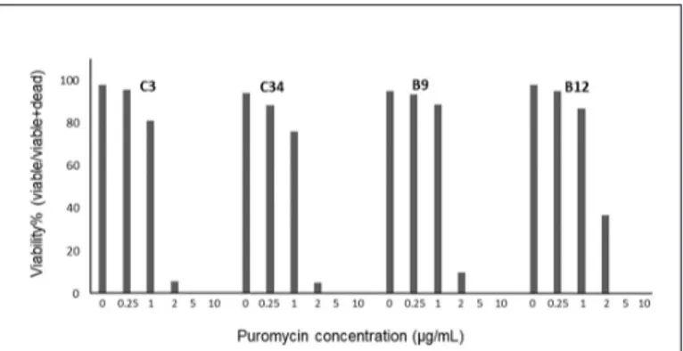
Viral transduced DLD-1 subclones were selected by puromycin (Thermo Fisher, Waltham, MA, USA). An antibiotic kill curve experiment was performed to determine the concentration of puromycin that kills all cells without a resistance gene incorporated in their DNA. 5x10⁴ cells of each DLD-1 subclone was seeded in 24-well plates as 3 replicates. Puromycin (Sigma) solutions were prepared as 0, 0.25, 1, 2, 5, and 10 µg/mL and given to cells after 24 hours. Cells were observed for 7 days. On the 7th day, cells were trypsinized, stained with trypan blue (Sigma-Aldrich, St. Louis, MO, USA) and counted using a Neubauer chamber, as 4 replicates.
The viability percentage of puromycin concentration that was lethal for non-transduced cells was determined. Transduced cells were given complete RPMI 1640 media with puromycin at this concentration for 7 days.
Epifluorescent imaging
At the end of 7 days epifluorescent imaging of stable lentiviral transduced DLD-1 subclones were performed using IX71 (Olympus, Hamburg, Germany) at 10x magnification, and U-MWG2 and U-MWB2 filters.
RESULTS
DLD-1 subclones sorted according to their DNA content via FACS were cultured and propagated. Each subclone was transduced with a lentivirus labeled with a florescent label which will enable the subclone to be specifically detected in a mix of cells. Lentiviral transduced DLD-1 subclones were selected by puromycin. To determine the optimum selection conditions of transduced cells, an antibiotic selection experiment was carried out and kill curves of DLD-1 subclones were plotted as shown in Figure 2. The lethal concentration of puromycin for the DLD-1 subclones was determined as 5 µg/mL. Puromycin at this concentration was prepared in complete RPMI 1640 and was used to select stably transduced cells for 7 days. The fluorescent labeling of four subclones, 2 of them near-diploid (C3 and C34) and 2 of them near-tetraploid (B9 and B12) via lentiviral transduction was performed successfully as confirmed by epifluorescent imaging (Figure 3). The reason 2N clones were both imaged red and 4N clones were both imaged green is the lack of appropriate filters for each color on inverted fluorescent microscope.
Figure 3. Epifluorescent images of lentiviral stable transduced DLD-1 subclones. Table 1. Lentiviruses and DLD-1 subclones
SUBCLONE LENTIVIRUS
rLV.EF1.ZsYellow1-9 C3
rLV.EF1.tdTomato-9 C34
rLV.EF1.ZsGreen1-9 B9
rLV.EF1.AmCyan1-9 B12
DISCUSSION
Intratumoral heterogeneity is one of the main reasons that cancer patients respond differently to similar treatments and is also responsible for observed therapy resistance by providing the means of the selective growth of cells that carry mutations and epigenetic patterns that provide advantage in adaptation to microenvironmental conditions altered by therapeutics (5). Identification of these subclones and analysis of the resistance-related genomic differences are uttermost important in order to develop efficient therapy methods against these cancer cells. Research performed in recent years have revealed important genomic signatures in solid tumors. It is very important to be able to detect and target these signatures in tumors that harbor many different subclones, as a result of clonal evolution. Chromosomal aneuploidies are observed in all sporadic carcinomas. Some aneuploid signatures occur early in cancer development. Cells bearing these signatures are continuously selected and those traits are preserved. As a consequence, tumor-specific special aneuploid patterns arise. There are mainly three generalizations that are accepted to apply to solid tumors: 1) Distribution of genetic imbalances are tumor-specific. 2) These genetic/ chromosomal imbalances are not observed in normal cells. 3) They emerge before the disease gain invasive ability (7). Based upon these basic implications, we can define novel markers for prognosis, progression, and transition from precancerous lesion to carcinoma. Copy number variations (CNVs) acquired somatically in solid tumors with epithelial origins can encompass whole chromosomes, chromosome arms or focal amplifications and deletions (7–9). By investigation of increasing cellular dysplasia steps during tumorigenesis in colorectum and cervix, it has been demonstrated that specific copy number changes occur before transition to invasive disease (8, 10–12). Chromosomal imbalances needed for clonal expansion of premalignant lesions are generally the only abnormalities can be detected at early stages. These tissue-specific aberrations are conserved in local and distant metastases (13, 14) and also in cell lines originated from primary tumors (15, 16). Despite chromosomal imbalances, centrosome amplifications, missegregation of chromosomes, and arising of different aneuploidies in subclones, conservation of cancer type-specific abnormalities such as 3q gain in cervix cancer indicates a continuous selective pressure.
Chromosomal aneuploidies affect chromosome-wide gene expression levels and thus cause a major change in transcriptome of cancer cells (17). This situation is observed in either mouse and human cells, or in cells where an artificial trisomy is induced by Microcell Mediated Chromosome Transfer (MMCT) (18). In addition, several studies have shown that majority of the genes deregulated by copy number increases at low levels are related to RNA or cellular metabolism (19, 20). Therefore, increased expression of these genes can provide the cells an early proliferative advantage. The advantage provided by aneuploidy is concordant with the observations that copy number increases at low levels occurring in early stages of tumorigenesis (8, 21).
Research is continuing to identify specific genomic and epigenetic changes in solid tumors that may be potential targets for new therapeutic strategies. Nevertheless, the cellular heterogeneity of tumors presents a challenge to overcome. How different clones of cancer cells interact with each other, how they respond to different stimuli and stress, and how these factors affect the cellular kinetics in the tumor, are areas in which our knowledge is limited. To expand research in these areas we need in vitro models for intratumoral heterogeneity.
Our aim in this study was to generate a model to enable the investigate tumor subclones with distinct genetic properties. With this model, it will be possible to study cellular behaviors under altered conditions –especially during therapy– and by this means to identify the effects of different variations at the genomic level. We isolated four genomically different subclones from the DLD-1 colon cancer cell line; and fluorescent labeled them by lentiviral transduction. Lentiviral transduction was chosen for fluorescent labeling because it is the most efficient stable integration method known today (22, 23). The lack of appropriate filters on our inverted microscope prevented us to observe the 4 different colors of the labeled clones. For this reason, the 2N clones were both imaged red and 4N clones were both imaged green. Prepared co-culture will be imaged with confocal microscope in our next study in order to distinguish each clone and to observe the changes in their properties over time and under different conditions.
In conclusion, in this study we have generated a model of colon cancer comprised of subclones originating from the same tumor but have distinct characteristics. Since the subclones that constitute this in vitro tumor heterogeneity model are stably fluorescent labeled in different colors, it makes possible to study the features of each subclone individually in a co-culture setting. Data obtained by the studies that will use this model will contribute to the illumination of the underlying mechanism of cancer therapy resistance and will play a substantial role in future therapy approaches, especially in personalized medicine.
ACKNOWLEDGEMENTS
Lentiviral transduction was performed in DEU ARLAB (Dokuz Eylül Üniversitesi Tıp Fakültesi Araştırma Laboratuvarı). Fluorescent microscopy was performed in iBG (İzmir Biomedicine and Genome Center).
Peer-review: Externally peer-reviewed.
Author Contributions: Concept - ZY, ÖGG; Design - ZY, ÖGG; Supervision - ZY; Fundings
- ZY; Materials - ZY; Data Collection and/or Processing - ÖGG, HE, SBK, ÖU; Analysis and/ or Interpretation - ÖGG, ZY, HE, SBK, ÖU; Literature Search - ÖGG, ZY ; Writing Manuscript - ÖGG, ZY, HE; Critical Review - ZY
Conflict of Interest: No conflict of interest was declared by the authors.
REFERENCES
1. Ruiz C, Lenkiewicz E, Evers L, et al. Advancing a clinically relevant perspective of the clonal nature of cancer. Proc Natl Acad Sci 2011;108:12054–12059. [CrossRef]
2. Nowell PC. The clonal evolution of tumor cell populations. Science 1976;194:23–28.
3. Naugler CT. Population genetics of cancer cell clones: possible implications of cancer stem cells. Theor Biol Med Model 2010;7:42. [CrossRef]
4. Salk JJ, Fox EJ, Loeb LA. Mutational Heterogeneity in Human Cancers: Origin and Consequences. Annu Rev Pathol 2010;5:51–75. [CrossRef] 5. Masramon L, Vendrell E, Tarafa G, et al. Genetic instability and
divergence of clonal populations in colon cancer cells in vitro. J Cell Sci 2006;119:1477–1482. [CrossRef]
6. Wangsa D, Quintanilla I, Torabi K, et al. Near-tetraploid cancer cells show chromosome instability triggered by replication stress and exhibit enhanced invasiveness. FASEB J 2018;32:3502–3517. [CrossRef]
7. Ried T. Homage to Theodor Boveri (1862–1915): Boveri’s theory of cancer as a disease of the chromosomes, and the landscape of genomic imbalances in human carcinomas. Environ Mol Mutagen 2009;50:593–601. [CrossRef]
8. Ried T, Heselmeyer-Haddad K, Blegen H, Schröck E, Auer G. Genomic changes defining the genesis, progression, and malignancy potential in solid human tumors: A phenotype/genotype correlation. Genes Chromosomes and Cancer 1999;25:195–204. [CrossRef]
9. Beroukhim R, Mermel CH, Porter D, et al. The landscape of somatic copy-number alteration across human cancers. Nature 2010;463:899–905. [CrossRef]
10. Ried T, Knutzen R, Steinbeck R, et al. Comparative genomic hybridization reveals a specific pattern of chromosomal gains and losses during the genesis of colorectal tumors. Genes Chromosomes Cancer 1996;15:234–245. [CrossRef]
11. Heselmeyer K, Schröck E, Du Manoir S, et al. Gain of chromosome 3q defines the transition from severe dysplasia to invasive carcinoma of the uterine cervix. Proc Natl Acad Sci U S A 1996;93:479–484. [CrossRef]
12. Habermann JK, Paulsen U, Roblick UJ, et al. Stage-specific alterations of the genome, transcriptome, and proteome during colorectal carcinogenesis. Genes Chromosomes Cancer 2007;46:10–26. [CrossRef]
13. Platzer P, Upender MB, Wilson K, et al. Silence of chromosomal amplifications in colon cancer. Cancer Res 2002;62:1134–1138. https:// cancerres.aacrjournals.org/content/canres/62/4/1134.full.pdf 14. Schwendel A, Langreck H, Reichel M, et al. Primary small-cell lung
carcinomas and their metastases are characterized by a recurrent pattern of genetic alterations. Int J Cancer 1997;74:86–93. [CrossRef] 15. Ghadimi BM, Schröck E, Walker RL, et al. Specific chromosomal
aberrations and amplification of the AIB1 nuclear receptor coactivator gene in pancreatic carcinomas. Am J Pathol 1999;154:525–536. [CrossRef] 16. Knutsen T, Padilla-Nash HM, Wangsa D, et al. Definitive molecular cytogenetic characterization of 15 colorectal cancer cell lines. Genes Chromosomes Cancer 2010;49:204–223. [CrossRef]
17. Ried T, Hu Y, Difilippantonio MJ, Ghadimi BM, Grade M, Camps J. The consequences of chromosomal aneuploidy on the transcriptome of cancer cells. Biochim Biophys Acta 2012;1819:784–793. [CrossRef] 18. Upender MB, Habermann JK, McShane LM, et al. Chromosome
transfer induced aneuploidy results in complex dysregulation of the cellular transcriptome in immortalized and cancer cells. Cancer Res 2004;64:6941–6949. [CrossRef]
19. Chin K, DeVries S, Fridlyand J, et al. Genomic and transcriptional aberrations linked to breast cancer pathophysiologies. Cancer Cell 2006;10:529–541. [CrossRef]
20. Neve RM, Chin K, Fridlyand J, et al. A collection of breast cancer cell lines for the study of functionally distinct cancer subtypes. Cancer Cell 2006;10:515–527. [CrossRef]
21. Heim S, Mitelman F, editors. Cancer Cytogenetics: Chromosomal and Molecular Genetic Aberrations of Tumor Cells, 4th ed. Wiley-Blackwell; 2015. 648 p. [CrossRef]
22. Elegheert J, Behiels E, Bishop B, et al. Lentiviral transduction of mammalian cells for fast, scalable and high-level production of soluble and membrane proteins. Nat Protoc 2018;13:2991–3017. [CrossRef] 23. Milone MC, O’Doherty U. Clinical use of lentiviral vectors. Leukemia
2018;32:1529–1541. https://www.nature.com/articles/s41375-018-0106-0