75
Abs tract
Malignancy is an important cause of mortality in acromegaly. Hematological malignancies are very rare in acromegaly. Here, we report an 80-year-old patient with acromegaly and multiple myeloma. Patient died within a month of diagnosis. Previous studies have shown that growth hormone and somatomedin-C activate B lymphocyte and somatomedin-C receptors are found in multiple myeloma cells. Possible effects of growth hormone and somatomedin-C on multiple myeloma progression are discussed in the light of the relevant literature. Turk Jem 2013; 17: 75-8
Key words: Acromegaly, multiple myeloma
Özet
Akromegali hastalarında malignite önemli bir mortalite nedenidir. Hematolojik maligniteler akromegali hastalarında oldukça nadiridir. Burada eş zamanlı akromegali ve multiple myeloma tanısı konulan 80 yaşında bir olgu sunuldu. Olgu tanıdan sonraki bir ay içinde kaybedildi. Büyüme hormonu ve somatomedin-C ‘nin B lenfositleri aktive ettiği ve somotomedin-C reseptörlerinin multiple myeloma hücrelerinde bulunduğu daha önceden gösterilmiştir. Büyüme hormonu ve somatomedin-C ‘nin myeloma progresyonu üzerine olan bu olası etkileri literatür eşliğinde tartışıldı. Turk Jem 2013; 17: 75-8
Anah tar ke li me ler: Akromegali, multiple myeloma
Murat Atmaca, Saliha Yıldız, Işılay Kalan, Mehmet Fatih Özbay**, İsmet Seven*, Mustafa Öztürk***
Yüzüncü Yıl University School of Medicine, Department of Endocrinology and Metabolism, Van, Turkey *Yüzüncü Yıl University School of Medicine, Department of Endocrinology and Metabolism, Van, Turkey **Van Education and Research Hospital, Department of Internal Medicine, Van, Turkey ***Medipol Yıl University School of Medicine, Department of Endocrinology and Metabolism, Istanbul, Turkey
Association of Acromegaly and Multiple Myeloma: A Case Report
Akromegali ve Multiple Myeloma Birlikteliği: Bir Olgu Sunumu
DOI: 10.4274/Tjem. 2182
Case Report Olgu Sunumu
Introduction
Acromegaly is a disorder characterized by excessive and disproportionate somatic growth. The prevalence and annual incidence are less than 70 in a million and 3-4 in a million, respectively (1,2). Almost all of the patients (98%) have a benign growth hormone (GH) secreting pituitary adenoma (3). Cardiovascular disease is the most common cause of death in patients with acromegaly. 20 to 25% of cases are lost due to various malignancies (4,5). Evidence for an effect on the development of malignancy in acromegaly is controversial. Hematological malignancies, especially multiple myeloma with acromegaly is rare in the literature and only found in the form of case reports. Here, we presented a case diagnosed with acromegaly and multiple myeloma simultaneously.
Case
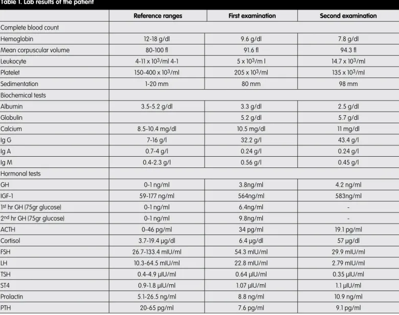
An eighty-year-old female patient admitted to our hospital clinic with the complaints of feet and tongue growth, widespread body pain and fatigue. The patients’ slurred speech, body aches, and weakness was continuing for the last 2 months. The patient’s personal and family history was unremarkable. The patient had typical acromegalic appearance as she had the growth of the hands and feet, prognathism, widening between the teeth and frontal proptosis; and the patient had 145/85mmhg arterial blood pressure, 84 per minutes pulse and 36.9 degrees C body temperature. Pale conjunctivae, and macroglossia were present in the system examination. Heart, lung and abdominal examinations were normal. The patient’s laboratory tests are shown in Table 1. The results showed high basal growth hormone (GH) and IGF-1
Turkish Journal of Endocrinology and Metabolism, published by Galenos Publishing.
Address for Correspondence/Yazışma Adresi: Murat Atmaca MD, Yüzüncü Yıl University School of Medicine, Department of Endocrinology and Metabolism, Van, Turkey GSM: +90 505 581 90 03 E-mail: [email protected] Recevied/Geliş Tarihi: 16.11.2012 Accepted/Kabul Tarihi: 14.02.2013
76
Atmaca et al. Turk Jem 2013; 17: 75-8 Association of Acromegaly and Multiple Myeloma: A Case Reportlevels. During 75 g glucose growth hormone suppression test, the lowest value of GH was 3.8 ng / ml and a paradoxical increase was observed during the test. In patient’s pituitary MRI, a 1 cm in diameter macroadenoma was detected. The patient got diagnosed with acromegaly with the current findings and her corrected calcium level was 11.1 mg / dl. The level of parathyroid hormone which was done for the differential diagnosis of hypercalcemia was 7.6 pg/ml. We suspected that the patient had malign hypercalcemia because she also had high globulin level and high erythrocyte sedimentation rate. Protein electrophoresis and immunoglobulin levels were requested to investigate the presence of multiple myeloma. IgG level was 32.2 g/l and monoclonal gammopathy was present in the protein electrophoresis. The bone marrow aspiration biopsy was performed. The result was consistent with plasma cell myeloma and kappa light chain was positive (Figure 2). Accordingly, the patient was diagnosed with acromegaly and multiple myeloma with the current findings. We
suggested transsphenoidal adenomectomy for acromegaly, and VAD (Vincritine, adriamycine, dexamethasone) chemotherapy for multiple myeloma, but the patient refused the treatment. A month later after her discharge, she re-admitted to our emergency department with complaints of cough and fever. On physical examination, the patient’s respiratory system examination showed right lower lobe crackles and 39.2 degrees C of fever. There was infiltration of the right baseline on chest radiograph of the patient. Laboratory tests of the patient with pneumonia are given in Table 1. On the second day of admission, the patient died due to respiratory failure although antibiotherapy was started.
Discussion
Increased levels of growth hormone in acromegaly often cause hypercalciuria with vitamin D activation (6). Whereas in acromegaly, hypercalcemia is rare and usually caused by parathyroid adenoma or hyperplasia, and this situation is often associated with multiple endocrine neoplasia type 1 (MEN-1) (7). In our case of hypercalcemia, we found suppressed levels of parathyroid hormone. Therefore, parathyroid adenoma, parathyroid hyperplasia, and MEN-1 diagnoses were abandoned. The common cause of hypercalcemia with suppressed parathyroid hormone level is malignancy. Usually hypercalcemia of malignancy develops due to two mechanisms that are humoral hypercalcemia of malignancy and local osteolytic hypercalcemia of malignancy (8). Our patient with hypercalcemia was diagnosed with multiple myeloma. Multiple myeloma is a hematologic malignancy and often causes hypercalcemia by lytic bone lesions (8). Malignancy is the third most common cause of death in patients with acromegaly (4,5). In acromegaly, an increase in the secretion and synthesis of growth hormone and insulin-like growth factor-1 (IGF-1) have potent growth stimulant and anabolic effects (9). The diagnosis of acromegaly due to pituitary disease typically gets delayed and usually found about 10 years after the onset of symptoms (10). This long-term exposure to GH and IGF-1 raises concerns in terms of secondary tumor development and progression. There are publications available about acromegaly increasing the incidence of the colon cancer associated with adenomatous polyps and especially increasing the incidence of the breast cancer in women (11-13). However, direct cause and effect relationship between acromegaly and the presence or absence of cancer has not been proven (14-16). A retrospective analysis of nine studies reported acromegaly did not increase the incidence of cancer (15). However, in acromegalic patients with the complication of cancer, IGF-1 can stimulate the growth of neoplastic tissue and accelerate the progression of cancer (15,17).
Acromegaly with hematological malignancies is a rare condition and is available in the form of case reports in the literature (18,19). It is not clear whether acromegaly has an effect on the development of multiple myeloma. It has been previously shown that GH and IGF-1 have activating effects on B-lymphocytes (20). IGF-1 receptors were identified on some plasma cell lines and it has been shown in vitro that IGF-1 stimulation of these receptors causes proliferation of myeloma cell. This supports a possible relationship between these two diseases (21). In a recent in vitro study of myeloma cells, it is reported that IGF-1 receptor expression is a poor prognostic factor
Figure 1.
Atmaca et al. Association of Acromegaly and Multiple Myeloma: A Case Report
Turk Jem 2013; 17: 75-8
77
(22). Also in animal studies, the application of IGF-1 on plasma tumor cell lines is shown to increase the growth rate approximately two times more (23). In a case presented by Tucci et al., a patient with monoclonal gammopathy of undetermined significance (MGUS) showed a rapid progression to multiple myeloma after getting diagnosed with acromegaly, which supports previous findings (24). In our patient diagnosed with acromegaly and multipl myeloma simultaneously, findings including deepening of anemia, development of thrombocytopenia, a 40% increase in the level of IgG can be explained by the rapid progression caused by IGF-1. In this rare combination of these two diseases, it is not clear which disease should be given the priority to in terms of treatment. Although the primary determinant of survival seems to be multiple myeloma, because of the acceleration of myeloma progression due to high levels of IGF-1; it is also convenient to give priority to the treatment of acromegaly. In fact, Kanazawa and colleagues reported a patient with MGUS associated with acromegaly and they show that a decrease in the level of IGF-1 after pituitary adenomectomy caused a decline in the level of IgG as well, which supports these findings (25).
As a result, simultaneous presentation of acromegaly and multipl myeloma is very rare and it is not clear whether acromegaly has an effect on the development of multiple myeloma. However, especially in acromegaly patients with hypercalcemia, multiple myeloma should be considered in the differential diagnosis even if it is a very rare situation.
References
1. Alexander L, Appleton D, Hall R, Ross WM, Wilkinson R. Epidemiology of acromegaly in the Newcastle region. Clin Endocrinol (Oxf) 1980;12:71-9.
2. Ritchie CM, Atkinson AB, Kennedy AL, et al. Ascertainment and natural history of treated acromegaly in Northern Ireland. Ulster Med J 1990;59:55-62.
3. Ben-Shlomo A, Melmed S. Acromegaly. Endocrinol Metab Clin North Am 2008;37:101-22.
4. Wright AD, Hill DM, Lowy C, Fraser TR. Mortality in acromegaly. Q J Med 1970;39:1-16.
5. Loeper S, Ezzat . Acromegaly: re-thinking the cancer risk. Rev Endocr Metab Disord 2008;9:41-58.
6. Lund B, Eskildsen PC, Lund B, Norman AW, Sørensen OH. Calcium and vitamin D metabolism in acromegaly. Acta Endocrinol (Copenh) 1981;96:444-50.
Table 1. Lab results of the patient
Reference ranges First examination Second examination Complete blood count
Hemoglobin 12-18 g/dl 9.6 g/dl 7.8 g/dl
Mean corpuscular volume 80-100 fl 91.6 fl 94.3 fl
Leukocyte 4-11 x 103/ml 4-1 5 x 10³/m l 14.7 x 10³/ml Platelet 150-400 x 103/ml 205 x 103/ml 135 x 103/ml Sedimentation 1-20 mm 80 mm 98 mm Biochemical tests Albumin 3.5-5.2 g/dl 3.3 g/dl 2.5 g/dl Globulin 5.2 g/dl 5.7 g/dl Calcium 8.5-10.4 mg/dl 10.5 mg/dl 11 mg/dl Ig G 7-16 g/l 32.2 g/l 43.4 g/l Ig A 0.7-4 g/l 0.24 g/l 0.24 g/l Ig M 0.4-2.3 g/l 0.56 g/l 0.45 g/l Hormonal tests GH 0-1 ng/ml 3.8ng/ml 4.2 ng/ml IGF-1 59-177 ng/ml 564ng/ml 583ng/ml 1st hr GH (75gr glucose) 0-1 ng/ml 6.4ng/ml -2nd hr GH (75gr glucose) 0-1 ng/ml 9.8ng/ml -ACTH 0-46 pg/ml 34 pg/ml 19.1 pg/ml Cortisol 3.7-19.4 µg/dl 6.4 µg/dl 57 µg/dl
FSH 26.7-133.4 mIU/ml 54.3 mIU/ml 29.9 mIU/ml
LH 10.3-64.5 mIU/ml 22.8 mIU/ml 2.79 mIU/ml
TSH 0.4-4.9 µIU/ml 0.64 µIU/ml 0.35 µIU/ml
ST4 0.9-1.8 µIU/ml 1.07 µIU/ml 1.1 µIU/ml
Prolactin 5.1-26.5 ng/ml 8.8 ng/ml 10.9 ng/ml
78
Atmaca et al. Turk Jem 2013; 17: 75-8 Association of Acromegaly and Multiple Myeloma: A Case Report7. Ueda M, Inaba M, Tahara H, et al. Hypercalcemia in a patient with primary hyperparathyroidism and acromegaly: distinct roles of growth hormone and parathyroid hormone in the development of hypercalcemia. Intern Med 2005;44:307-10.
8. Rosol TJ, Capen CC. Mechanisms of cancer-induced hypercalcemia. Lab Invest 1992;67:680-702.
9. Clayton PE, Banerjee I, Murray PG, Renehan AG. Growth hormone, the insulin-like growth factor axis, insulin and cancer risk. Nat Rev Endocrinol 2011;7:11-24.
10. Jadresic A, Banks LM, Child DF, et al. The acromegaly syndrome. Relation between clinical features, growth hormone values and radiological characteristics of the pituitary tumours. Q J Med 1982;51:189-204. 11. Ron E, Gridley G, Hrubec Z, et al. Acromegaly and gastrointestinal cancer.
Cancer 1991;68:1673-7.
12. Barzilay J, Heatley GJ, Cushing GW. Benign and malignant tumors in patients with acromegaly. Arch Intern Med 1991;151:1629-32.
13. Nabarro JD. Acromegaly. Clin Endocrinol (Oxf) 1987;26:481-512.
14. Jenkins PJ, Mukherjee A, Shalet SM. Does growth hormone cause cancer? Clin Endocrinol (Oxf) 2006;64:115-21.
15. Melmed S. Acromegaly and cancer: not a problem? J Clin Endocrinol Metab 2001;86:2929-34.
16. Jenkins PJ, Besser M. Clinical perspective: acromegaly and cancer: a problem. J Clin Endocrinol Metab 2001;86:2935-41.
17. Orme SM, McNally RJ, Cartwright RA, Belchetz PE. Mortality and cancer incidence in acromegaly: a retrospective cohort study. United Kingdom Acromegaly Study Group. J Clin Endocrinol Metab 1998;83:2730-4. 18. Hägg E, Asplund K, Holm J. Acromegaly and multiple myeloma. Ann Intern
Med 1988;8:437-38.
19. Barbosa FR, Vieira Neto L, Lima GA, et al. Hematologic neoplasias and acromegaly. Pituitary 2011;14:377-81.
20. Murphy WJ, Rui H, Longo DL. Effects of growth hormone and prolactin immune development and function. Life Sci 1995;57:1-14.
21. Georgii-Hemming P, Wiklund HJ, Ljunggren O, Nilsson K. Insulin-like growth factor I is a growth and survival factor in human multiple myeloma cell lines. Blood 1996;88:2250-8.
22. Sprynski AC, Hose D, Caillot L, et al. The role of IGF-1 as a major growth factor for myeloma cell lines and the prognostic relevance of the expression of its receptor. Blood 2009;113:4614-26.
23. Ge NL, Rudikoff S. Insulin-like growth factor I is a dual effector of multiple myeloma cell growth. Blood 2000;96:2856-61.
24. Tucci A, Bonadonna S, Cattaneo C, et al. Transformation of a MGUS to overt multiple myeloma: the possible role of a pituitary macroadenoma secreting high levels of insulin-like growth factor 1 (IGF-1). Leuk Lymphoma 2003;44:543-5.
25. Kanazawa I, Yamaguchi T, Yamane Y, et al. Acromegaly associated with monoclonal gammopathy of undetermined significance (MGUS). Endocr J 2006;53:687-91.