1 2 3 4 5 6 7 8 9 10 11 12 13 14 15 16 17 18 19 20 21 22 23 24 25 26 27 28 29 30 31 32 33 34 35 36 37 38 39 40 41 42 43 44 45 46 47 48 49 50 51 52 1 2 3 4 5 6 7 8 9 10 11 12 13 14 15 16 17 18 19 20 21 22 23 24 25 26 27 28 29 30 31 32 33 34 35 36 37 38 39 40 41 42 43 44 45 46 47 48 49 50 51 52 1 Indian Journal of Dermatology, Venereology, and Leprology | January-February 2015| Vol 81 | Issue 1
Lack of effectiveness of keratin
dressings in epidermolysis
bullosa
Dear Editor,
Keratin is the fundamental protein that creates cell structure in many tissues, particularly in the skin, hair, and nails.[1‑4] In recent years, emphasis has been placed on the role of keratin in healing wounds. The effects of keratin on cell multiplication and replacement, epithelisation, and wound closure have been observed in many cases. Molecular analysis shows that keratin proteins have a positive effect on wound healing via increasing or accelerating epithelization. This is caused by keratinocyte activation and migration, which is, in turn, caused by increasing keratin gene expression. The synthesis of fundamental proteins such as collagen 4, collagen 7, and collagen 17 increases in active and stimulated keratinocytes.[1] Keratin 17 production increases when the skin is damaged. It has been observed that wound healing does not occur properly in patients with low amounts of keratin 17. As a result of this keratin, wound healing is hastened, and the frequency of blistering is reduced.[2]
Taking the aforementioned into consideration, the effects of applying keratin to the skin as an exogen during wound healing have been studied, and keratin‑based care products have been developed in which the activity of keratin protein has been retained. Bandages with keratin content help provide keratins to wounds. They are well‑tolerated by patients’ tissues during rehabilitation due to their non‑immunogenic characteristics.[3] A gel manufactured containing keratin obtained from sheep’s wool, has been used in the treatment of EB patients.[4]
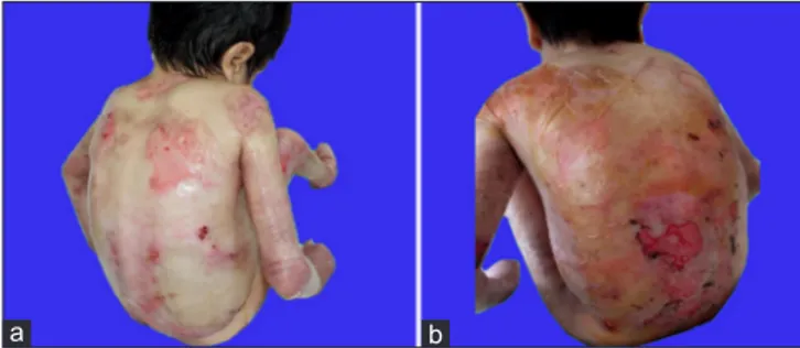
A total of five epidermolysis bullosa (EB) patients, including three dystrophic EB (DEB) and two EB simplex (EBS) patients (case 1 = 9‑year‑old male with DEB; case 2 = 17‑year‑old female with DEB; case 3 = 3‑month‑old female with DEB; case 4 = 6‑year‑old male with EBS; case 5 = 2‑month‑old female with EBS), were regularly subjected to application of a thick hydrogel rich in keratin (Keragel T™, Keraplast Technologies, Lincoln, New Zealand). The gel was applied every other day. The bodies of the patients were entirely washed with lukewarm sterile water, and the gel was rubbed on after the skin dried. The skin was then wrapped with a non‑stick bandage (paraffin gauze bandage, Smith Medical Limited, Hull, England) after waiting for 15 minutes. The treatment was abandoned in case 3 at the fourth month of the treatment upon the request of the family because the number of blisters had increased significantly. Clinical photos of the fourth and fifth patients were taken before and after the treatment [Figures 1 a and b, 2 a and b]
A significant decrease in the number of blisters was observed in all of our patients within the first two months of treatment. This result boosted the morale of the patients, their relatives, and the clinic employees. However, relapses in the form of additional blistering were observed in all of the patients beginning from the third month of the treatment, and no changes were noticed in the healing period of the existing eroded areas throughout the treatment. Case 3 gave up the treatment during the fourth month upon request of her family because the treatment was ineffective. The treatment was stopped in all of the remaining patients after six months of treatment due to the requests of the patients and their relatives.
IJDVL_155_14R9
Letters to
the Editor
Figure 1: (a) Pre-treatment appearance; There were eroded area in the arm, left knee ext mid-lateral tibial and ankle extensor (Patient 2). (b) Post-treatment appearance; Worsening of erosions after three months of treatment
Figure 2: (a) Pre-treatment appearance; There were eroded area on the back, right thigh extensor (Patient 1). (b) Post-treatment appearance; Worsening of erosions after three months of treatment
b
1 2 3 4 5 6 7 8 9 10 11 12 13 14 15 16 17 18 19 20 21 22 23 24 25 26 27 28 29 30 31 32 33 34 35 36 37 38 39 40 41 42 43 44 45 46 47 48 49 50 51 52 1 2 3 4 5 6 7 8 9 10 11 12 13 14 15 16 17 18 19 20 21 22 23 24 25 26 27 28 29 30 31 32 33 34 35 36 37 38 39 40 41 42 43 44 45 46 47 48 49 50 51 52
Indian Journal of Dermatology, Venereology, and Leprology | January-February 2015| Vol 81 | Issue 1 2
Letters to the Editor
In another study, a keratin‑based care product was applied directly to the damaged areas of the left hand and left foot, as well as the right hand and foot, of a patient with dystrophic epidermolysis bullosa as a standard‑care treatment four times a week for a period of 6 weeks.[5] After treatment, it was observed that the number of blisters and the epithelisation period of the eroded areas significantly decreased in areas subjected to keratin application. Increases in the hand‑based skills and quality of life of the patient were observed after keratin gel treatment. The hypothesis stating that keratin‑based products affect the keratinocytes present at wounds and speed up healing is supported by these favorable results.It was also noted that the application of keratin‑based products for long periods of time increases their quality of life and reduces their treatment costs.[5]
In contrast, our patients were disappointed with the results of keratin dressings.
Hacer Altun Sürücü, Yavuz Yeşilova,
Enver Turan, Mustafa Aksoy
1, Osman Tanrıkulu,
Naime Eroglu
Departments of Dermatology, Harran University School of Medicine, Sanliurfa, Fish Lake Sanliurfa State Hospital, Sanliurfa, Turkey
Address for correspondence: Dr. Yavuz Yesılova, Department of Dermatology, 63300, Sanliurfa, Turkey. E‑mail: yavuzyesilovagmail.com
REFERENCES
1. Rogel MR, Jaitovich A, Ridge KM. The role of theubiquitinproteasomepathway in keratin intermediate filament protein degradation. Proc Am Thorac Soc 2010;7:71‑6. 2. Kim S, Wong P, Coulombe PA. A keratin cytoskeletal protein
regulates protein synthesis and epithelial cell growth. Nature 2006;441:362‑5
3. De Guzman RC, Merrill MR, Richter JR, Hamzi RI, Greengauz‑Roberts OK, Van Dyke ME. Mechanical and biological properties of keratosebiomaterials. Biomaterials 2011;32:8205‑17.
4. Pechter PM, Gil J, Valdes J, Tomic‑Canic M, Pastar I, Stojadinovic O, et al. Keratin d ressings speed epithelialization of deep partial‑thickness wounds. Wound Repair Regen 2012;20:236‑42.
5. Kirsner RS, Cassidy S, Marsh C, Vivas A, Kelly RJ. Use of a keratin‑based wound dressing in the management of wounds in a patient with recessive dystrophic epidermolysis bullosa. Adv Skin Wound Care 2012;25:400‑3.
Access this article online Quick Response Code: Website:
www.ijdvl.com
DOI:
*****
PMID: