Fingolimod for the Treatment of Relapsing-Remitting Multiple Sclerosis
Relapsing Remitting Multipl Skleroz Tedavisinde Fingolimod Kullanımı
Burcu Altunrende1, Erkingül Birday2, Mithat Kasap3, Gülşen Akman Demir1 1Istanbul Bilim University Faculty of Medicine, Department of Neurology, Istanbul, Turkey 2Medipol Mega University Hospital, Department of Neurology, Istanbul, Turkey 3Novartis Pharmaceuticals Turkey, Istanbul, Turkey
Multiple sclerosis (MS) is a chronic autoimmune disease of the central nervous system and is characterized by inflammation, demyelination, and axonal loss. Fingolimod is the first oral drug for the treatment of MS approved by the United States Food and Drug Administration, European Union countries, and various other countries. The compound exerts its effect via interaction with lysophospholipid receptors known as sphingosine-1 phosphate receptors. Although fingolimod has a very convenient daily oral dosing, it may cause development of bradycardia at the first dose, macular edema, infection, all of which require attention. Randomized double-blind clinical trials have shown that fingolimod significantly reduces relapse rates and is beneficial in brain magnetic resonance imaging measures when compared with both placebo and intramuscular interferon β-1a. This review describes the characteristics of fingolimod concerning its efficacy, safety, and tolerability in the clinical context of the management of MS.
Keywords: Multiple sclerosis, fingolimod, sphingosine-1 phosphate, efficacy, safety
Multipl skleroz (MS) merkezi sinir sisteminin enflamasyon, demiyelinizasyon ve akson kaybı ile karakterize kronik otoimmün, nörodejeneratif bir hastalığıdır. Fingolimod Amerikan İlaç ve Gıda Dairesi ve Avrupa Birliği üyesi ülkelerin de bulunduğu 80’den fazla ülke tarafından onaylanmış ilk oral MS ilacıdır. Bu bileşik, sfingozin-1 fosfat olarak bilinen lizofosfolipid reseptörleri aracılığı ile etkilerini gösterir. Günlük oral yolla kullanımı kolaylık sağlamakla birlikte bazı hastalarda ilk doz sırasında bradikardi gelişmesi, maküler ödem, enfeksiyon gibi bazı dikkat edilmesi gereken durumlar ile karşılaşılabilmektedir. Randomize çift-kör klinik çalışmalar, plasebo ve intramüsküler interferon β-1a tedavilerine kıyasla fingolimodun atak sıklığını anlamlı derecede azalttığını ve beyin manyetik rezonans ölçütleri üzerine yararlı etkiler gösterdiğini ortaya koymuştur. Bu derlemede, fingolimodun klinikte MS hastalarının yönetimindeki etkinliği, güvenliliği ve tolerabilitesi ele alınmıştır.
Anahtar Kelimeler: Multipl skleroz, fingolimod, sfingozin-1 fosfat, etkinlik, güvenlilik
Abstract
Öz
Ad dress for Cor res pon den ce/Ya z›fl ma Ad re si: Burcu Altunrende MD, Istanbul Bilim University Faculty of Medicine, Department of Neurology, Istanbul, Turkey
Phone: +90 505 442 45 17 E-mail: [email protected] ORCID ID: orcid.org/0000-0003-1383-3557
Re cei ved/Ge lifl Ta ri hi: 05.01.2017 Ac cep ted/Ka bul Ta ri hi: 15.04.2017
Introduction
Multiple sclerosis (MS) is a chronic, autoimmune, neurodegenerative disorder of the central nervous system (CNS) characterized by inflammation, demyelination, and axonal loss (1). It is more prevalent in women and is the leading cause for neurologic dysfunction in young adults (2).
Clinically, the condition is often associated with recurrence and remission of neurologic symptoms, and hence it is referred to as relapsing-remitting MS (RRMS). Characteristic inflammation and demyelination attacks are followed by complete or partial functional remission; sometimes re-myelination may also be observed early in the disease course. This form of the disease can convert in time into a form that is characterized by progressive deterioration in functions. This form of the disease, with a reduced number of attacks, is referred to as secondary progressive MS (SPMS). Early repair mechanisms fail and axonal loss worsen in patients with SPMS. About 15-20% of all patients with MS display a course that is progressive from onset, which is referred to as primary progressive MS (3). The signs of MS vary depending on the type of MS and severity of attacks. The most common symptoms include fatigue, depression, visual disturbances, sensory symptoms such as loss of sense or pain, spasticity, weakness, ataxia, and fecal or urinary incontinence. These symptoms may also be accompanied by cognitive disorders.
The etiology of MS remains unknown. There is a complex interaction at play, which involves carrying a genetic risk against autoimmunity, impaired immune system, and environmental factors (exposure to viruses, vitamin D deficiency, and smoking) (4). Immunologically, the inflammatory process is believed to take place through T-cell and B-cell responses against autoantigens in the CNS. Axonal loss and damage sustained by oligodendrocytes resulting from disturbed pro-inflammatory and anti-inflammatory balance leads to permanent damage in the CNS.
For the last two decades, the therapeutic approach in treating MS has involved reduction in the frequency of symptoms and slowing down the disease progression. The first agents that were shown to be able to positively alter the disease course in RRMS, including interferon β (IFN β) and glatiramer acetate, are referred to as classic immunomodulator therapies and are acknowledged as the first-line therapies. They reduce annual attack rates by approximately 30% (5). Natalizumab, a second-line treatment choice, acts by targeting α4β1integrin expressed on lymphocytes and is more effective than first-line therapies. All these treatments target the immune system; they are, however, parenteral treatments and are associated with patient adherence issues as well as undesirable adverse effects. Moreover, whether the currently available therapies alter the neurodegenerative outcomes in MS is disputable (5).
The advent of FTY720/fingolimod, which can be used orally, has been a significant step in this context. Fingolimod (Gilenya®) was the first oral MS drug approved by the United States Food and Drug Administration (FDA), European Union countries, and several other countries. It was first authorized and made available in Turkey in April 2011. This compound acts through lysophospholipid receptors, also known as sphingosine-1 phosphate (S1P) receptors. This review addresses the structure of fingolimod, and its efficacy and safety in MS.
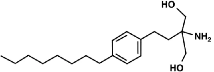
Structure of Fingolimod
Fingolimod (2-amino-2-(2-(4-octylphenyl)ethyl)propan-1,3-diol hydrochloride) is a small, lipophilic molecule, which is derived from a metabolite of the fungus Isaria sinclairii, and is structurally similar to the endogenous sphingosine (Figure 1). Sphingolipids contained in the construction of the cell membrane are involved in several intracellular signaling pathways. Sphingosine kinase enzyme ensures phosphorylation of sphingosine and mediates the dislocation of the molecule from the cell wall and form S1P. Fingolimod is rapidly phosphorylated by the endogenous sphingosine kinase 2 enzyme to fingolimod phosphate because it is structurally similar to sphingosine, and binds with high affinity to other S1P receptors except S1P2 (0.3-3.1 nM) (6).
S1P receptors are expressed in the CNS and peripherally in several cell types including endothelial cells, lymphocytes, smooth muscle cells, cardiac myocytes, and nerve cells. There are five S1P receptor sub-types that have key roles in the regulation of several biologic functions including leukocyte circulation, neural cell proliferation, neural cell function, cell migration, endothelial cell functioning, and vascular hemostasis: S1P1, S1P2, S1P3, S1P4, and S1P5. Of these receptors, S1P1, S1P2, and S1P3 are found extensively in the immune system, cardiovascular system, and CNS. S1P1 is highly expressed on T and B lymphocytes. In adults, S1P4 is mainly found in lymphoid and hematopoietic tissues, whereas S1P5 is located in white matter of the CNS (Table 1).
The Mechanism of Fingolimod’s Therapeutic
Effect in Multiple Sclerosis
One of the primary mechanisms leading to the beneficial effect of fingolimod in MS is the prevention of auto-aggressive lymphocytes from penetrating into the CNS by blocking the dissociation of T-cells from lymphatic tissues through the modulation of S1P1 on lymphocytes. Following antigenic stimulation, naïve T-cells differentiate into diverse storage cells, i.e. central storage T (CST) cells in lymph nodes and effector storage T-cells (EST). In patients with MS, most T-cells in the CNS are CST cells that have migrated from lymph nodes.
S1P1 is mainly expressed on lymphocytes and is the key target for the therapeutic efficacy of fingolimod in MS. The S1P1 pathway controls the circulation of lymphocytes that carry the CCR7 receptor, particularly storage T-cells, T-17 cells, and B-cells, in the blood and lymphatic systems. Fingolimod phosphate, the active form of fingolimod, binds to S1P1 on T-cells by acting as a potent antagonist of S1P1, and triggers internalization of the receptor, which renders the cell irresponsive to the signal of detachment of S1P from the lymph node, thereby trapping it inside the lymph node. Thus,
fingolimod therapy traps CCR7-negative EST in the circulation, and by acting on CCR7-positive lymphocytes, inhibits passage of autoreactive CST cells to blood, trapping them in the lymph node, thereby decreasing migration of cells to the CNS (7,8,9,10).
Experimental data have shown that fingolimod, besides its effects on T-cell retention in lymph nodes, had protective effects on the CNS through direct targeting of CNS cells. Fingolimod displays lipophilic properties due to its structural similarity to sphingosine. It therefore readily crosses the blood-brain barrier and penetrates into the CNS, and may accumulate in the myelin (11). In vitro experiments demonstrated that the drug was effective on astrocytes, neurons, and oligodendrocytes (12). It has been shown in in vivo animal MS models that fingolimod reduced
demyelination in the brain and medulla spinalis, and was thus efficient in reducing disease severity (13).
Efficacy of Fingolimod in Multiple Sclerosis
The efficacy of fingolimod in RRMS was evaluated in phase 2 and phase 3 studies. The phase 3 studies were placebo-controlled FREEDOMS (FTY720 Research Evaluating Effects of Daily Oral therapy in MS) (14) and FREEDOMS II studies, and TRANSFORMS (Trial Assessing Injectable Interferon vs. FTY720 in RRMS) (15,16), which compared fingolimod with intramuscular IFN β-1a (16), and their extensions. Table 2 provides an overview of the FREEDOMS, FREEDOMS II, and TRANSFORMS trials.
Table 1. Expression of sphingosine-1 phosphate receptor subtypes and their primary functions Receptor
subtype Site of expression Function
S1P1 Lymphocytes Nerve cells Vessels
Excitation of lymphocytes from secondary lymphoid organs Neural cell migration
Embryonic development of cardiovascular and nervous system Angiogenesis
Protection of endothelial barrier
S1P2 Diffuse
Vascular tonus
Protection of endothelial barrier Protection of hearing and balance Nerve conduction
S1P3 Diffuse (neural cells, astrocytes, endothelial cells) Protection of endothelial barrier Neural cell migration
S1P4 Lymphocytes Not known
S1P5 Brain (white matter)Oligodendrocytes Oligodendrocytes’ functioningNatural killer cell migration
S1P: Sphingosine-1 phosphate
Table 2. Overview of FREEDOMS, FREEDOMS II and TRANSFORMS
Study Study plan Treatment groups Primary endpoint Key findings
FREEDOMS (14) n=1272, 24-month, double-blind, parallel-group, comparative, multi-centered Fingolimod 0.5 mg/day Fingolimod 1.25 mg/day Placebo Reduction in annual relapse rate
Annual relapse rate: 0.16-0.18 vs. 0.40 (fingolimod vs. placebo; p<0.001) Relapse-free patients: 70-75% vs. 46% (fingolimod vs. placebo; p<0.001) FREEDOMS II (15) n=1083, 24-month, double-blind, parallel-group, placebo-controlled, multi-centered Fingolimod 0.5 mg/day Fingolimod 1.25 mg/day Placebo Reduction in annual relapse rate
Annual relapse rate: 0.20-0.21 vs. 0.40 (fingolimod vs. placebo; p<0.001)
Relapse -free patients: 72-73% vs. 53% (fingolimod vs. placebo; p<0.0001) TRANSFORMS (16) n=1292, 12-month, double-blind, parallel-group, placebo-controlled, multi-centered Fingolimod 0.5 mg/day Fingolimod 1.25 mg/day IFN β-1a 30 μg/week intramuscular
Reduction in annual relapse rate
Annual relapse rate: 0.16-0.20 vs. 0.33 (fingolimod vs. IFN β-1a; p<0.001)
Relapse -free patients: 80-83%vs. 69% (fingolimod vs. IFN β-1a; p<0.0001)
FREEDOMS and FREEDOMS II were randomized, double-blind, placebo-controlled, multi-national, multi-center 24-month studies (14,15). In the randomized, double-blind TRANSFORMS trial, in which the primary and secondary endpoints were the same as in FREEDOMS, patients were randomized into fingolimod or intramuscular IFN β-1a (30 μg/week) groups and were treated for 12 months (16). In a study that pooled the populations in FREEDOMS, FREEDOMS II, and TRANSFORMS studies, and compared the annual relapse frequency between placebo and IFN β-1a, patients had a median age of 37.8 years, 70.3% were female, the time from the first symptom to randomization was about 5.2 years, and the median Expanded Disability Status Scale (EDSS) score was 2.3 (17).
The Effect of Fingolimod on Relapses
In FREEDOMS, the frequency of relapse with fingolimod treatment was reduced significantly by 54% compared with placebo at the end of 24 months. During the 24-month study period, the proportion of relapse-free subjects was 70-75% in the fingolimod group versus 46% in the placebo group. In FREEDOMS II, the annual frequency of relapse was decreased by 48% in the group receiving fingolimod compared with placebo. In TRANSFORMS, the annual frequency of relapse over the 12-month treatment period was 52% lower in the group receiving fingolimod 0.5 mg/day than in the group treated with IFN β-1a.
The Effect of Fingolimod on Disability
Progression
In FREEDOMS, both 3- and 6-month confirmed disability progression was slower in fingolimod-recipients compared with placebo. EDSS scores and MS Functional Composite (MSFC) z scores in the fingolimod groups remained stable or were slightly improved. The primary results of FREEDOMS II showed no significant differences in disability progression between the groups (15). The analysis of the subjects with baseline EDSS scores of ≥1 demonstrated that a statistically significant reduction was achieved in the risk of 3- and 6-month confirmed disability progression. This result was consistent across FREEDOMS and FREEDOMS II studies (18,19). FREEDOMS II showed a significant improvement in the MSFC score at month 24 versus placebo.
In TRANSFORMS, MSFC scores displayed significantly more favorable changes in both fingolimod groups compared with IFN β-1a. However, the proportion of patients without disability progression was similarly high across IFN β-1a and fingolimod groups at the study end at month 12.
The Effects of Fingolimod on Magnetic
Resonance Imaging (MRI) Lesion Activity
In FREEDOMS, there were fewer new T2-weighted lesion occurrences in MRI at the end of 6, 12, and 24 months in subjects in the fingolimod group compared with the placebo group. The proportion of gadolinium-enhancing T1 lesions at month 24 was 82% decreased in the group of subjects receiving fingolimod 0.5 mg/day compared with placebo (p<0.001) (14). The median volume of lesions in the fingolimod groups was lower compared
with the placebo subjects. Similarly, the changes in the volume of hypodense lesions in T1-weighted images favored the fingolimod groups (14). In TRANSFORMS, new T2-weighted lesion occurrences in MRI and the proportion of gadolinium-enhancing T1 lesions at the end of 1 year was significantly reduced in the fingolimod 0.5 mg/day group compared with IFN β-1a.
The Effect of Fingolimod on Brain Atrophy
MS is a condition caused by inflammation and progressive neuroaxonal damage. No Evidence for Disease Activity (NEDA) is described as a new therapeutic target and defined as the absence of disease activity. It is termed NEDA-4 when there are no attacks, no disability progression, no MR lesion activity, and no brain atrophy. Current evidence indicates that patients whose treatment targets were set as NEDA had better disease courses compared with those whose only clinical disease activity was monitored. Therefore, brain volume loss was defined as an endpoint in all fingolimod phase 3 studies. In fingolimod studies, changes in brain volume loss were investigated using the Structural Image Evaluation using Normalization of Atrophy method. This method allows demonstrating the percent change in brain volume between two MRI taken at different time points. In FREEDOMS and FREEDOMS II, the percentage (%) change in brain volume (PBVC) in subjects receiving fingolimod 0.5 mg/ day was significantly reduced compared with placebo at months 6, 12, and 24 (14,15). During the 24-month study period, the percentage changes in brain volume with fingolimod 0.5 and 1.25 mg/day were -0.84% and -0.89%, respectively, compared with -1.31% reported for placebo (p<0.001). The TRANSFORMS study demonstrated that subjects receiving fingolimod 0.5 mg/day had 31% less mean reduction in brain volume at the end of the first year compared with subjects receiving IFN β-1a (p<0.001), (p<0.001) (16).
In addition, the reduction in mean brain volume was evaluated separately in patient groups with and without gadolinium-enhancing lesions at baseline in all phase 3 studies. Accordingly, the achieved effect was maintained regardless of the presence of Gd-enhancing lesions in the group of patients treated with fingolimod 0.5 mg/day compared with both placebo and IFN β-1a. This result can be considered as an indication that the effect of fingolimod on brain atrophy is independent from the anti-inflammatory activity (20,21).
Real-World Evidence Studies with Fingolimod
Studies in patient populations in European countries demonstrated that switching patients treated with the approved first-line injectable drugs or with the second-line treatment agent natalizumab to fingolimod had favorable outcomes (22). A real-world study with three healthcare centers from Italy demonstrated that 88.1% of patients were relapse-free, 69.0% had no disability progression, 68.5% had no de novo or expanded T2 lesions, and 81.7% had no Gd+ T1 lesions at the end of 3 years after switching from IFN β or glatiramer acetate to fingolimod. Based on these results, the proportion of patients with no disease activity for 3 years was approximately 42% (23). The results of the interim analysis of the 5-year Post-Authorization Non-interventional German Safety
Study of Gilenya in RRMS (PANGAEA) study also demonstrated an 80% average reduction in the annual frequency of attacks at year 4 after switching the patients from IFN β or glatiramer acetate to fingolimod (24,25). The proportion of subjects whose EDSS remained stable at the end of 4 years was more than 90%. In the MSBase Registry study, the time to first attack in a 12-month period was significantly prolonged in patients who were switched from IFN β/glatiramer acetate to fingolimod. There was also a significant decrease in treatment discontinuation among these patients (26). Another similar study from the MSBase Registry demonstrated that disability progression in patients switching from IFN β/glatiramer acetate to fingolimod treatment was 47% less compared with patients switching from another injectable treatment, and disability regression doubled when patients were switched to fingolimod (27).
A study on real-world data on fingolimod treatment in Middle Eastern countries demonstrated that the annual frequency of attacks regressed from 1.16 to 0.29 in patients who switched from injectable treatments to fingolimod, and also showed that the proportion of patients with no clinically or MRI-confirmed disease activity at the fourth year among patients who switched to fingolimod was approximately 45% (28). Based on the results of a Turkish real-world evidence study that included data of 1361 patients, the annual frequency of attacks was reduced by 88% at year 2 after switching to fingolimod treatment (29). In addition, 85.9% of the patients achieved ‘no MR lesion activity’ outcomes. These results were also consistent with previous real-world studies and clinical studies.
The Safety of Fingolimod
In FREEDOMS, adverse events that led to the discontinuation of the study drug were reported by 7.5% and 7.7% in the fingolimod 0.5 mg and placebo groups, respectively, and the incidence of serious adverse events was 10.1% and 13.4%, respectively (14). The most common adverse events in fingolimod 0.5 mg/day patients were headache, increase in alanine aminotransferase (ALT)/aspartate aminotransferase, influenza, diarrhea, back pain, and cough. The most common serious adverse event was bradycardia at the first dose, MS attack, and basal-cell carcinoma (14).
According to safety data of the TRANSFORMS study, 5.6% and 3.7% of the patients in the fingolimod 0.5 mg and IFN β-1a groups, respectively, discontinued the study drug over the 12-month study period due to an adverse event. In the same groups, serious adverse events were observed in 7.0% and 5.8% of the patients, respectively (16).
Of the adverse events observed in ≥10% of the subjects, headache was more common in the fingolimod group, whereas influenza-like symptoms, pyrexia, and myalgia were more common in the IFN β-1a group.
The Effect of Fingolimod on Other Systems
Effects on the Cardiovascular System
Fingolimod has an effect on the heart rate and blood pressure because of the expression of the S1P receptors on the surface of smooth muscle walls including heart muscles and cardiac vein walls, particularly on the expression of S1P1 in the atrial myositis,
sinoatrial node, and atrioventricular (AV) node cells (30). S1P1 receptor activates G-protein-mediated gating of inward rectifier potassium channels, leading to penetration of potassium ions into the cell. Penetration of potassium ions in cells in the sinoatrial node and AV nodes results in slowed heart rate and conduction rate. Therefore, a transient and mostly asymptomatic reduction in heart rate and, more rarely, a slowing down of AV conduction can be observed in most patients at the start of fingolimod treatment as a result of the mechanism of action.
In the STudy to Validate TelemetRic ECG Systems for FirsT Dose Administration of Fingolimod, the prospective, 1-week, open-label study investigating the cardiac safety on fingolimod at the start of treatment, bradycardia was observed only in one patient 6 hours after the first dose. The proportion of patients who developed a cardiac event during one week was 1.4% (bradycardia <0.8%, AV block <0.5%, and QT prolongation <0.3%) (31). Likewise, according to the results of the German National PANGAEA study, 0.3% and 1.3% of the patients experienced second-grade AV block and bradycardia, respectively (25). These results are consistent with the results of phase 3 studies and cardiac safety studies (32,33). Fingolimod can also alter blood pressure due to its interactions with the S1P1 receptors located in the blood vessel smooth muscle cells. Clinical studies on patients with MS have reported small, transient decreases in blood pressure following administration of the first dose. These small decreases were followed by small increases in blood pressure one month after the initiation of treatment with fingolimod. Treatment with fingolimod 0.5 mg/day lead to a 3 mmHg increase in systolic and a 1 mmHg increase in diastolic blood pressure, and occurrence of hypertension as an adverse event was reported in 6.1% and 3.8% of the patients in fingolimod 0.5 mg/day and placebo arms, respectively. Based on these clinical data, blood pressure monitoring during fingolimod treatment is recommended.
Effects on the Retina
The S1P signaling pathway has an important role in the regulation of vascular permeability and in maintaining endothelial barrier integrity. Regulation of vascular permeability is dependent on the balance among S1P1, S1P2, and S1P3 signaling pathways. S1P1 activation decreases, whereas S1P2 and S1P3 stimulation increases permeability (34). Patients with MS develop progressive reduction in retinal fiber thickness and macula volume as a typical finding. In FREEDOMS, macular edema was observed in three patients (14) and according to the pooled analysis of FREEDOMS and TRANSFORMS, macular edema occurred in 0.7% of patients. It’s noteworthy that 26% of patients who had macular edema had previous history of uveitis (35).
These data suggest that a fundus examination including the macula should be performed 3-4 months after the start of treatment in all patients receiving fingolimod. Given the higher risk of developing macular edema in patients with diabetes or previous uveitis history, it is recommended that a fundus examination be performed prior to the initiation of fingolimod treatment as well, and follow-up examinations be performed at 3 to 4-month intervals thereafter.
Effects on the Pulmonary System
S1P receptors are expressed on bronchial smooth muscle cells. In the phase 3 studies with patients with MS using fingolimod,
slight decreases (1.6-3.5%) were observed in mean forced expiratory volumes in the first second, although there were no further decreases observed during the later stages of the study (14,15). Respiratory tests should be monitored during fingolimod use in individuals with serious respiratory disease, lung fibrosis or chronic obstructive pulmonary disease.
Effects on the Liver
In clinical studies, 8.0 and 1.8% of the patients treated with fingolimod 0.5 mg experienced symptomatic increases of ≥3x the upper limit or normal (ULN) and ≥5x ULN, respectively, in serum ALT levels compared with 1.9 and 0.9%, respectively, in patients on placebo (14,15,36). Most of these increases occurred within the first 6 to 9 months and returned to normal within approximately 2 months following discontinuation of fingolimod.
Effects on Lymphocyte Count and Infection
As an expected consequence of the mechanism of action, there was a 73% mean reduction in lymphocyte count at the end of the first month of treatment in the fingolimod 0.5 mg/day, which persisted 24 months later. Treatment should be interrupted if lymphocyte levels decrease below 200 cells/L in follow-up visits. Lymphocyte levels are expected to return to normal after approximately 2 months because the effect of fingolimod on lymphocytes is reversible.
The rapid and reversible decrease in the lymphocyte count caused by fingolimod was not associated with an increase in infection rate compared with placebo. This is because fingolimod redistributes lymphocytes in the secondary lymph organs and increases the blood levels of lymphocytes (37). In another study evaluating the fluctuations in lymphocyte levels over a period of 4 to 7 years in patients receiving fingolimod, the lymphocyte changes seen with fingolimod treatment were not associated with clinical disease activity or serious adverse effects (38). Similar rates of infection were observed among IFN β-1a and placebo groups and fingolimod in phase 3 studies of fingolimod. In patients receiving fingolimod, influenza, bronchitis, and sinusitis infections were more frequent, with other types of infection being comparable between fingolimod and placebo groups. The rate of serious infections was also similar between fingolimod 0.5 mg and placebo groups (14,16).
In FREEDOMS, herpes infections were seen in 6% to 9% of patients (22). In FREEDOMS II, herpes virus infections were more frequent in groups receiving fingolimod compared with placebo (15). In TRANSFORMS, herpes virus infections were seen in 2.1% of patients in the fingolimod 0.5 mg/day arm compared with 2.8% in the IFN β-1a arm (16). Patients should be tested for VZV IgG positivity before initiating fingolimod therapy because primary herpes zoster infection may be fatal in patients receiving fingolimod.
The estimated risk of progressive multifocal leukoencephalopathy (PML) is very low, i.e. 0.056/1,000, with fingolimod, which has been used by 160,000 patients worldwide, giving rise to 368,000 patient-years of use according to August 2016 data. Therefore, no additional recommendations in treatment management have been provided by the EMA or FDA regarding PML (39). During routine MRI (in accordance with national and local recommendations), physicians should be vigilant towards lesions suggestive of PML. Close monitoring with frequent MRIs
may be performed in patients for whom there may be an increased risk of PML. In case PML is suspected, diagnostic MRI should be performed immediately and treatment with fingolimod should be withheld as long as PML cannot be ruled out.
Effects on Tumor Development
Cases of basal cell carcinoma have been reported in patients treated with fingolimod. In FREEDOMS, five patients in the fingolimod arm and three patients in the placebo arm developed basal cell carcinoma (14). In FREEDOMS II, development of basal cell carcinoma was reported in fewer patients receiving fingolimod compared with placebo (15). Vigilance against skin lesions is warranted and medical examination of the skin should be performed at the start of treatment, at least after one year, and then at least every year based on clinical judgment (36).
Based on these data, it is important to perform appropriate monitoring prior to fingolimod use and during treatment in patients with RRMS in order to avoid potential adverse effects (Table 3). Active Varicella-zoster virus (VZV) vaccination is also recommended for patients who are not immune to herpes zoster. Questioning prior to medication during prescreening is important with regards to drug interactions. Cardiac dysrhythmias (bradycardia or conduction blocks) may develop in individuals who are taking medicinal products that prolong QT interval (e.g., chlorpromazine, haloperidol, methadone, erythromycin) or medicinal products that affect conduction (e.g., beta-adrenergic blockers, diltiazem, verapamil, digoxin).
Clinic Use of Fingolimod
Fingolimod was approved in 2010 by the FDA as the first-line treatment of patients with RRMS, and as the second line treatment in the same population in the European Union and Turkey. The approved indications listed in the updated prescribing information of fingolimod in Turkey as of 26.08.2016 are as follows:
1. Patients with RRMS with progressive disease defined as 2 or more relapsed and 1 or multiple gadolinium-enhancing lesions in cranial MRI at one year or significantly increased T2 lesions compared with a previous recent MRI,
2. RRMS patients with highly active disease despite a full or sufficient course of therapy with at least one disease-modifying treatment,
3. Adult patients with RRMS who are unable to tolerate first-line treatments due to adverse effects to parenteral administration or the substance used, in whose bodies no site suitable for injection remains due to long-term treatment with injections.
The contraindications of fingolimod are as follows: - Known immune deficiency syndrome.
- Patients with increased risk for opportunistic infections, including immunocompromised patients (including patients who are currently receiving immunosuppressive therapies or those who are immunocompromised due to prior therapies).
- Severe active infections, active chronic infections (hepatitis, tuberculosis).
- Known active malignancies.
- Severe liver impairment (Child-Pugh class C).
- Hypersensitivity to the active substance or to any of the excipients.
- Myocardial infarction, unstable angina, stroke, transient ischemic attack, decompensated heart failure requiring hospitalization or class 3/4 heart failure in the past 6 months.
- History or presence of Mobitz type 2 second-degree or third-degree AV block or sick sinus syndrome (unless the patient has a pacemaker).
- Baseline QTc interval ≥500 ms.
- Treatment with class 1a or class 3 anti-arrhythmic drugs. Generally, patients treated with IFN β or glatiramer acetate can switch to fingolimod without a wash-out period. A switch from teriflunomide to fingolimod requires an accelerated elimination procedure or, alternatively, a wash-out period of no less than 3.5 months. Due to the long half-life of natalizumab, a wash-out period of 2 to 3 months is recommended following natalizumab withdrawal and before switching to fingolimod in order to avoid simultaneous exposure and thus simultaneous occurrence of immune effects (40,41,42). Resolution of lymphopenia should be waited before switching from any of these agents to fingolimod. There is very limited experience with the use of fingolimod following monoclonal antibodies (alemtuzumab or rituximab), which lead to reduced lymphocytes.
Immunosuppressive medicinal products should not be used in patients discontinuing fingolimod treatment until their lymphocyte counts returns to normal (generally 2 months). There have been cases consistent with rebound clinical and MRI activity reported following discontinuation of fingolimod, which is an effective choice of treatment. Caution is required although there are no comprehensive prospective studies evaluating rebound effects after fingolimod treatment (43). Fingolimod has not been approved for use in children and adolescents. Experience with the elderly is limited. Fingolimod has been shown to be teratogenic in rats (44). Therefore, although some pregnancies involving fingolimod treatment have resulted in healthy deliveries, women
of childbearing potential are recommended to practice effective contraception during fingolimod use and for 2 months following treatment discontinuation.
Long-term Efficacy and Safety Data
The long-term safety and efficacy data of fingolimod is being investigated in the LONGTERMS study, comprising the ongoing extension phase of open-label, single-arm phase 2/3/3b fingolimod studies (45,46,47). Based on the results of the interim analysis of this study, the efficacy in reducing the annual attack rates achieved with fingolimod was maintained, to a lower extent, for 7 years, and the mean EDSS change over 7 years remained stable. The long-term safety data of fingolimod were also examined in an interim analysis of the same study, and demonstrated that there was no increase in the rate of adverse events and serious adverse events within a 7-year fingolimod treatment period, as compared with those reported in the main study. EMA reviewed the 5-year post-marketing data in September 2015, and described that the agent as having a favorable benefit-risk profile (48). The long-term effect of fingolimod treatment on brain volume loss was also evaluated; the rate of mean annual brain atrophy remained stable.
Interaction of Fingolimod with Other Medicinal
Products
An additional suppression of the immune system is possible if fingolimod is co-administered with other neoplastic and immunosuppressive medicinal products. Caution should therefore be exercised when fingolimod and these agents are co-administered or follow each other within very short periods of time. The efficacy and safety of fingolimod, when combined with other disease-modifying agents, have not been established. Fingolimod does not
Table 3. Patient monitoring before and after fingolimod use
Before treatment
Whole blood count, hepatic function tests Varicella history or varicella zoster antibody test
In the absence of immunity against varicella zoster, varicella vaccination and postponement of the treatment for 1 month Electrocardiogram before the first dose and at the sixth hour following the first dose
In case of any abnormalities, referral to the cardiology unit before treatment
Ophthalmological examination and/or optic coherence tomography in patients with history of diabetes mellitus and uveitis Pulmonary function tests and/or referral to chest diseases unit for patients with asthma, chronic obstructive pulmonary disease, smoking history or other pulmonary system conditions
Pregnancy test for women of childbearing potential
Brain magnetic resonance imaging (contrasted and non-contrasted)
During treatment
Ophthalmological examination and/or optic coherence tomography (after 3-4 months) Whole blood count, hepatic function tests (after 3-6 months)
Brain magnetic resonance imaging (after 1 year)
For respiratory symptoms, pulmonary function tests and/or referral to chest diseases unit Monitoring of signs and symptoms of infection
interact to a significant extent with other drugs that are used to treat MS symptoms (e.g., fluoxetine, paroxetine, carbamazepine, baclofen, gabapentin, oxybutynin, amantadine, modafinil, amitriptyline, pregabalin, and corticosteroids). In studies on patients with MS, short-term use of fingolimod with corticosteroids did not lead to an infection risk. Due to a potentially increased risk of infections, attenuated live vaccine administration should be avoided in patients receiving fingolimod. Vaccination can be less effective during treatment, and for up to two months following treatment with fingolimod.
Pharmacodynamic interactions are likely to occur if fingolimod and drugs with similar cardiac effects are co-administered (49). A study that evaluated whether antihypertensive drugs potentiated the effects of fingolimod on heart rate used atenolol or diltiazem with fingolimod 5 mg/day. The heart rate was 56 bpm in the group receiving fingolimod with diltiazem (240 mg, for 5 days) and 56 bpm in the group receiving fingolimod alone. In the group receiving fingolimod with atenolol (50 mg, for 5 days), there was a 15% decrease in heart rate compared with the group receiving fingolimod alone. However, no clinically relevant changes in mean blood pressure and ECG parameters were observed in the group receiving the agents concomitantly (47). Nevertheless, co-administration of fingolimod with agents that induce bradycardia (e.g., beta adrenergic blockers, calcium-channel blockers, digoxin, anticholinesterase inhibitors, and pilocarpine) is not recommended.
Conclusion
Based on the results of the clinical studies summarized here, it appears that fingolimod significantly reduces relapse rate, suppresses inflammatory activity in the brain, and slows down brain atrophy compared with placebo and intramuscular IFN β treatments. Although the favorable effect on disability progression was only shown in the FREEDOMS study, it was concluded that a consistent efficacy was obtained in patients with baseline EDSS scores higher than 1 in the FREEDOMS II study. In addition, the effects on disability progression and reduced relapse rate were shown to be maintained both in late phase 3 studies, in 7-year outcomes of the LONGTERMS study, and in real-world studies. It has been concluded that the efficacy obtained in MRI lesion activity and brain atrophy parameters with fingolimod was consistent across phase 3 and long-term studies. In addition, fingolimod was shown to be effective when switching from injectable treatments in clinical and real-world studies. There are data indicating that disease activity remained stable following switch from natalizumab.
In conclusion, tolerability and treatment compliance is generally quite good with fingolimod but it is important to clearly define comorbidities and concomitant medications, and to institute appropriate monitoring during fingolimod treatment due to the risk of potential development of adverse events including decreased heart rate associated with the administration of the first dose, macular edema, and viral infections.
Fingolimod stands out as a new and efficient oral treatment choice in reducing the relapse rate, brain inflammation, and brain atrophy in patients with RRMS who are treatment-naïve or in patients with RRMS who have received therapy with
first-line treatment choices. Fingolimod is an important choice for the treatment of patients with active RRMS.
Ethics
Peer-review: Externally and internally peer-reviewed. Authorship Contributions
Surgical and Medical Practices: B.A., E.B., G.A.D., Concept: B.A., E.B., M.K., Design: G.A.D., M.K., B.A., Data Collection or Processing: B.A., E.B., M.K., Analysis or Interpretation: G.A.D., B.A., M.K., Literature Search: M.K., B.A., Writing: M.K., B.A., E.B.
Conflict of Interest: No conflict of interest was declared by the authors.
Financial Disclosure: The authors declared that this study received no financial support.
References
1. Compston A, Coles A. Multiple sclerosis. Lancet 2008;372:1502-1517. 2. Hauser SL, Goodin DS. Multiple Sclerosis and Other Demyelinating
Diseases. In: Longo D, Fauci AS, Kasper DL, Hauser S, Jameson JL, Loscalzo J (eds). Harrison’s Principles of Internal Medicine. 18th ed. McGraw-Hill Global Education Holdings, LLC, 2012:33.
3. Lublin FD, Reingold SC. Defining the clinical course of multiple sclerosis: Results of an international survey. National Multiple Sclerosis Society (USA) Advisory Committee on Clinical Trials of New Agents in Multiple Sclerosis. Neurology 1996;46:907-911.
4. Ascherio A, Munger KL. Environmental risk factors for multiple sclerosis. Part II: Noninfectious factors. Ann Neurol 2007;61:504-513.
5. No authors listed. Randomised double-blind plasebo-controlled study of interferon beta-1a in relapsing/remitting multiple sclerosis. PRISMS (Prevention of Relapses and Disability by Interferon beta-1a Subcutaneously in Multiple Sclerosis) Study Group. Lancet 1998;352:1498-1504.
6. Mehling M, Johnson TA, Antel J, Kappos L, Bar-Or A. Clinical immunology of the sphingosine 1-phosphate receptor modulator fingolimod (FTY720) in multiple sclerosis. Neurology 2011;76(Suppl 3):20-27.
7. Brinkmann V, Billich A, Baumruker T, Heining P, Schmouder R, Francis G, Aradhye S, Burtin P. Fingolimod (FTY720): discovery and development of an oral drug to treat multiple sclerosis. Nat Rev Drug Discov 2010;9:883-897. 8. Chiba K, Hoshino Y, Suzuki C, Masubuchi Y, Yanagawa Y, Ohtsuki M,
Sasaki S, Fujita T. FTY720, a novel immunosuppressant possessing unique mechanisms. I. Prolongation of skin allograft survival and synergistic effect in combination with cyclosporine in rats. Transplant Proc 1996;28:1056-1059.
9. Webb M, Tham CS, Lin FF, Lariosa-Willingham K, Yu N, Hale J, Mandala S, Chun J, Rao TS. Sphingosine 1-phosphate receptor agonists attenuate relapsing-remitting experimental autoimmune encephalitis in SJL mice. J Neuroimmunol 2004;153:108-121.
10. Mandala S, Hajdu R, Bergstrom J, Quackenbush E, Xie J, Milligan J, Thornton R, Shei GJ, Card D, Keohane C, Rosenbach M, Hale J, Lynch CL, Rupprecht K, Parsons W, Rosen H. Alteration of lymphocyte trafficking by sphingosine-1-phosphate receptor agonists. Science 2002;296:346-349. 11. Foster CA, Howard LM, Schweitzer A, Persohn E, Hiestand PC, Balatoni B,
Reuschel R, Beerli C, Schwartz M, Billich A. Brain penetration of the oral immunomodulatory drug FTY720 and its phosphorylation in the central nervous system during experimental autoimmune encephalomyelitis: consequences for mode of action in multiple sclerosis. J Pharmacol Exp Ther 2007;323:469-475. 12. Di Menna L, Molinaro G, Di Nuzzo L, Riozzi B, Zappulla C, Pozzilli C,
Turrini R, Caraci F, Copani A, Battaglia G, Nicoletti F, Bruno V. Fingolimod protects cultured cortical neurons against excitotoxic death. Pharmacol Res 2013;67:1-9.
13. Balatoni B, Storch MK, Swoboda EM, Schönborn V, Koziel A, Lambrou GN, Hiestand PC, Weissert R, Foster CA. FTY720 sustains and restores neuronal function in the DA rat model of MOG-induced experimental autoimmune encephalomyelitis. Brain Res Bull 2007;74:307-316.
14. Kappos L, Radue EW, O’Connor P, Polman C, Hohlfeld R, Calabresi P, Selmaj K, Agoropoulou C, Leyk M, Zhang-Auberson L, Burtin P; FREEDOMS Study Group. A placebo-controlled trial of oral fingolimod in relapsing multiple sclerosis. N Engl J Med 2010;362:387-401.
15. Calabresi PA, Radue EW, Goodin D, Jeffery D, Rammohan KW, Reder AT, Vollmer T, Agius MA, Kappos L, Stites T, Li B, Cappiello L, von Rosenstiel P, Lublin FD. Safety and efficacy of fingolimod in patients with relapsing-remitting multiple sclerosis (FREEDOMS II): a double-blind, randomised, placebo-controlled, phase 3 trial. Lancet Neurol 2014;13:545-556. 16. Cohen JA, Barkhof F, Comi G, Hartung HP, Khatri BO, Montalban X,
Pelletier J, Capra R, Gallo P, Izquierdo G, Tiel-Wilck K, de Vera A, Jin J, Stites T, Wu S, Aradhye S, Kappos L; TRANSFORMS Study Group. Oral fingolimod or intramuscular interferon for relapsing multiple sclerosis. N Engl J Med 2010;362:402-415.
17. Derfuss T, Ontaneda D, Nicholas J, Meng X, Hawker K. Relapse rates in patients with multiple sclerosis treated with fingolimod: Subgroup analyses of pooled data from three phase 3 trials. Mult Scler Relat Disord 2016;8:124-130.
18. Bergvall N, Sfikas N, Chin P, Tomic D, von Rosenstiel P, Alsop J, Cutter G, Calabresi P, Jeffery D, Lublin F, Kappos L. Effect of Fingolimod on Disability Progression in Multiple Sclerosis Patients With and Without Disability at Baseline: Post Hoc Analyses of FREEDOMS and FREEDOMS II. Poster presented at the 65th American Academy of Neurology (AAN) Annual Meeting 2013:P04;128.
19. Sfikas N, Bergvall N, Chin P, Tomic D, von Rosenstiel P, Alsop J, Cutter G, Calabresi P, Jeffery D, Lublin F, Kappos L. Effect of Fingolimod on Clinical and Magnetic Resonance Imaging Outcomes in Multiple Sclerosis Patients with no Disability at Baseline: Post Hoc Analyses of FREEDOMS and FREEDOMS II. Poster presented at the 65th American Academy of Neurology (AAN) Annual Meeting 2013:P07;124.
20. Radue EW, O’Connor P, Polman CH, Hohlfeld R, Calabresi P, Selmaj K, Mueller-Lenke N, Agoropoulou C, Holdbrook F, de Vera A, Zhang-Auberson L, Francis G, Burtin P, Kappos L; FTY720 Research Evaluating Effects of Daily Oral therapy in Multiple Sclerosis (FREEDOMS) Study Group. Impact of fingolimod therapy on magnetic resonance imaging outcomes in patients with multiple sclerosis. Arch Neurol 2012;69:1259-1269.
21. Cohen J, Radue EW, Barkhof F, Kappos L, Calabresi P, Häring DA, Sfikas N, von Rosenstiel P, Francis G. Fingolimod-effect on brain atrophy and clinical/ MRI correlations in three phase 3 studies - TRANSFORMS, FREEDOMS and FREEDOMS II. American Academy of Neurology (AAN) 2013:S51.006. 22. Fernandez O. Best practice in the use of natalizumab in multiple sclerosis.
Ther Adv Neurol Disord 2013;6:69-79.
23. Totaro R, Di Carmine CD, Costantino G, Fantozzi R, Bellantonio P, Fuiani A, Mundi C, Ruggieri S, Marini C, Carolei A. Fingolimod treatment in relapsing-remitting multiple sclerosis patients: a prospective observational multicenter postmarketing study. Mult Scler Int 2015;2015:763418. 24. Ziemssen T, Albrecht H, Haas J, Klotz L, Lang M, Lassek C, Schmidt S,
Tackenberg B, Cornelissen C. 4 years PANGAEA: effectiveness update of a 5 year non-interventional study on the daily use of fingolimod in Germany. 32nd ed. Congress of the European Committee for Treatment and Research in Multiple Sclerosis; London, United Kingdom, 2016:14-17.
25. Ziemssen T, Vollmar P, Diaz-Lorente M, Fuchs A, van Lokven T. 18-month interim results of a registry study to establish long-term safety and pharmaco-economic data on fingolimod (Gilenya®) in multiple sclerosis patients in Germany (PANGAEA). 29th ed. Congress of the European Committee for Treatment and Research in Multiple Sclerosis; Copenhagen, Denmark, 2013:2-5.
26. Spelman T, Bergvall N, Tomic D, Trojano M, Izquierdo G, Lugaresi A, Alroughani R, Grammond P, Grand’Maison F, Duquette P, Havrdova E, Lechner-Scott J, Verheul F, Butzkueven H. Real-world comparative effectiveness of fingolimod and interferon/glatiramer therapies in a switch population using propensity-matched data from MSBase. Mult Scler 2013;19(Suppl 1):514.
27. He A, Spelman T, Jokubaitis V, Havrdova E, Horakova D, Trojano M, Lugaresi A, Izquierdo G, Grammond P, Duquette P, Girard M, Pucci E, Iuliano G, Alroughani R, Oreja-Guevara C, Fernandez-Bolaños R, Grand’Maison F, Sola P, Spitaleri D, Granella F, Terzi M, Lechner-Scott J, Van Pesch V, Hupperts R, Sánchez-Menoyo JL, Hodgkinson S, Rozsa C, Verheul F, Butzkueven H, Kalincik T; MSBase Study Group. Comparison of switch to fingolimod or
interferon beta/glatiramer acetate in active multiple sclerosis. JAMA Neurol 2015;72:405-413.
28. Yamout BI, Zeineddine MM, Tamim H, Khoury SJ. Safety and efficacy of fingolimod in clinical practice: The experience of an academic center in the Middle East. J Neuroimmunol 2015;289:93-97.
29. Terzi M, Kurtuncu M, Eraksoy M, Karabudak R, Tuncer A, Altunrende B, Akcalı A, Boz C, Sevim S, Nur Y, Tamam Y, Bitnel M, Turan OF, Soysal A, Ozerden M, Terzi Y. Real-life data from efficacy of fingolimod treatment in multiple sclerosis patients in Turkey, P651, Poster presented at the 32nd ed. Congress of the European Committee for Treatment and Research in Multiple Sclerosis; London, United Kingdom, 2016:14-17.
30. Mazurais D, Robert P, Gout B, Berrebi-Bertrand I, Laville MP, Calmels T. Cell type-specific localization of human cardiac S1P receptors. J Histochem Cytochem 2002;50:661-670.
31. Limmroth V, Haverkamp W, Dechend R, Lang M, Haas J, Wagner B, Richter S, Schieb H, Ziemssen T. Interim analysis of the START study - extensive electrocardiographic monitoring confirms the good cardiac safety profile of Fingolimod. ECTRIMS; Barcelona, Spain, 2015:7-10.
32. DiMarco JP, O’Connor P, Cohen JA, Reder AT, Zhang-Auberson L, Tang D, Collins W, Kappos L. First-dose effect of fingolimod: pooled safety data from three phase 3 studies. Mult Scler Rel Dis 2014;3:629-638.
33. Gold R, Comi G, Palace J, Siever A, Gottschalk R, Bijarnia M, von Rosenstiel P, Tomic D, Kappos L; FIRST Study Investigators. Assessment of cardiac safety during fingolimod treatment initiation in a real-world relapsing multiple sclerosis population: a phase 3b, open-label study. J Neurol 2014;261:267-276.
34. Singleton PA, Dudek SM, Ma SF, Garcia JG. Transactivation of sphingosine 1-phosphate receptors is essential for vascular barrier regulation. Novel role for hyaluronan and CD44 receptor family. J Biol Chem 2006;281:34381-34393.
35. Zarbin MA, Jampol LM, Jager RD, Reder AT, Francis G, Collins W, Tang D, Zhang X. Ophthalmic evaluations in clinical studies of fingolimod (FTY720) in multiple sclerosis. Ophthalmology 2013;120:1432-1439.
36. Fingya Kısa Ürün Bilgisi (26.08.2016). Accessed date: 2016 Oct 9. Available from: https://www.novartis.com.tr/sites/www.novartis.com.tr/files
37. Francis G, Kappos L, O’Connor P, Collins W, Tang D, Mercier F, Cohen JA. Temporal profile of lymphocyte counts and relationship with infections with fingolimod therapy. Mult Scler 2014;20:471-480.
38. Henault D, Galleguillos L, Moore C, Johnson T, Bar-Or A, Antel J. Basis for fluctuations in lymphocyte counts in fingolimod-reated patients with multiple sclerosis. Neurology 2013;81:1768-1772.
39. Novartis data on file (Data as of 31st May 2016).
40. Jokubaitis VG, Li V, Kalincik T, Izquierdo G, Hodgkinson S, Alroughani R, Lechner Scott J, Lugaresi A, Duquette P, Girard M, Barnett M, Grand’Maison F, Trojano M, Slee M, Giuliani G, Shaw C, Boz C, Spitaleri DL, Verheul F, Haartsen J, Liew D, Butzkueven H; MSBase Study Group. Fingolimod after natalizumab and the risk of short-term relapse. Neurology 2014;82:1204-1211.
41. Ziemssen T, Albrecht H, Haas J, Klotz L, Lang M, Lassek C, Schmidt S, Tackenberg B, Cornelissen C. 4 Years PANGAEA: Long Term Data on Effectiveness and Safety from Patients on Natalizumab Switching to Fingolimod in Real World (P6.188). Neurology 2016:86(Suppl);16. 42. Mallada JJ, Barrero F, Martínez M, Marzo E, Meca VM, Ricart FJ, Garcia
E. Interim Analysis Results of an Observational, Retrospective, Multicenter Study to Assess the Effectiveness of Fingolimod Treatment in Clinical Practice in Patients with Relapsing-Remitting Multiple Sclerosis in Spain: The NEXT Study (P3.096). Neurology 2016:86(Suppl);16.
43. Hatcher SE, Waubant E, Nourbakhsh B, Crabtree-Hartman E, Graves JS. Rebound syndrome in patients with multiple sclerosis after cessation of fingolimod treatment. JAMA Neurol 2016;73:790-794.
44. Kovarik JM, Lu M, Riviere GJ, Barbet I, Maton S, Goldwater DR, Schmouder RL. The effect on heart rate of combining single-dose fingolimod with steady-state atenolol or diltiazem in healthy subjects. Eur J Clin Pharmacol 2008;64:457-463.
45. Cohen J, Pradhan A, Gottschalk R, Chen Y, Kappos L. Ongoing safety and effectiveness: An interim analysis of long-term fingolimod treatment. Poster 3.057 presented at the 68th Congress of the American Academy of Neurology; Vancouver, Canada, 2016:15-21.
46. Khatri BO. Fingolimod in the treatment of relapsing-remitting multiple sclerosis: long-termexperience and an update on the clinical evidence. Ther Adv Neurol Disord 2016;9:130-147.
47. Radue EW, Barkhof F, Cohen J, Gottschalk R, Zhang Y, Cappiello L, von Rosenstiel P, Kappos L. Sustained low rate of brain volume loss under long-term fingolimod treatment in relapsing multiple sclerosis: results from the LONGTERMS study. Mult Scler 2014;20(Suppl 1):67-284.
48. Summary of opinion1 (post authorisation). Gilenya Fingolimod. Accessed date: 09.11.2016. Available from: http://www.ema.europa.eu/docs/en_GB/ document_library/Summary_of_opinion/human/002202/WC500194180. pdf
49. Gajofatto A, Benedetti MD. Treatment strategies for multiple sclerosis: when to start, when to change, when to stop? World J Clin Cases 2015;3:545-555.