BIOMEDICAL APPLICATIONS OF PEPTIDE NANOSTRUCTURES
A DISSERTATION SUBMITTED TO
THE GRADUATE SCHOOL OF ENGINEERING AND SCIENCE OF BILKENT UNIVERSITY
IN PARTIAL FULFILLMENT OF THE REQUIREMENTS FOR THE DEGREE OF
DOCTOR OF PHILOSOPHY IN
MATERIALS SCIENCE AND NANOTECHNOLOGY
By
MELİS ŞARDAN EKİZ April 2016
ii
BIOMEDICAL APPLICATIONS OF PEPTIDE NANOSTRUCTURES By Melis Şardan Ekiz
April 2016
We certify that we have read this thesis and that in our opinion it is fully adequate, in scope and in quality, as a thesis for the degree of Doctor of Philosophy.
Mustafa Özgür Güler (Advisor)
Adil Denizli
Ayşe Begüm Tekinay
Bilge Baytekin
Erhan Bat
Approved for the Graduate School of Engineering and Science:
Levent Onural
iii
ABSTRACT
BIOMEDICAL APPLICATIONS OF PEPTIDE NANOSTRUCTURES
Melis Şardan Ekiz
PhD in Materials Science and Nanotechnology Advisor: Mustafa Özgür Güler
April, 2016
Nature as an important inspirational source for scientists presents complex and elegant examples for adaptive and intelligent systems created by self-assembly. There has been a great endeavour for understanding these sophisticated systems and self assembly gives us an opportunity to emulate these systems by customized molecular designs. In this thesis, next-generation biomaterials were developed by integrating peptide amphiphile molecules into sugar, lipid or inorganic based materials through covalent or noncovalent interactions and their biomedical applications were investigated. Peptide amphiphiles can form morphologically different supramolecular nanostructures based on the amino acid sequence they bear. Also, a bioactive amino acid sequence can be inserted into the peptide backbone not only to enhance the biocompatibility of the material but also to direct cellular responses. On the other hand, amphiphilic character of peptide amphiphiles enables to interact with other amphiphilic molecules or hydrophobic surfaces via hydrophobic interactions. In the scope of the present thesis, peptide based systems were utilized in three different biomedical applications: drug delivery, bioimaging and regenerative medicine. In the first chapter, the self-assembly as one of the
iv
bottom-up approaches, peptide self-assembly, the forces and factors triggering the peptide assembly, the importance of peptide amphiphile design in the resulting nanostructure and the biomedical applications of peptide amphiphiles are shortly introduced. In the second chapter, a new drug delivery system is devised by incorporating peptide amphiphile molecules, which are synthesized using arginine residues that are known to enhance cell penetration, into the liposomal system. Beside the investigation of model dye and anticancer drug encapsulation capacities of the prepared formulation, cellular uptake profiles and therapeutic effects of liposomal systems are also examined. In the third chapter, mesoporous silica nanoparticles, which are anticipated to be used in drug delivery and theranostic applications, are individually functionalized with two distinct peptide amphiphile molecules with different overall charges and their biocompatibilities and cellular uptake profiles are examined. In the fourth chapter, superparamagnetic iron oxide nanoparticles that have also potential to be utilized in clinical applications are shown to be applicable as negative contrast agents in magnetic resonance imaging after coating with peptide amphiphiles. The last chapter covers the development of glycopeptides nanofibers that can mimic natural hyaluronic acid abundantly found in native cartilage tissue and its effect on the cartilage regeneration.
Keywords: Peptide amphiphiles, self-assembly strategy, liposomes, mesoporous silica nanoparticles, superparamagnetic iron oxide nanoparticles, glycopeptides, biomedical applications
v
ÖZET
PEPTİT NANOYAPILARIN BİYOMEDİKAL UYGULAMALARI
Melis Şardan Ekiz
Malzeme Bilimi ve Nanoteknoloji, Doktora Tez Danışmanı: Mustafa Özgür Güler
Nisan, 2016
Bilim adamları için önemli bir ilham kaynağı olan doğa, kendiliğinden bir araya gelerek oluşan uyarlanabilir akıllı sistemler yaratabilmek için mükemmel ve kompleks örnekler sunmaktadır. Bu sofistike sistemleri anlamak için büyük çaba harcanmaktadır. Kendiliğinden bir araya gelme yaklaşımı farklı moleküler dizaynlar kullanarak bu sistemleri taklit etmemize olanak sağlamaktadır.Bu tez çalışmasında, peptit amfifil molekülleri şeker, lipit ya da inorganik içerikli yapılara kovalent ya da nonkovalent etkileşimler sayesinde entegre edilerek yeni nesil biyomalzemeler geliştirilmiştir ve bu malzemelerin biyomedikal uygulamaları test edilmiştir. Peptit amfifiller, içerdikleri farklı kimyasal özellikteki amino asitler sayesinde değişik morfolojilerde supramoleküler nanoyapılar oluşturabilmektedirler. Amaca uygun olarak eklenen biyoaktif amino asit dizileri malzemenin biyouyumluluğunu arttırırken aynı zamanda hücresel faaliyetleri de yönlendirme özelliğine sahiptir. Bunun yanısıra, amfifilik karakterleri hidrofobik etkileşimler sonucunda diğer amfifilik yapılarla ya da hidrofobik yüzeylerle etkileşime girmelerini mümkün kılar. Bu tez çalışması kapsamında üç uygulama alanı için farklı hibrit peptit amfifil sistemleri incelenmiştir. İlk bölümde aşağıdan yukarı fabrikasyon yaklaşımlarından
vi
biri olan kendiliğinden bir araya gelme stratejisi, bu stratejinin genel olarak peptitlerde kullanımı, peptitlerin kendiliğinden birleşmelerini tetikleyen etkileşimler ve faktörler, oluşan yapılarda peptit amfifil dizaynının önemi ve geliştirilen peptit amfifil sistemlerin biyomedikal uygulamaları hakkında bilgiler verilmiştir. İkinci kısımda hücre penetrasyonunu arttırdığı bilinen arginine amino asitleri kullanılarak sentezlenen peptit amfifil molekülleri lipozomal sisteme dahil edilerek yeni bir ilaç taşıma sistemi geliştirilmiştir. Bu sistemin farklı model boya ve antikanser ilaçlarını enkapsüle etme kapasitesinin yanında yüklenen aktif moleküllerin hücreye alınma profilleri ve terapötik etkileri incelenmiştir. Üçüncü kısımda, ilaç taşıma ve teranostik amaçlar için kullanılması hedeflenen mezoporoz silika nanoparçacıklar farklı yüklerde sentezlenmiş peptit amfifiller ile fonksiyonelleştirilerek biyouyumlulukları ve hücreye alınma eğilimleri test edilmiştir. Dördüncü bölümde de klinik olarak da kullanılma potansiyeli olan süperparamanyetik demir oksit nanoparçacıkların peptit amfifil molekülleri ile kaplandıktan sonra manyetik rezonans görüntülemede negatif kontrast ajanı olarak kullanılabileceği gösterilmiştir. Son bölümde ise kıkırdak dokusunda yoğunlukla bulunan hyaluronik asit yapısını taklit edebilen ve doğal olarak kendiliğinden bir araya gelebilen glikopeptit nanofiber yapıları geliştirilerek kıkırdak rejenerasyonundaki etkisi hücre kültürü ve hayvan deneyleri ile incelenmiştir.
Anahtar kelimeler: Peptit amfifiller, kendiliğinden bir araya gelme stratejisi, lipozomlar, mezoporoz silika nanoparçacıklar, süperparamanyetik demir oksit nanoparçacıklar, glikopeptitler, biyomedikal uygulamalar
vii
ACKNOWLEDGEMENT
I would like to express my deepest gratitude to my PhD advisor Prof. Mustafa Özgür Güler for his invaluable guidance, encouragement, knowledge, patience and support throughout my PhD thesis study. I am also sincerely grateful to Prof. Ayşe Begüm Tekinay for her knowledge, guidance and perpective. I would like to thank my jury members Prof. Dr. Adil Denizli, Prof. Bilge Baytekin and Prof. Erhan Bat for their contribution to my thesis. I would like to thank Prof. Dr. Semra İde for her scientific contribution to my thesis.
I would like to express my special thanks to Dr. Adem Yildirim, Dr. Seher Üstün Yaylacı, Elif Arslan, Ayşe Özdemir, Murat Kılınç, Didem Mumcuoğlu, Ilghar Orijalipoor, Alper Devrim Özkan, Egemen Deniz Eren, Gökhan Günay and Aygül Zengin for their fruitful collaboration. Another special thank to Dr. Rükan Genç and Dr. Ashif Shaikh who shared their knowledge and experience in liposome and carbohydrate chemistry, respectively.
I would also like to acknowledge my old and present lab and ofis members Gülistan Tansık, Gülcihan Gülseren, Melis Göktaş, Dr. Handan Acar, Dr. Hakan Ceylan, Dr. Ruslan Garifullah, M. Aref Khalily, Turan Selman Erkal, Hatice Kübra Kara, Melike Sever, Dr. Özlem Erol, Dr. Gözde Uzunallı, Meryem Hatip, Hepi Susapto, Begüm Dikeçoğlu, Oya İlke Şentürk, Samet Kocabey, Dr. Rashad Mammadov, Dr. Büşra Mammadov, Elif Arslan, Çağla Eren, Nuray Gündüz, Berna Şentürk, Aslı Çelebioğlu, Zeynep Aytaç, Yelda Ertaş, İrem Gürbüz, Seylan Ayan, Yasin Tümtaş, Öncay Yaşa, Ceren Garip, Ahmet Emin Topal, Mustafa Beter, Zeynep Orhan, Fatih Yergöz, Merve Şen, Canelif Yılmaz, İbrahim Çelik, Idil Uyan,
viii
Şehmus Tohumeken, Nurcan Haştar, Oğuz Tuncay and Özge Uysal for creating such a warm working environement. My special thanks to Mrs. Zeynep Erdoğan, Mr. Mustafa Güler and Dr. Gökçe Çelik for their immense technical help.
I would like to acknowledge The Scientific and Technological Research Council of Turkey (TÜBİTAK BIDEB-2211A, 113T045 and 114S913) for funding my PhD research and financially supporting me in one international conference (in the form of 2224-A). In addition, I would like to thank to European Cooperation in Science and Technology (COST CM1102 Action) for giving me an opportunity to visit Prof. Bruce Turnbull’s laboratory and be in their research environment.
I especially thank to my dearest friends Göksu Çinar, Bihter Dağlar, Fulya Karahan Dağ, Merve Kaplan, Sena Cüce Köse, Selen Güven, İnci Günler, Oya Ustahüseyin, Gülsu Şener, Başak Aysin, Adem Yildirim, Sinan Baysal, Burcu Barca, Cansu Beşiroğlu, Ongun Bilgin, Çağlar Beşiroğlu, Seren Hamsici, Özüm Şehnaz Günel and Barış Çiftçi for their endless support. Their friendship deserves all compliments. I am so lucky having you.
I wish to give special thanks to my family for their unquestionable belief in me. Without their love, support and trust, this thesis would not be done.
Last but not least, I owe my sincere gratitude to my life partner, Okan Öner Ekiz, who stood by me for all those times. None of this could have happened without his continuous support, patience and encouragement.
ix
CONTENTS
ABSTRACT ... iii ÖZET... v ACKNOWLEDGEMENT ... vii CONTENTS ... ix LIST OF FIGURES ... xvLIST OF TABLES ... xxi
Abbreviations ... xxii
Chapter 1 ... 1
INTRODUCTION ... 1
1.1 Self-Assembly ... 2
1.2 Self-Assembly of Peptides ... 3
1.2.1 Forces Driving the Peptide Self-Assembly ... 4
1.2.2 Factors Triggering the Peptide Self-Assembly ... 7
1.3 Synthetic Approaches for the Synthesis of Peptides ... 10
1.4 Design-Nanostructure Relation in Peptide-Based Systems ... 14
1.5 Peptide Amphiphiles ... 17
1.6 Biomedical Applications of Peptide Amphiphiles ... 26
Chapter 2 ... 29
CELL PENETRATING PEPTIDE AMPHIPHILE INTEGRATED LIPOSOMAL SYSTEMS FOR ENHANCED DELIVERY OF ANTICANCER DRUGS TO TUMOR CELLS ... 29
x
2.1 INTRODUCTION ... 30
2.2 EXPERIMENTAL SECTION ... 32
2.2.1 Materials ... 32
2.2.2 Synthesis and Characterization of Peptide Amphiphiles ... 33
2.2.3 Liposome Preparation...…….33
2.2.4 Particle Size and Zeta-Potential by Dynamic Light Scattering ... 34
2.2.5 Transmission Electron Microscopy ... 34
2.2.6 Spectroscopic Analysis for the Determination of Nile Red Integration into Liposomes...34
2.2.7 Quantification of the Amount of Integrated Peptide Amphiphile Molecules into Liposomal Membrane ... 35
2.2.8 Encapsulation of Liposomes with Model Reagents and Anticancer Drugs ... 36
2.2.8.1 Hydrophilic Dye and Drug Encapsulation ... 36
2.2.8.2 Hydrophobic Dye and Drug Encapsulation... 36
2.2.9 Release Study for Rhodamine B ... 37
2.2.10 In Vitro Studies ... 38
2.2.11 Statistical Analysis ... 39
2.3. RESULTS AND DISCUSSION ... 40
2.3.1 Synthesis and Characterization of Liposomes ... 40
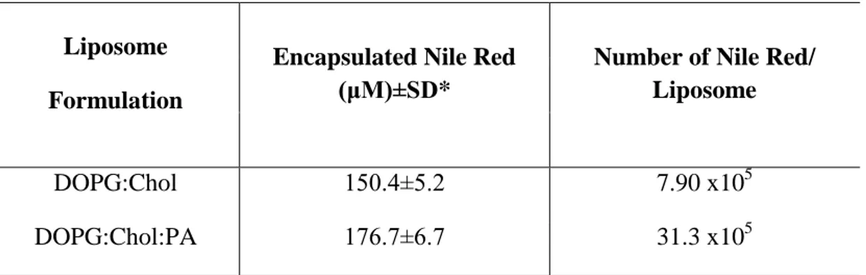
2.3.2 Encapsulation of Dyes and Anticancer Drugs ... 48
2.3.2.1 Encapsulation of Dyes: Rhodamine B and Nile Red ... 49
2.3.2.2 Encapsulation of Anticancer Drugs: Doxorubicin-HCl and Paclitaxel ... 53
xi
2.3.3 Release of Rhodamine B ... 56
2.3.4 Uptake of Dye Encapsulated Liposomes by MCF7 Cells ... 57
2.3.5 Therapeutic Effects of Anticancer Drug Encapsulated Liposomes on MCF7 Cells ... 60
2.4 CONCLUSION ... 64
Chapter 3 ... 66
NONCOVALENT FUNCTIONALIZATION OF MESOPOROUS SILICA NANOPARTICLES WITH AMPHIPHILIC PEPTIDES ... 66
3.1 INTRODUCTION ... 67
3.2 EXPERIMENTALSECTION....…..………....…………69
3.2.1 Materials ... 69
3.2.2 Synthesis of MSN and OMSN ... 70
3.2.3 Synthesis and Characterization of Peptide Amphiphile Molecules ... 71
3.2.4 Coating the OMSNs with Peptide Amphiphiles ... 72
3.2.5 Characterization of the Particles ... 72
3.2.6 In Vitro Studies ... 73
3.3 RESULTS and DISCUSSION ... 75
3.3.1 Synthesis and Characterization of Peptide Functionalized MSNs ... 75
3.3.2 In Vitro Cell Compatibility of MSNs ... 85
3.3.3 Cellular Uptake Studies ... 86
3.4 CONCLUSION ... 89
Chapter 4 ... 90
AMPHIPHILIC PEPTIDE COATED SUPERPARAMAGNETIC IRON OXIDE NANOPARTICLES FOR IN VIVO MR TUMOR IMAGING ... 90
xii
4.1 INTRODUCTION ... 91
4.2 EXPERIMENTAL SECTION ... 95
4.2.1 Materials ... 95
4.2.2 Synthesis of Superparamagnetic Iron Oxide Nanoparticles ... 96
4.2.3 Synthesis of Peptide Amphiphile (PA) ... 96
4.2.4 Surface Coating of SPIO Nanoparticles ... 97
4.2.5 Characterization of SPIO Nanoparticles and Hybrid SPION-PA System ... 98
4.2.5.1 Morphological Characterization ... 98
4.2.5.2 X-Ray Diffractometry Analysis ... 98
4.2.5.3 Chemical Characterization by Infrared Spectroscopy ... 98
4.2.5.4 Thermogravimetric Analysis ... 99
4.2.5.5 Particle Size and Zeta-Potential Analysis ... 99
4.2.5.6 Iron Content Determination ... 100
4.2.5.7 PA Content Determination ... 100
4.2.6 In Vitro Studies ... 101
4.2.7 In Vivo Studies ... 102
4.2.8 Statistical Analysis ... 103
4.3 RESULTS AND DISCUSSION ... 104
4.3.1 Synthesis and Characterization of the Nanoparticles ... 104
4.3.2 In Vitro Cytotoxicity and Uptake Studies ... 115
4.3.3 In Vivo Magnetic Resonance Imaging Studies ... 118
xiii
Chapter 5 ... 122
SUPRAMOLECULAR GAG-LIKE SELF-ASSEMBLED GLYCOPEPTIDE NANOFIBERS INDUCE CHONDROGENESIS AND CARTILAGE REGENERATION ... 122
5.1 INTRODUCTION ... 123
5.2 EXPERIMENTAL SECTION ... 127
5.2.1 Materials ... 127
5.2.2 Synthesis of Acetylated β-D-Glucose Linked Serine Glyco Amino Acid...128
5.2.2.1 Protection of the Carboxylic Acid of Ser-OH to Yield Fmoc-Ser-OAllyl...128
5.2.2.2 D-Glucose Pentaacetate Coupling to Ser-OAllyl to Yield Fmoc-Ser[β-D-Glc(OAc4)]-OAllyl...129
5.2.2.3 Deprotection of Allyl Group to Yield Fmoc-Ser[β-D-Glc(OAc4)]-OH.. ...130
5.2.3 Synthesis and Characterization of Glycopeptide and Peptide Amphiphiles ... 130
5.2.4 Hydrogel Preparation ... 133
5.2.5 Chemical, Physical and Mechanical Characterization of Self-Assembled Nanofiber Networks ... 134
5.2.5.1 Circular Dichroism ... 134
5.2.5.2 Determination of Zeta-Potential of Nanofiber Systems at Dilute Concentrations ... 135
xiv
5.2.5.4 Scanning Electron Microscopy Imaging ... 135
5.2.5.5 Small X-Ray Scattering (SAXS) ... 136
5.2.5.6 Investigation of Viscoelastic Behavior of Nanofiber Systems by Oscillatory Rheology ... 138
5.2.6 In Vitro Studies ... 138
5.2.7 In Vivo Studies ... 140
5.2.8 Statistical Analysis ... 141
5.3 RESULTS AND DISCUSSION ... 141
5.3.1 Synthesis and Characterization of Glucose Linked Serine Amino Acid ... 141
5.3.2 Design, Synthesis and Characterization of Glycopeptide Nanofibers 147 5.3.3 Characterization of HA/K-PA Assembly ... 158
5.3.4 In Vitro Studies ... 160
5.3.5 In Vivo Study ... 163
5.4 CONCLUSION ... 164
Chapter 6 ... 166
CONCLUSION AND FUTURE PERSPECTIVES ... 166
Bibliography ... 171
xv
LIST OF FIGURES
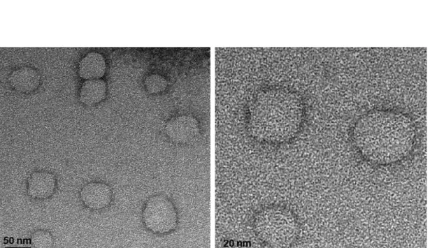
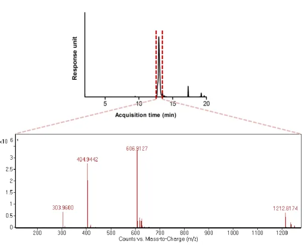
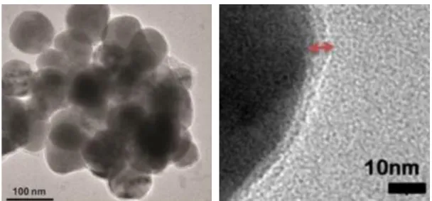
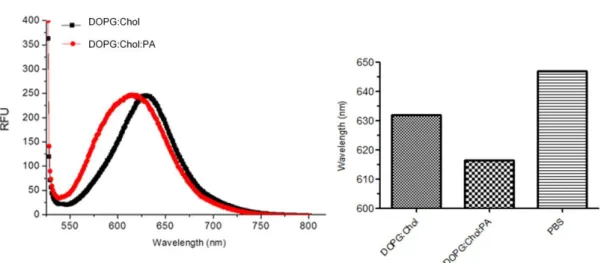
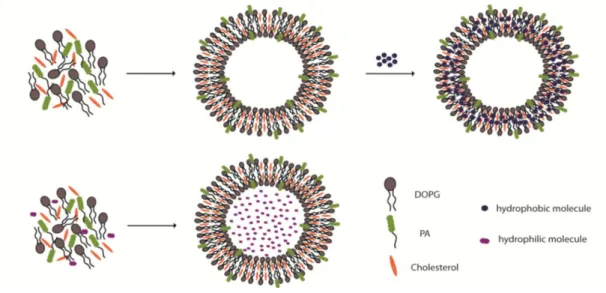
Figure 1.1 Examples of self-assembly.. ... 3 Figure 1.2 Strength and properties of noncovalent interactions involved in self-assembly ... 5 Figure 1.3 Schematic representation of the molecular self-assembly of peptides involved in the formation of different secondary structures ... 7 Figure 1.4 Responsive self-assembled peptide systems...9 Figure 1.5 Schematic illustration of solid phase peptide synthesis ... 12 Figure 1.6 Examples of nanostructures achieved by peptide/protein self-assembly..16 Figure 1.7 Self assembled peptide amphiphiles used in biomedical applications…..28 Figure 2.1 Transmission electron microscope images of liposomes composed of 1:1 molar ratio of DOPG:Chol (scale bars= 50 nm and 20 nm, respectively). ... 41 Figure 2.2 Chemical structures of peptide amphiphile (PA) Lauryl-PPPPRRRR-Am, negatively charged phospholipid 1,2-dioleoyl-sn-glycero-3-phospho-(1’-rac-glycerol) (DOPG) and cholesterol. ... 43 Figure 2.3 Liquid chromatogram of Lauryl-PPPPRRRR-Am and mass spectrum of corresponding peptide molecule ... 44 Figure 2.4 Transmission electron microscope images of liposomes composed of 7:8:1 molar ratio of DOPG:Chol:PA (scale bars= 100 nm and 10 nm, respectively) ... 45 Figure 2.5 Fluorescence measurements performed with Nile Red for the investigation of PA integration into the liposomal membrane. ... 47 Figure 2.6 Schematic illustration for the preparation of PA integrated liposomes loaded with hydrophobic (Nile Red, Paclitaxel) or hydrophilic (Rhodamine B, Doxorubicin-HCl) molecules. ... 48
xvi
Figure 2.7 Chemical structures of hydrophobic and hydrophilic dyes and cancer drugs used for liposomal encapsulation. ... 49 Figure 2.8 Rhodamine B calibration in (a) PBS and (b) ethanol (λex= 540 nm, λem
= 575 nm).. ... 50 Figure 2.9 Nile Red calibration in ethanol (λex= 474 nm, λem= 633 nm). ... 52
Figure 2.10 Doxorubicin-HCl calibration in ethanol (λex= 480 nm, λem= 588 nm). .. 54
Figure 2.11 a) Calibration curve of Paclitaxel plotted from HPLC results, b) HPLC chromatogram of 0.16 mg/mL Paclitaxel in PBS. ... 55 Figure 2.12 Rhodamine B calibration in 10% FBS containing PBS and ethanol (λex=
540 nm, λem: 575nm) ... 57
Figure 2.13 Release profile of DOPG:Chol and DOPG:Chol:PA liposomes at pH 7.4 and pH 5.5 for Rhodamine B...57 Figure 2.14 Cellular viability of MCF7 cells after a) 4 h and b) 24 h. Colours of the bars represent the final peptide concentration, green: 250 µM, blue: 25 µM and red: 12.5 µM. Results were normalized to cells treated with PBS, (n=4).. ... 58 Figure 2.15 Analyses of uptaken model dyes by MCF7 breast cancer cells... 60 Figure 2.16 a) Dose and b) time response of MCF7 cells to the treatment of Doxorubicin-HCl loaded liposomes or free drug ... 62 Figure 2.17 a) Dose and b) time response of MCF cells to the treatment of Paclitaxel loaded liposomes or free drug ... 63 Figure 3.1 a) Schematic illustration for the preparation of fluorescent peptide functionalized MSNs, b) Chemical structures of the peptide amphiphile molecules used in this study ... 69 Figure 3.2 Fluorescence emission spectra of bare and modified MSNs ...76
xvii
Figure 3.3 Liquid chromatogram of Lauryl-PPPPGE and mass spectrum of corresponding amphiphilic peptide molecule ... 77 Figure 3.4 Liquid chromatogram of Lauryl-PPPPGK-Am and mass spectrum of corresponding amphiphilic peptide molecule. ... 78 Figure 3.5 (a) Photographs of OMSNs before and after coating with peptide amphiphiles and (b) Photograph of MSN dispersion in water ... 79 Figure 3.6 Morphological characterization of the mesoporous silica nanoparticles. TEM images of (a) MSN, (b) OMSN, (c) E-OMSN and (d) K-OMSN ... 80 Figure 3.7 FTIR spectra (a) and TGA spectra (b) of particles and peptide amphiphiles.. ... 81 Figure 3.8 Number (%) distribution of MSN, E-OMSN and K-OMSN ... 84 Figure 3.9 Cellular viability results of (a) A10 and (b) HUVECs treated with bare and peptide functionalized MSNs ... 86 Figure 3.10 Uptake results of bare and peptide functionalized MSNs. ... 88 Figure 4.1 Images of hydrophobic iron oxide nanoparticles in the absence and presence of magnetic field when they are dispersed in hexane (10 mg/mL, concentration based on the iron amount)...105 Figure 4.2 a) HR-TEM image of SPIONs, b) lattice fringe corresponds to the (400) lattice plane of magnetite nanoparticles, c) TEM image of SPIONs d) SAED pattern of SPIONs. ... 106 Figure 4.3 Number distribution of hydrodynamic diameter of SPIONs dispersed in hexane...107 Figure 4.4 a) XRD patterns and b) FTIR spectra of bare and modified nanoparticles ... 108
xviii
Figure 4.5 Liquid chromatogram of Lauryl-PPPGK-Am and mass spectrum of corresponding peptide amphiphile molecule. ... 110 Figure 4.6 a) Photograph of modification of hydrophobic SPIONs with peptide amphiphile molecules, b) Chemical structure of peptide amphiphile molecule, K-PA . ... 111 Figure 4.7 Gravimetric analysis of a) SPIONs before assembly with PA, b) SPION/K-PA nad PA samples ... 113 Figure 4.8 TEM images of PA coated SPIONs, scale bars = 10 nm and 20 nm, respectively.. ... 114 Figure 4.9 a) Dynamic light scattering (DLS) analysis to determine the dispersion stability of PA coated SPIONs prepared in two different media, b) photographs taken after a week dispersion in corresponding media ... 115 Figure 4.10 Calibration curve plotted for iron content determination...116 Figure 4.11 a) Live-dead assay for HUVECs treated with K-PA (855 µg/ml) and SPION/K-PA (75 µg/ml) for 24 h. scale bar = 100 µm, 10x magnification b) Quantification of live-dead assay c) Light microscopy of HUVECs after 6 h of incubation with and w/o SPION/K-PA. ... 117 Figure 4.12 a) In vivo MRI of rats treated with and w/o SPION/K-PA b) Signal intensity changes in T2 relaxations at pre-, immediately after injection (post), 1 h, 4 h
and 15 d post administration in breast, liver, spleen, kidney and breast tumor of rats. ... 119 Figure 5.1 Schematic representation of the sequential synthesis of amphiphilic glycopeptide...133 Figure 5.2 Synthesis of compound 3, Fmoc-Ser [β-D-Glc(OAc)4]-OH...142
xix
Figure 5.4 Mass spectrum of compound 1.. ... 144
Figure 5.5 1H-NMR spectrum of compound 2. ... 144
Figure 5.6 13C-NMR spectrum of compound 2 ... 145
Figure 5.7 Mass spectrum of compound 2.. ... 145
Figure 5.8 1H-NMR spectrum of compound 3. ... 146
Figure 5.9 1H-NMR spectrum of compound 3.. ... 146
Figure 5.10 Mass spectrum of compound 3 ... 147
Figure 5.11 Liquid chromatograms and mass spectra of protected amphiphilic glycopeptide ... 148
Figure 5.12 Chemical structures of amphiphilic glycopeptide (Glc-PA) and two peptide amphiphiles (E-PA and K-PA) ... 150
Figure 5.13 Liquid chromatograms and mass spectra of PAs………..151
Figure 5.14 CD spectra of peptide amphiphile solutions and nanofiber systems ……….…..152
Figure 5.15 Temperature dependent CD spectra of a) Glc-PA/E-PA and c) K-A/E-PA nanofibers. Change in molar ellipticity at certain wavelengths with respect to temperature b) Glc-PA/E-PA at 202 nm and d) K-PA/E-PA at 222 nm………...153
Figure 5.16 STEM images of a) Glc-PA/E-PA and b) K-PA/E-PA nanofibers, scale bar = 100 nm ………....154
Figure 5.17 a) SAXS profile and fit curve for the scattering data of Glc-PA/E-PA and PA nanofibers, b) Pair distance distributions of Glc-PA/E-PA and K-PA/E-PA nanofiber systems ………..155
Figure 5.18 SEM micrograph of Glc-PA/E-PA nanofibers (a, c), and K-PA/E-PA nanofibers (b, d), scale bar = 2 μm (a, b) and 1 μm (c, d).………...157
xx
Figure 5.19 Time sweep test for Glc-PA/E-PA and K-PA/E-PA nanofibers prepared a) in water at pH 7 and b) in 0.25 M sucrose/DMEM medium. Equilibrium storage and loss moduli of Glc-PA/E-PA and K-PA/E-PA nanofibers prepared c) in water at pH 7 and d) in 0.25 M sucrose/DMEM medium………..158 Figure 5.20 Hyaluronic acid (HA) and its self-assembly with K-PA molecule. a) Chemical structure of HA. b) TEM image of self-assembled HA/K-PA nanostructures, scale bar = 1 µm. c) Circular dichroism spectra of HA and HA/K-PA. d) SEM micrograph of HA/K-PA assembly, scale bar = 2 µm………...…….159 Figure 5.21 Zeta potentials of glycopeptide and peptide nanofiber systems…..…..160 Figure 5.22 a) Cellular viability of mMSC cultured on Glc-PA/E-PA, K-PA/E-PA, HA/K-PA and TCP at days 1, 2 and 3. b) Hyal-1 gene expression after 36 h of anti-CD44 incubation ………..160 Figure 5.23 a)Safranin-O stainings and b) DMMB assay for quantification of GAGs (normalized to DNA amount) produced by mMSCs cultured on Glc-PA/E-PA and HA/K-PA on days 3, 7 and 14 in chondrogenic medium. c) Sox 9 expression analysis performed at day 1 using mMSCs cultured in chondrogenic medium……...……..162 Figure 5.24 Staining of tissue sections with a) Safranin-O and b) Collagen II immunostain………..164
xxi
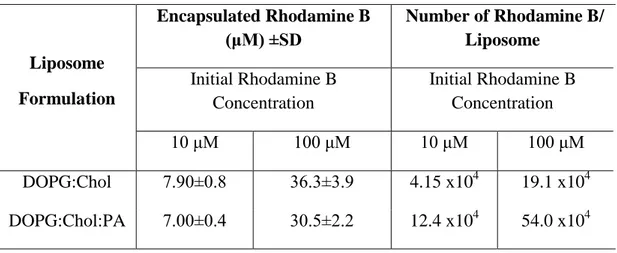
LIST OF TABLES
Table 1.1 Recently published studies using peptide amphiphile architectures.. ... 19 Table 2.1 Physical properties of liposomes composed of DOPG:Chol ... 41 Table 2.2 Physical properties of PA integrated liposomes……… . ...45 Table 2.3 Encapsulation of Rhodamine B by PA free and PA integrated liposomes. 50 Table 2.4 Encapsulation of Nile Red by PA free and PA integrated liposomes ... 52 Table 2.5 Doxorubicin-HCl encapsulation capacities of two liposomal formulations……… ...54 Table 2.6 Paclitaxel encapsulation capacities of two liposomal formulations ... 56 Table 3.1 Physical properties of bare, octyl modified and PA functionalized MSNs 84 Table 4.1 The d-spacing values (nm) calculated from peak positions in XRD pattern inserted in Figure 5.4 and standard atomic spacing for magnetite together with respective Miller indices (hkl) from the JCPDS card (19-0629)…………..…...108 Table 4.2 Physical properties of fatty acid/amine capped SPIONs and PA functionalized SPIONs (a: in water, b: in 5% dextrose, c: in hexane) ... 115 Table 5.1 Results obtained from the fitting process of Glc-PA/E-PA and K-PA/E-PA scattering data using flexible cylinder-polydisperse length model………...155
xxii
Abbreviations
A10 Vascular smooth muscle cell line
ANOVA Analysis of variance
APTES Aminopropyl triethoxysilane
ATRP Atom transfer radical polymerization
Boc Tert-butoxycarbonyl
CD Circular dichroism
CPP Critical packing parameter
CS Chondroitin sulfate
CTAB Cetyltriammoniumbromide
CTLP Curvature tuned liposome preparation
DAB Diaminobenzidine
DCM Dichloromethane
DIEA N,N-diisopropylethylamine
DMEM Dulbecco's modified Eagle's medium
DMF N,N-Dimethylformamide
DMMB Dimethylmethylene blue
DMSO Dimethyl sulfoxide
DOPG 1,2-dioleoyl-sn-glycero-3-[phosphor-rac-(1-glycerol)]
ECM Extracellular matrix
FBS Fetal bovine serum
FITC Fluorescein isothiocyanate
Fmoc 9-Fluorenylmethoxycarbonyl
FTIR Fourier transform infrared spectroscopy
GAG Glycosaminoglycan
GAPDH Glyceraldehyde 3-phosphate dehydrogenase
HAADF High-angle annular dark field
HBTU N,N,N′,N′-Tetramethyl-O-(1H-benzotriazole-1-yl)
uronium hexafluorophosphate
HPLC High pressure liquid chromatography
xxiii
HUVEC Human umbilical vein endothelial cell
IC50 Half maximal inhibitory concentration
ICP-MS Inductively coupled plasma-mass spectrometry
LC-MS Liquid chromatography-mass spectroscopy
MCF7 Michigan Cancer Foundation-7
mMSC Mouse mesenchymal stem cells
MSN Mesoporous silica nanoparticle
NCA N-carboxyanhydrides
NCL Native chemical ligation
NMR Nuclear magnetic resonance
OD Optical density
OMSN Octyl modified mesoporous silica nanoparticle
OTS Octyltriethoxysilane
PA Peptide amphiphile
PBS Phosphate buffered saline
PDD Pair distance distributions
qRT-PCR Quantitative real-time polymerase chain reaction
QTOF Quadrapole time of flight
ROI Region of interest
RT Room temperature
SAXS Small angle X-ray spectroscopy
SD Standard deviation
SEM Standard error of mean
SEM Scanning electron microscopy
SI Signal intensity
SPION Superparamagnetic iron oxide nanoparticle
SPPS Solid phase peptide synthesis
STEM Scanning transmission electron microscopy
TCP Tissue culture plate
TEM Transmission electron microscopy
TEOS Tetraetyl orthosilicate
xxiv
TGA Thermal gravimetric analysis
THF Tetrahydrofuran
TIS Triisopropyl silane
VSM Vibrating sample magnetometer
1
CHAPTER 1
2
1.1 Self-Assembly
Molecular self-assembly, as one of the bottom-up approaches, is emerging powerful tool in the fabrication of functional nanoscale structures. It is defined as the spontaneous organization of molecules into stable, well-defined structures under equilibrium conditions through noncovalent interactions. The self-assembly is ubiquitous in many biological systems and results in the formation of a variety of complex biological structures found in Nature. These complex and elegant examples inspire scientists to create similar artificial systems by using building blocks of life such as peptides, lipids, nucleic acids, and saccharides. Naturally, many key developments in last century have paved the way for the progress of this sophisticated field such as the finding of close-packed form of amphiphilic molecules, the arrangement of monolayers by long chain hydrocarbon amines and the distribution of ordered monolayers of alkanethiolate molecules on gold substrate.1
In most cases, thermodynamically stable structures are formed from the assembling subunits utilized in biomaterials where both the molecular species and the solvent molecules contribute enthalpically and entropically. Since the mobility of the components involved in self-assembly is crucial, fluid phases or smooth surfaces are preferred for the assembly. In the case of the self-assembly of more complex systems yielding meso- or macroscopic structures, gravitational attraction, external electromagnetic fields, entropic interactions and magnetic capillary are also taken into consideration beside noncovalent or weak covalent interactions.2
3
The reason of the rapid growth of this field is that self-assembly allows seeking insight into the structural features of many important biological compounds and makes possible to synthesize larger structures ( > 1000 atoms) that cannot be feasible with covalent bonds due to the poor stability.3 Synthesized molecules can be organized on larger scales by means of self-assembly (Figure 1.1).4
Figure 1.1 Examples of self-assembly. (i) Crystal structure of a ribosome. (ii)
Selfassembled peptide-amphiphile nanofibers. (iii) Electrostatic self-assembly (ESA) of polymeric microspheres on a charge-exchanging substrate modified by wet stamping. (iv) ESA of macroscopic 2D crystals whose formation is mediated by charges developed by contact electrification. (v) Capillary SA of polymeric plates at an interface between two liquids. (vi) Self-assembled polymeric microspheres of complex internal structures. (Reproduced from Ref. 4 with permission from American Chemical Society)
1.2 Self-Assembly of Peptides
Amino acids serve a diverse pool for the construction of peptide-based materials through distinct physicochemical properties, regarding the charge, hydrophobicity,
4
size and polarity, of their side chains that increases the possibility of creating peptidic materials with different amino acid sequences and lengths. This diversity is further enriched by the introduction of non-proteinogenic groups into the peptide backbone. Since peptides are the biomolecules whose synthesis, structure and properties are well understood, significant effort is directed toward the research on self assemblies using peptide-based elements.5 Peptide self assembly is mainly governed by noncovalent interactions. This type of interactions not only drive the self-assembly but also stabilize the secondary structure of peptides and proteins.6
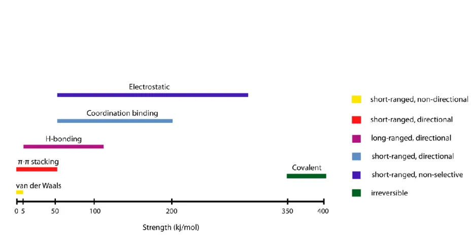
1.2.1 Forces Driving the Peptide Self Assembly
Noncovalent interactions such as hydrogen bonding, hydrophobic, electrostatic, and van der Waals interactions, π-π stacking and coordination bonds are the main contributors of molecular self assembly (Figure 1.2).7-8 While nonpolar amino acids, including aliphatic (eg. alanine, leucine, valine) and aromatic (eg. tyrosine, phenylalanine) amino acids, are mostly responsible for hydrophobic clustering through hydrophobic interactions and π-π stacking, respectively, polar amino acids result in either hydrogen bonding or electrostatic interactions depending on the fact that they have uncharged (eg. serine, asparagine) or charged (eg. lysine, histidine, glutamic acid) residues.9 In addition to individual amino acids, the peptide backbone itself provides considerable stability through hydrogen bonds. Although these highly dynamic interactions are weak, when considered individually, compared to covalent interactions, concomitant interactions can elicit stable assemblies. Multiplicity in the noncovalent interactions leads to obtaining specificity in the self-assembling system.10
5
6
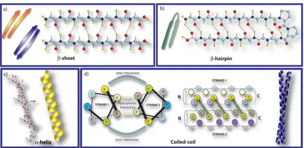
Naturally occurring hydrogen bonding patterns such as those found in β-sheet, α-helix, and coiled coils, are utilized in the design of a number of peptide sequences to form higher order structures as a result of self assembly (Figure 1.3).11 The stabilization of the multiple peptide backbone arrangements by hydrogen bonding interactions between the backbone amide and carbonyls results in the formation of β-sheets. The sheet is either parallel or anti-parallel depending on the direction of the strands. Peptides composed of simple repeat sequences for hydrophobic and hydrophilic amino acids are organized in such a way that hydrophilic and hydrophobic faces are produced and hydrophobic face is buried while its hydrophilic face is exposed to aqueous environment.12 Unlike β-sheets, the α-helices are formed by individual peptide chains where backbone amide components are intramolecularly hydrogen bonded. It leads to the presentation of the amino acid side chains on the surface of each helix and furtherly facilitates the accessibility of amino acid side chains to solvent. In some cases, these single α- helices can assembly by coiling together and form so called coiled coils. Peptide sequences responsible from coiled coil formation generally bear a repeat motif consisting of seven residues. This heptad motif is derived by utilizing from a wheel diagram labelled with specific letters (a-g). So far, the rules required to create coiled coil structure were well studied and conceived. In this section of the review, we give a relatively brief account for the H-bonding patterns exploited in peptide self-assembly; however, there are comprehensive reviews in the literature for more detailed information.13-14
7
Figure 1.3 Schematic representation of the molecular self-assembly of peptides
involved in the formation of different secondary structures (Reproduced from Ref. 11 with permission from Royal Society of Chemistry)
1.2.2 Factors Triggering the Peptide Self Assembly
There is a growing interest in creating systems that assemble and disassemble in response to the external cues. Since the noncovalent interactions are delicate interactions, they have great tendency to respond to the alterations in the environmental conditions such as pH, light, temperature, ionic strength, and solvent polarity (Figure 1.4).15 Among them, pH switch is the most facile approach to control and direct the self-assembly.16-20 A number of peptides are inherently sensitive to pH change due to the introduced charged amino acids, leading to a structural transformation. Apart from pH stimuli, light triggered systems designed by van Hest and Stupp groups were developed by using photocleavable group bearing peptide amphiphiles and morphological transitions were observed for both cases in response to light.21-23 Furthermore, polymerizable diacetylene unit containing peptide molecules were shown to respond to UV light irradiation to acquire one dimensional
8
nanostructures by Tovar group.24 On the other hand, thermally triggered peptide-based materials causing a change in the self-assembly of the nanostructure were reported by several groups.25-30 Alternative to the thermal energy, Ulijn group demonstrated that ultrasound energy can assist the reorganization of supramolecular nanostructures due to the alteration in the contribution of noncovalent interactions, especially of H-bonding and π- stacking, to the resulting supramolecular assembly.31 Differently, the effect of ionic strength and metal ions on the self-assembly mechanism of peptide molecules were examined by the addition of different type of cations into the peptide-based systems. While charged amino acids are mainly responsible for the salt-induced self-assembly as in the case of ionic-complementary peptides32, beta-hairpin peptides33, ultrashort peptides34 and peptide amphiphiles35-36, histidine residues have been predominantly used as metal-binding domain due to the affinity of imidazole ring to several coordination metal ions.37-40 In addition to the above-mentioned external stimulus factors, solvent polarity can also exert a profound effect on the supramolecular nanostructures. Solvent controlled structural transition was observed for several peptides due to the changed interactions between the hydrophobic domain of peptide and solvent molecules.41-44 Self assembly can be alternatively induced by enzyme catalyzed reactions. Structural switches can be employed in a controllable manner by using enzymes specific to peptide sequences.45-49
9
Figure 1.4 Responsive self-assembled peptide systems (Reproduced from Ref. 15
10
1.3 Synthetic Approaches for the Synthesis of Peptides
Peptides consisting of natural or synthetic amino acids are interesting building blocks for the construction of supramolecular assemblies. These simpler structures aid us to understand more complex systems present in nature. Scientists employ a variety of approaches in the synthesis of peptide building blocks and strive for the synthesis of the peptide of interest while minimizing or eliminating the other possible by-products. The synthetic approaches utilized to afford peptide can be classified in three main classes: solid-phase strategies, ring-opening polymerization techniques and genetic engineering.50-51
Most convenient methodology for the synthesis of small to medium-sized peptides is solid phase peptide synthesis (SPPS). In this technique, peptide is grown step by step on an insoluble polymeric resin as a result of iterative amino acid couplings where the deprotection of N-terminus of solid support bound amino acid is followed by the attachment of the subsequent amino acid to it after washing and filtering the unreacted reagents and by-products left from previous reaction (Figure 1.5).52 Amino acids used for the couplings bear orthogonal protecting groups. Orthogonal chemistry approach enables two or more protecting groups to be used concurrently without affecting each other when one of the functional group requires manipulation. General features associated with these protecting groups can be considered as i) the ease of introduction into the functional group; ii) the stability in different reaction conditions; and iii) the safe removal at the end of the synthetic process.53 Prior to the coupling, there is a need to activate the carboxylic group of amino acid by using phosphonium, aminium, uronium or carbodiimide-based reagents for the formation of amide bond. Although carbodiimide derivatives lead to an increase in the degree
11
of racemization during the activation of amino acids, it can be minimized by using additives, especially hydroxy derivatives, which suppress the formation of N-acylurea by shifting reaction towards the formation of active ester form of amino acid.54 At the end of the synthesis, the peptide is easily cleaved from the resin with an acidic treatment.
Nowadays, microwave-assisted SPPS is another option to synthesize longer peptides with increased reaction rates and purities. Typically, coupling reagents and additives used in microwave-assisted SPPS are identical with those applied in conventional SPPS and peptide couplings are carried out at temperatures in the range 50-80°C unless amino acids carry a potential risk of epimerization.55 Setbacks encountered in solution phase synthesis, such as time consuming isolation and purification of intermediate peptides are overcome with SPPS. With SPPS strategy, it is possible to control not only the amino acid sequence but also the C and N terminal functionality of the created peptide.56 Therefore, this technique is eligible for the synthesis of linear, branched, dendritic or cyclic peptides.57-58 In addition, different chemical moieties, such as fatty acids, lipids, saccharides, nucleotides, polymers, drugs, aromatic units, and dyes, can be incorporated into peptide backbone with proper linkage chemistry while peptide is still on the resin.
12
Figure 1.5 Schematic illustration of solid phase peptide synthesis
Activation Deprotection Coupling Final deprotection Repeat steps for next coupling Cleavage and deprotection Free peptide
13
A further progress in peptide synthesis, which allows eliminating the limitations of SPPS with respect to the size and solubility of the peptides is the native chemical ligation (NCL) where two unprotected peptides, a α-thioester containing peptide and an N-terminal cysteine ended peptide, are chemoselectively conjugated through a thioester linkage. While individual peptides are synthesized using SPPS, they are linked to each other in solution phase to create large-sized peptides and even complex proteins.59
On the other hand, the preparation of high molecular weight monodisperse polypeptides with precisely defined primary structure can be achieved by protein engineering, also called as genetic engineering. This strategy allows to synthesize not only structural proteins but also de novo designed proteins as a result of the bacterial expression of artificial genes.60 This technology can be considered as the molecular tool for the preparation of peptide-based materials which relies on the fact that the synthetically designed gene incorporated into the circular DNA plasmid encodes for a specific polypeptide or protein.61 One of the advantages of this methodology is that broad range of nonproteogenic amino acids or functional groups can be also integrated for the preparation of the artificial proteins.62
Polypeptides with high molecular weight, narrow polydispersity and retained chiral integrity can be synthesized by the ring-opening polymerization of amino acid N-carboxyanhydrides (NCA) in a controlled manner. For the polymerization, there are two extensively accepted mechanisms, namely, normal amine (NAM) and the activated monomer (AMM) mechanisms. In both mechanisms, a range of nucleophiles and bases such as amines and metal alkoxides are used for the initiation of the polymerisation.63 Although NCA polymerization has the lack of control over
14
the exact primary peptide sequence, in contrast to SPPS or genetic engineering, it allows to prepare polypeptides in high yield and large quantity.64 In some cases, side reactions occurred during NCA polymerization may lead to the failure in the formation of homo and block polypeptides which have been mostly prevented by introducing various metal- and organo-catalysts.65 In addition, some applications necessitate using high purity monomers to obtain optically pure polypeptides with predictable molecular weights and low polydispersities.
Several other chemical methodologies have been developed to synthesize peptide- and protein-based hybrids. Anchorage of two components has been achieved through thiol-maleimide or alkyne-azide couplings as well as by imine, hydrazone linkages, atom transfer radical polymerization (ATRP) and chemoselective peptide ligation methods.51, 66-67 Although each strategy has its own limitations, with recent advances in synthetic strategies, sophisticated peptidic architectures can be created by using these versatile chemical tools.
1.4 Design-Nanostructure relation in peptide-based systems
The number, type, and sequence of amino acids can be manipulated to design self-assembled peptides. When amino acids are considered individually, they have their own specific characteristics. For instance, while glycine provides a high level of flexibility due to the absence of steric hindrance stemming from the side chains, locked conformation of proline results in a conformational rigidity.68-69 Cysteine, on the other hand, can be chemically or intermolecularly modified via thiol-maleimide chemistry or disulphide bringing, respectively. Moreover, tyrosine, serine and threonine are utilized in further chemical and enzymatic modifications through their
15
hydroxyl groups. Depending upon the chemical modifications of C- or N-terminus, properties of peptide building blocks can also be modulated. There are two other major parameters affecting the structure of peptide-based systems, the secondary structure and noncovalent interactions, which were mentioned in the previous section. Peptides supported by α-helical or β-sheet features allow constructing various different nanostructures.70-71 Apart from the secondary structure, to confer kinetically and thermodynamically stable structures, noncovalent interactions can also be enriched by conjugating functionalized organic linkers to peptide building blocks.72 Figure 1.6 demonstrated examples of different nanostructures obtained as a result of peptide self-assembly.73
16
Figure 1.6 Examples of nanostructures achieved by peptide/protein self-assembly
17
1.5 Peptide Amphiphiles
Peptide amphiphiles (PAs), also called lipidated peptides or lipopeptides, are composed of two main regions, hydrophobic alkyl tail and hydrophilic peptide sequence. This type of molecules are naturally found in living organisms that have important roles especially as an initiator in the signal transduction pathways74-75 and in the host defense mechanisms of bacteria.76 The lipidic parts of these molecules are believed to take part in protein-protein and protein-lipid interactions and provide a linkage to the cellular membrane.75
Amphiphilicity is the main triggering factor for the self-assembly of PA monomers into well-defined supramolecular nanostructures and the stability of the resulting assemblies can be further improved by enhancing the amphiphaticity of PAs. The self-assembly is mediated by a variety of different noncovalent interactions such as electrostatic interactions between charged amino acids, hydrogen bonding, π-π stacking and hydrophobic interactions, leading to the formation of nanostructures with diverse morphologies (fibers, micelles, nanotapes, nanotubes, nanosheets).69 As described by Israelachvili, the balance between hydrophobic and electrostatic interactions is needed to be taken into account while determining the geometry of amphiphiles with minimum free energy.77 Since an alteration in the hydrophilic segment can lead to a change in the critical packing parameter (CPP)78, it can affect the morphology of the overall assembly.79-80 In addition, Velichko et.al. investigated the contribution of hydrophobic interactions and hydrogen bonding to the morphology of the resulting PA assemblies by molecular simulation techniques and created a phase diagram displaying distinct morphologies with respect to the corresponding variables.81 Experimental results were also in good agreement with
18
those obtained by computational analysis. While high content of intermolecular hydrogen bonds between peptide blocks favored the formation of cylindrical PA nanofibers in solution, PA molecules lacking of hydrogen bonds had a tendency to form micellar structures.82-83 Tsonchev et.al performed Monte Carlo simulations to demonstrate the role of electrostatic interactions in the self-assembly of PA nanofibers.84 Later, their self-assembly mechanism was further studied by Lee et.al. and Fu. et.al. through molecular dynamic simulations and it was revealed that individual PAs initially formed spherical micelles and they transformed into long thin fibers by merging with one another due to hydrophobic interactions.85-86 Considering the diversity in the molecular design, including the type of alkyl tail, the choice of amino acids and their order in the β-sheet domain, exploration of distinct PA assemblies is certainly fruitful and rewarding. Table 1.1 summarizes the sequences of the peptide amphiphiles discussed in this section, the resulting nanostructures and the additional information regarding the structural features. Stupp and coworkers demonstrated that C16H31O-V3A3E3 PA, in which hydrophilicity
of the peptide domain increased towards the C-terminus, self-assembled into elongated fibers after neutralization by pH adjustment or by the salt-mediated screening of charged glutamic residues. Although both formulations exhibited similar morphologies, salt induced PA nanofibers formed stronger intra- and interfiber crosslinks through calcium mediated ionic bridges.87
19
Table 1.1 Recently published studies using peptide amphiphile architectures
Peptide amphiphile Structure Details Refs
C16H31O-V3A3E3 Nanofiber Salt induced nanofiber formation 87-88
C16H31O-F3E3 Twisted/helical ribbon
Time/sequence dependent morphological transformation
89
C16H31O-(VE)2, C16H31O-V2E2, C16H31
O-(EV)2, C16H31O-E2V2
Nanobelt/nanofiber/nanoribbon Structural variation in isomeric peptide amphiphiles
90
C16H31O-(VE)n (n=2,4 and 6) Nanobelt
Dimensions of belt structure vs. number of amino acid
91
C16H31O–WA4KA4KA4KA Wormlike micelle/Nanofibers Time dependent morphological transition 92
C16H31O-A4LSQETFSDLWKLLPEN Wormlike micelle
Secondary structure-supramolecular structure relation
93
C16H31O-KTTKS
Flat tape-like/twisted
structure/spherical micelle pH tuned morphology
16
C12H23O-GAGAGAGY Nanofiber/twisted nanoribbon pH tuned morphology 94
C16H31O-IAAAEEEE-Am Nanofiber
pH and concentration dependent self-assembly
20 C16H31O-KKFFVLK
Nanotube-helical ribbon/twisted
tapes Thermo-reversible transition
26
C12H23O-VFDNFVLK-Am and C12H23
O-VVAGE (mixture) Nanofiber Nanofiber diameter ≈10-20 nm
96
C12H23O-P4R4-Am, C12H23O-P4K2R8-Am Nanosphere Diameter ≈15-45 nm 97
KLWVLPKCK2A2V2K(-OC12H23)-Am
KLWVLPKCK2K(-OC22H41)-Am
Nanofiber/nanosphere Importance of β-sheet forming region on self-assembly
98
CnH(2n-1)O-VRGDV (n=10, 12, 14, 16) Nanofiber
Tail length vs. self assembly at different pH
99
C16H31O-H6-(OEG)4-Am,
(OEG)4-H6K(-OC12H23)-Am
Nanofiber/nanosphere Position of aliphatic tail-control on morphology
20
C24H47O-GANPNAAG (diacetylene units
on C24 chain)
Nanofiber Temperature vs. fiber stability 100
diC16H31O-EQLESIINFEKLTWE-Am Cylindrical micelle
8.0 ± 2.3 nm in diameter, polydisperse in length
101
21
Additionally, highly aligned fibrils were obtained with the same design by either injecting heated PA solution into a saline solution or dragging the same solution through a film of calcium salt solution.88 The lamellar plaque structure generated during the heat treatment transformed into bundles of nanofibers upon cooling. Same group also observed an unexpected morphological transformation from twisted ribbons into energetically more stable helical ribbons in C16H31O-F3E3 PAs after
ageing them at room temperature for a month.89 To clarify the reason behind this phenomenon, similar peptide was synthesized by replacing phenylalanine residues with alanine residues and it was observed that newly designed peptide formed 7-9 nm diameter of cylindrical fibrils and did not exhibit any change after being aged indicating that this conformational change is due to the aromatic stacking rearrangement and stems from the existence of the phenyalanine residues. Another critical parameter causing the morphological change in the nanostructure is the concentration. Cui et al. designed an alternating hydrophobic and hydrophilic amino acids containing PA, C16H31O-VEVE.103 In the customized molecular design,
laterally grown PAs self-assembled into one dimensional ultralong and wide nanobelts with widths on the order of 150 nm and heights between 10-20 nm presenting peptide epitopes at the surfaces. With a decrease in concentration, a morphological transition from large nanobelts to twisted nanoribbons was observed by TEM. Same peptide domain was also used in another study together with its isomeric counterparts to investigate the importance of peptide side chain interactions in the morphology of resulting supramolecular assembly.90 Each peptide amphiphile isomer displayed different one-dimensional nanostructure such as nanobelts, nanofibers, twisted and helical ribbons, and nanotubes due to the switch in the amino
22
acid order. While alternating sequence bearing molecules, as in VEVE and EVEV, exhibited flat morphologies as nanobelts and twisted ribbons, respectively, VVEE and EEVV peptide segments led to the formation of two distinct types of nanofibers. In addition to the amino acid order, the effect of the number of valine-glutamic acid (VE) dimeric repeats on the shape and dimension of the supramolecular nanostructures was also systematically studied.91 As the length of the peptide sequence increased, a shift from flat to cylindrical structures was observed due to the higher tendency of inducing twisting in β-sheets.
Apart from the discussed nanostructures, Tirrell group designed two different peptides self assembling into worm-like micelles due to their ɑ-helix propensities.
92-93
In the first design, lysine residues were symmetrically distributed around the helical structure to provide individual helices in the micellar state.92 The results acquired from cryo-TEM, IR and CD revealed that time dependent morphological transition from spherical to worm-like micelles was observed after days and eventually they were transformed into long nanofibrillar structures with outer diameter of ∼10 nm. In the second one, they explored the influence of hydrophobic amino acid residues on the self-assembly of PA by inserting four alanine residues between the palmitic acid and oligopeptide sequence.93 Interposing of hydrophobic alanine residues gave rise to a change in the morphology of corresponding PA from nanoribbons to worm-like micelles, further being confirmed by a secondary structural change from α-helices to β-sheets.
Nanostructural features of peptide amphiphiles can be manipulated by varying the external parameters such as pH, temperature or ionic strength. The effect of pH and temperature on the self-assembly behavior of C16H31O-KTTKS was investigated in
23
two separate studies.16, 104 While an increase in the temperature gave rise to the formation of micellar structures rather than extended tape structure due to the disruption of hydrogen bonding, a gradual decrease in pH transformed nanotapes into twisted fibrils at pH 4 and spherical micelles at pH 2. Similarly, pH dependent morphological alteration was reported for C12H23O-GAGAGAGY PA.94 Cylindrical
nanofibers observed at pH 9 transformed into twisted ribbons at pH 4, most likely due to the neutralization of carboxylic acid and subsequently the weakening of electrostatic interactions, leading to the stacking of the beta sheet laminates. Furthermore, Ghosh et al. demonstrated that the tendency of PA to respond to pH change can be programmed by changing the position of single hydrophobic amino acid residue in a short amphiphilic peptide.95 As isoleucine moved away from the alkyl tail, nanofiber formation was favored over spherical micelles due to the enhanced propensity for the beta sheet formation. In another study carried out by Hamley et.al., thermo-responsive structural change was reported in PAs decorated with a KLVFF core motif that induced self-assembly through π-π stacking and hydrophobic interactions.26 A reversible unwinding transition was observed between twisted tapes and nanotubes/ribbons in a temperature dependent manner.
Coassembly of two oppositely charged PAs can also lead to some modifications in the self-assembled nanostructures due to the electrostatic interactions occurring between charged amino acid residues. Cylindrical nanofibers were observed by Stupp group upon mixing of oppositely charged palmitoylated peptides.105-106 Our group also reported that lauric acid anchored lysine and glutamic acid bearing peptides self-assembled into high aspect ratio, one dimensional fibrillar nanostructures.35 Also, Guler group observed that short bioactive sequences
24
exploited in the PA design did not alter the shape of resulting supramolecular assemblies.96, 107-108 Hamley group studied the co-assembly of oppositely charged PAs by mixing them at different ratios.109 Coassembly driven mainly by electrostatic interactions brought about enhanced beta-sheet formation compared to samples prepared with a single component and resulted in the formation of nanotapes.
The shape of the nanostructure can be tuned by either varying or omitting the beta-sheet forming domain.103, 110 Guler et al. examined the importance of hydrogen bonding among the peptide segment in supramolecular assemblies by replacing trileucine residues with triproline one.110 Due to the beta sheet breaking nature of proline residues, spherical aggregates were observed instead of cylindrical nanofibers obtained with the former system. Mammadov111 et.al. and Mumcuoglu97 et al. also used proline residues to form micellar structures by disrupting beta sheet secondary structure to study the importance of nanostructural feature in tuning immune response and to develop an efficient delivery vehicle for the transfection of oligonucleotides, respectively. Recently, Moyer et al. demonstrated that nanofiber formation was disrupted when beta sheet forming region (A2V2) was removed from
the peptide sequence and corresponding beta-sheet region removed sequence self-assembled into 5-10 nm diameter of nanospheres depending on the length of the tail.98
The insertion of an alkyl tail to a peptide segment was shown to enhance the thermal stability of the corresponding structure.57, 75 The influence of alkyl tail length, its position and number on the self-assembly of PAs was investigated in detail by several research groups. The van Hest group examined the effect of alkyl tail length on the stability of β-sheet assemblies of GANPNAAG112
25
the length of the tail increased, the thermal stability of the PAs improved and the transition from beta sheet to random coil was observed at higher temperatures. Furthermore, Xu et al. synthesized four peptide amphiphiles with different tail lengths by using VRGDV sequence as the peptide domain and showed that PAs with shorter tail lengths did not maintain the stability at higher pHs due to weaker hydrophobic interactions.99 On the other hand, the relation between the site at which hydrophobic segment are attached to the peptide domain and the resulting nanostructure shape was investigated by incorporating an alkyl tail on either N- or C- terminus of an oligo-histidine peptide sequence and it was revealed that cylindrical and spherical morphologies were obtained at physiological conditions, respectively.20 He et al. reported that while two aliphatic tails attached to one side of the peptide sequence formed long fibrils, peptide being conjugated from either side self-assembled into short twisted fibrils.114 Differently, van del Heuvel et al. showed that stability of the self-assembled nanofibers can be controlled by changing the position of the diacetylene moiety on the hydrophobic tail without playing with the length of the tail.100 Tirrell and colleagues introduced dialkyl chain containing peptides 115 and investigated the effect of the length of double tail on the thermal stability of collagen like structure in the peptide amphiphile.116 They further studied the effects of both the number and the length of alkyl tail on the nanostructural feature of model collagen peptide amphiphiles by using the combination of small-angle neutron scattering and cryo-TEM.117 In their recent design, dialkyl tail conjugated T-cell epitope bearing peptide self-assembled into 8 nm diameter of cylindrical micelles.101 In addition to dialkyl tail bearing peptide amphiphiles, Cui group designed three Tat peptide conjugates with different numbers of octanoic acid tails.102 It was reported
26
that while single and double tail containing peptides could not form any well-defined nanostructure, four aliphatic tails attached one self-assembled into fibrillar structure with diameter of 15 nm, most likely due to enhanced intermolecular hydrogen bonding.
1.6 Biomedical Applications of Peptide Amphiphiles
Peptide amphiphile molecules combining the structural features of amphiphilic surfactants with the bioactive peptides have gained immense interest not only due to their unique nanostructural features but also because they are biocompatible and biodegradable.7, 83 Their self-assembly process is governed by various noncovalent interactions resulting in the generation of one-dimensional nanofibers under certain solution conditions.118 Taken together, they are attractive candidates to be used in diverse biomedical applications as drug delivery and antimicrobial agents, tissue scaffolds for regenerative medicine, and imaging probes (Figure 1.7).119-124
PAs were shown to be used as local or systemic therapeutic delivery vehicles for the delivery of hydrophobic drugs to a target. While their hydrophobic tail facilitates the translocation through cell membrane and enhances bioavailability, the bioactive sequence specifically interacts with the cell of interest.125 They can either carry the therapeutic agent or, in most cases, encapsulate it for sustained drug release. Since the self-assembly of PAs generally resulted in the formation of nanofibers, they formed hydrogels which is more suitable for sustained release applications. While Cui group designed hydrophobic drug camptothecin conjugated peptide amphiphile whose peptide segment was derived from tau protein,126 Stupp group encapsulated the same drug into palmitoyl-A4G3E3 peptide nanofibers to treat breast cancer.127
27
PA nanofibrils was demonstrated through enzymatic degradation.128 Moreover, anti-inflammatory drug, dexamethasone, conjugated palmitoyl-V2A2E2K nanofiber gel
was administered into mice, exhibiting minimal burst release compared to drug-peptide mixture and leading to a remarkable decrease in the number of infiltrating inflammatory cells.129 Alternatively, our group demonstrated that positively charged fibrillar and micellar peptide-based nanostructures are proper delivery vehicles for negatively charged antisense oligonucleotide drug.97, 130
Tirrell and coworkers reported that antimicrobial properties of peptides can be improved by the incorporation of a lipid tail into the peptide segment.131 Antibacterial activities of several PAs were investigated against some clinical bacteria even though the exact mechanism of action is still contentious. The results revealed that cationic-rich peptide amphiphiles exhibited enhanced antimicrobial activities.132-133
Apart from biocompatible nature of PAs, the capability of forming three-dimensional nanofibrous network makes them proper candidates to be utilized in tissue engineering and regenerative medicine as cell culture matrices or scaffolds. Peptide scaffolds exhibited superior adaptabilities to cellular environment compared to polymeric alternatives due to their ability to present bioactive signals and to mimic native extracellular matrix (ECM). Different bioactive domains resulting in specific binding to, such as TGFβ-1, VEGF, heparin, BMP-2 or integrin, were incorporated into peptide amphiphiles to develop scaffolds for articular cartilage regeneration,120 angiogenesis,122, 134 bone135 and spinal cord136 regeneration. Unlike linear peptide amphiphiles, branched RGDS containing peptide amphiphiles were also reported as suitable scaffolds to grow human bladder smooth muscle cells.137
28
Finally, PAs were also shown to be used as magnetic resonance imaging contrast agents. To obtain positive contrast in MRI, Gd(III) chelation was achieved by the integration of 1,4,7,10-tetraazacyclododecane-1,4,7,10 tetraacetic acid (DOTA) moiety into peptide backbone.138 On the other hand, iron oxide nanoparticles were decorated with lauryl-VVAGE and lauryl-VVAGK PAs to give negative contrast in MRI.139
Figure 1.7 Self assembled peptide amphiphiles used in biomedical applications. a)
VEGF-mimetic peptide amphiphile presenting high density of biological signals induces angiogenesis. b) Calcein-labeled cells are aligned on the peptide amphiphile nanofiber bundles. c) Peptide amphiphile is conjugated to a drug through reversible covalent bond. (Reproduced from Ref. 124 with permission from Royal Society of Chemistry)
29
CHAPTER 2
CELL PENETRATING PEPTIDE AMPHIPHILE INTEGRATED
LIPOSOMAL SYSTEMS FOR ENHANCED DELIVERY OF
ANTICANCER DRUGS TO TUMOR CELLS
Part of this chapter of thesis is published in the following article140; Reprinted from “Cell penetrating peptide amphiphile integrated liposomal systems for enhanced delivery of anticancer drugs to tumor cells”; Sardan, M., Kilinc, M., Genc, R., Tekinay, A. B., and Guler, M.O., Faraday Discussions, 2013, 166, 269-283, with permission from Royal Society of Chemistry