DEVELOPMENT OF A HISTOTRIPSY
AGENT USING HOST-GUEST
INTERACTION
a thesis submitted to
the graduate school of
engineering and natural sciences
of istanbul medipol university
in partial fulfillment of the requirements for
the degree of
master of science
in
biomedical engineering and bioinformatics
By
Tanzeel Ur Rehman
December, 2017
ABSTRACT
DEVELOPMENT OF A HISTOTRIPSY AGENT USING
HOST-GUEST INTERACTION
Tanzeel Ur Rehman
M.S. in Biomedical Engineering and Bioinformatics Advisor: Assoc. Prof. Dr. Yasemin Yuksel Durmaz
December, 2017
Histotripsy is a mechanical cell ablation technique, which works on the mech-anism of acoustic cavitation using microsecond-long, high-frequency ultrasound (US) pulses that can generate a bubble cloud (cavitation) using the already ex-isting gas pockets in the tissue. Once the bubble cloud gains enough energy, it collapses resulting in the cellular destruction/ablation of the surrounding tis-sue. Histotripsy requires extremely high pressures to initiate cavitation in the tissue. Recently developed Nanodroplet Mediated Histotripsy (NMH) addresses this limitation by lowering the cavitation threshold using perfluorocarbon filled nanodroplets as a histotripsy agent. Despite the fact that these nanodroplets work perfectly for NMH, the synthesis of these nanodroplets is complex, and re-quires expertise in the field of polymer chemistry. Thus, this work aims to address the need for a new histotripsy agent that can work as effectively as nanodroplets, but have better potential in terms of being more user-friendly, straightforward, and economical. Two currently available Food and Drug Administration (FDA) approved and commercial compounds, β-cyclodextrin (BCD) and perfluorohex-ane (PFH), were used to obtain an inclusion complex (IC) through host-guest interaction, where hydrophobic cavity of BCD accommodates hydrophobic per-flurorcarbon that might act as cavitation nuclei, and lower the threshold during histotripsy treatment.
PFH was successfully encapsulated in the cavity of BCD with an encapsulation efficiency of 98%. Physiochemical characterization of the IC supported the com-plex formation, and indicated the potential of having more than one PFC in the cavity, depending on PFH/BCD ratio. The size of the complex was measured at 48 nm, which is smaller than the size of nanodroplets. However, it has a tendency to form a bigger self-assembly depending on the dispersion concentration, which may affect cavitation behavior. Hemolytic activity and cytotoxicity experiments
v
revealed that the inclusion complex is biocompatible at the concentration as high as 1 mg/mL. Finally, the ability to lower cavitation threshold for histotripsy was tested, and it showed that it acts as desired nuclei sites for cavitation at low pressures than histotripsy, indicating that this new agent can be effectively used as a histotripsy agent.
¨
OZET
EV SAH˙IB˙I-M˙ISAF˙IR ETK˙ILES
¸ ˙IM˙I KULLANILARAK
H˙ISTOT˙IR˙IPS˙I AJANI GEL˙IS
¸T˙IR˙ILMES˙I
Tanzeel Ur Rehman
Biyomedikal M¨uhendisli˘gi ve Biyoinformatik, Y¨uksek Lisans Tez Danı¸smanı: Do¸c. Dr. Yasemin Y¨uksel Durmaz
Aralık, 2017
Histotripsi, mikrosaniye boyunda, y¨uksek frekanslı ultrason (US) sinyallerini kul-lanarak, akustik kavitasyon mekanizmasını ile mekanik olarak h¨ucre par¸calama tekni˘gidir. Bu US sinyalleri vucutta hˆalihazırda ¸c¨oz¨unm¨u¸s olarak bulunan gaz baloncuklarından bir baloncuk bulutu olu¸stururlar. Bu bulutun yeteri kadar en-erji kazanarak par¸calanması sonucu i¸cerisinde bulundukları dokuda da mekanik bir par¸calanma/hasar olu¸sur ve bu prosesin ger¸cekle¸smesi i¸cin y¨uksek basın¸c gerekmektedir. Yakın zamanlarda, i¸cerisine perflorokarbon doldurulmu¸s nan-odamlacıkların histotoripsi ajanı olarak kullanıldı˘gı nanodamlacık ortamlı histo-toripsi (NMH) bu y¨uksek basın¸c sınırlamasına ¸c¨oz¨um olmu¸stur. Nanodamlacıklar histotripsi i¸cin m¨ukemmel ajanlar olmalarına ra˘gmen, sentezleri karma¸sıktır ve polimer kimyası alanında tecr¨ube gerektirmektedir. Bu ¸calı¸smanın amacı, his-totripsi i¸cin nanodamlacıklar kadar etkin ¸calı¸sacak, ama ¨uretimi ¸cok daha kolay, b¨uy¨uk miktarlarda ¨uretime uygun ve daha kolay ticarile¸sebilme potansiyeli olan yeni bir histortripsi ajanı geli¸stirmektir. Amerikan Gıda ve ˙Ila¸c Kurumu (FDA) tarafından onaylamı¸s ve ticari olarak temin edilebilen β-siklodekstrin (CD) ve perflorohekzan (PFH) bile¸siklerinin ev sahibi-misafir etkile¸simi ile inkl¨usyon kom-pleksin olu¸sturması ile, hidrofobik karakterdeki perflorokarbon siklodekstrinin hidrofobik kavitesine girerek, histotripsi sırasında kavitasyon olu¸sturacak ¸cekirdek g¨orevi ¨ustelenebilir. Bu durum histotripsi uygulaması sırasında kavitasyon i¸cin gerekli e¸sik basıncının d¨u¸s¨ur¨ulmesini sa˘glayacaktr.
PFH ba¸sarılı bir ¸sekilde %98 etkinlikle siklodekstrinin hidrofobik kavitesine yerle¸stirilmi¸stir. Karakterizasyon ¸calı¸smalar inkl¨usyon kompleksinin olu¸sumunu desteklemi¸s ve PFH/CD oranına ba˘glı olarak bir CD kavitesine 2 tane PFH‘nin girme olasılı˘gını desteklemi¸stir. Bu inklusyon kompleksinin boyutu nan-odamlacıklardan daha k¨u¸c¨uk bir de˘ger olan 48 nm olarak ¨ol¸c¨ulm¨u¸st¨ur, fakat
vii
elde edilen komleksin suda da˘gıtılma konsantrasyonuna ba˘glı olarak kendi kendini daha b¨uy¨uk boyutlu par¸cacıklar halinde d¨uzenledi˘gi g¨ozlenmi¸stir, bu durum histo-toripside kavitasyon davranı¸sını etkileyen bir parametre olabilir. Ayrıca hemoli-tik aktivitelerinin ve h¨ucre i¸ci sitotoksisitelerinin incelenmesi sonucu bu kom-plekslerin 1mg/mL gibi y¨uksek konsantrasyonlarda bile toksik etki g¨ostermedi˘gi g¨ozlenmi¸stir. Son olarak, histotripsi ajanı olarak kavitasyon e¸sik basıncını d¨u¸s¨urebilme kapasitesi test edildi˘ginde, beklenen etkiyi ger¸cekle¸stirdi˘gi, nan-odamlacıklarda oldu˘gu gibi kavitasyon e¸sik basıncını d¨u¸s¨urd¨u˘g¨u g¨ozlenmi¸stir.
per-Acknowledgement
First and foremost, I would like to express my sincere gratitude to my advisor Assoc. Prof. Dr. Yasemin Yuksel Durmaz for the continuous support of my M. Sc. study and research, for her patience, motivation, enthusiasm, and immense knowledge. I could not have asked for better guidance in this educational journey. I would also like to thank the rest of my committee: Asst. Prof. Dr. Mehmet Ucisik and Assoc. Prof. Dr. Bunyamin Karagoz for their encouragement, in-sightful comments, and inspiring questions.
In addition, I thank my fellow labmate and friend Erhan Demirel for the stim-ulating discussions, hard work, and selfless knowledge shared during our research work.
Last but not the least, I would like to thank my family for always supporting my dreams and aspirations. They are the pillar of my life, and I am forever grateful to them for guiding me to a bright future.
Contents
1 Introduction 1
2 Imaging and therapy agents for ultrasound applications 5
2.1 Imaging agents for ultrasound applications . . . 5
2.2 Drug delivery using ultrasound . . . 7
2.3 Ultrasound based theranostics . . . 8
2.4 Therapy using mechanical ablation techniques . . . 9
2.4.1 Radiofrequency ablation . . . 10
2.4.2 Laser thermal ablation . . . 11
2.4.3 High intensity focused ultrasound ablation . . . 11
2.4.4 Histotripsy . . . 13
2.4.5 Nanodroplet mediated histotripsy . . . 14
2.5 Host—guest inclusion complex . . . 17
CONTENTS x 2.5.2 Perfluorocarbons . . . 20 2.6 Hypothesis . . . 22 3 Experimental section 23 3.1 Materials . . . 23 3.2 Methods . . . 24
3.2.1 Preparation of β—cyclodextrin and perfluorohexane inclu-sion complex . . . 24 3.2.2 Methylation of β—cyclodextrin . . . 24 3.2.3 Preparation of methylated β—cyclodextrin and
perfluoro-hexane inclusion complex . . . 25 3.2.4 Hemolysis . . . 25 3.2.5 Cell toxicity studies . . . 26 3.2.6 Fluorescence probe method (Investigation of critical micelle
concentration) . . . 30 3.2.7 Ablation of agarose tissue phantom using inclusion complex
as a histotripsy agent . . . 30 3.3 Characterization . . . 31 3.3.1 Hydrogen nuclear magnetic resonance spectroscopy . . . . 31 3.3.2 Fluorine nuclear magnetic resonance spectroscopy . . . 32 3.3.3 Fourier transform infrared spectroscopy . . . 32
CONTENTS xi
3.3.4 Thermal gravimetric analysis . . . 32
3.3.5 Gas chromatography . . . 32
3.3.6 Dynamic light scaterring . . . 34
3.3.7 Scanning electron microscopy . . . 34
4 Results and discussions 35 4.1 Preparation of inclusion complex . . . 36
4.2 Characterizations of inclusion complex . . . 42
4.2.1 Nuclear magtenic resonance spectroscopy . . . 42
4.2.2 Fourier transform infrared spectroscopy . . . 45
4.2.3 Thermal gravimetric analysis . . . 46
4.2.4 Dynamic light scattering . . . 47
4.2.5 Scanning electron microscopy . . . 49
4.3 Toxicity studies of the inclusion complex . . . 51
4.3.1 Interaction with Red Blood Cells (hemolytic activity) . . . 51
4.3.2 Cell viability . . . 53
4.4 Critical micelle concentration . . . 55
4.5 Scanning electron microscopy of MIC at 0.1 mg/mL and 1.0 mg/mL concentrations . . . 56 4.6 Measurement of histotripsy threshold in agarose tissue phantoms
CONTENTS xii
5 Conclusion 60
List of Figures
2.1 A comparison of an US image before and after the addition of the contrast agents. . . 6 2.2 The working of the RFA (left). The needle electrode used in RFA
(right). . . 10 2.3 HIFU delivering a focused US beam to the tumor from outside the
skin and a thin layer between dead and alive cells can be seen. . . 12 2.4 An imaging probe and a transducer used for HIFU. . . 13 2.5 The letter ’M’ drawn on a tissue phantom using histotripsy. . . . 14 2.6 The effect of 2-cycle histotripsy pulse (20.7 MPa) with a pulse
repetition frequency of 10 MHz on the nanodroplets with different PFP loading and embedded with RBCs in agarose gels. . . 15 2.7 The effect of 2-cycle histotripsy pulse (11.0 MPa) with a pulse
repetition frequency of 10 MHz on nanodroplets containing 1 —2 % PFP. . . 16 2.8 A PFP encapsulated nanodroplet with a cross-linked tri-block
LIST OF FIGURES xiv
2.9 Comparison of the inner diameter of alpha, beta and gamma cy-clodextrins, also showing the structural shape of the cyclodextrins. 19
2.10 A 3D structure of BCD showing its hydrophobic cavity. . . 20
2.11 A 2D and 3D structure of PFH. . . 22
4.1 Visual representation of the formation of an IC . . . 35
4.2 The calibration curve made using GC for the quantitative analysis of amount of PFH in IC and MIC. . . 37
4.3 1H NMR spectrum of BCD and MCD. A clear peak shift and a decrease in the intensities of a, b, and c can be seen. . . 39
4.4 Comparison of reaction flask before and after addition of PFH at room temperature. There is a clear visual difference in the solution color proving the formation of the MIC. . . 40
4.5 Picture on the left shows the prediction of one PFH filling the cavity of BCD or MCD(side and top view). On the right, the prediction of two PFH filling one cavity is shown (side and top view) 42 4.6 1H NMR spectrum of MCD and MIC recorded in D2O and DMSO, respectively. . . 43
4.7 19F NMR spectrum of MIC and PFH. Clear shift of peaks of CF 3 and CF2 can be seen. . . 44
4.8 FTIR spectrum of BCD, MCD, PFH and MIC. . . 45
4.9 Thermal gravimetric analysis of BCD, MCD, and MIC. . . 47
4.10 Size comparison of BCD, MCD, and MIC. . . 48
LIST OF FIGURES xv
4.12 Comparison of SEM images of BCD, MCD, IC, and MIC. (a)BCD at 5000X magnification. (b)BCD at 25000X magnification. (c)MCD at 5000X magnification. (d)MCD at 10000X magnifi-cation. (e)IC at 5000X magnifimagnifi-cation. (f)IC at 25000X magni-fication. (g)MIC at 5000X magnimagni-fication. (h) MIC at 25000X magnification. . . 50 4.13 A representation of percentage hemolysis caused by BCD, MCD,
and MIC. . . 52 4.14 Cell viability of HEK-293T cells after incubation with different
concentrations of BCD, MCD, MIC, and PFH. . . 54 4.15 Fluorescence intensity of pyrene mixed with the different
concen-tration of MIC. The two tangents cut at 0.295 mg/mL concentra-tion which reveals the CMC for the MIC . . . 56 4.16 SEM images of freeze dried MIC at a concentration of 0.1 mg/mL
and 1.0 mg/mL in PBS and water. (a) 0.1 mg/mL in PBS, (b) 1.0 mg/mL in PBS, (c) 0.1 mg/mL in water, and (d) 1.0 mg/mL in water. . . 57 4.17 Bubble cloud was generated using MIC as historipsy agent in the
agarose phantom. . . 58 4.18 Bubble cloud was generated using MIC as historipsy agent in the
agarose phantom at voltages of 100 V and 150 V using a 700 kHz transducer with 5 cycles per pulse. . . 59
List of Tables
2.1 A comparison of the physical properties of the CDs. . . 20
4.1 % yield and EE of different ICs produced using different molar ratios. 38 4.2 List of % yield and EE of different MICs produced using different
molar ratios. . . 41 4.3 Peak assignments for 1H NMR spectrum of MCD and MIC and
their change in ppm . . . 43 4.4 Peak assignments for 19F NMR spectrum of PFH and MIC and
Chapter 1
Introduction
Cancer is a set of diseases caused by the abnormal and uncontrolled division of cells [1]. The cells divide without stopping, and continue spreading and surround-ing the area around the tissue. The whole body is made up of trillions of cells, and cancer can start in any of them because it is not specific to a particular body part. Normally, a cell grows, divides, and when its purpose is served, it ages and dies. When it comes to cancer cells, this process is altered. When a cell becomes abnormal, it gets old, but it does not die as it is expected. Additional cells are created as well, even though they are not needed as the existing ones did not die. These extra cells, once they start proliferating, do not stop, and cause excessive growths known as tumors. In 2017 only, more than 1.5 Million new cancer cases are expected with more than 0.6 Million deaths [2]. In 2016, approximately 23% of the total deaths in USA were caused by cancer [3].
Cancer staging, a process which provides information about the location and the size of the tumor, is one of the initial steps in the therapy. It also gives infor-mation about whether the tumor has spread to other organs or not [4] .Treatment decisions are made based on the information provided by staging. Although, removal of cancer by surgery, also known as oncological surgery, is known to be the oldest treatment for cancer, but it involves complex surgical procedures.
Most of these surgeries are followed either by chemotherapy or by radiation ther-apy, in which exhausts the patients too much, and includes affects like hair loss and intense weakness. Depending upon the cancer type and size, chemother-apy and radiation therchemother-apy are sometime used on their own without the surgery. Chemotherapy, basically, is just the use of any drug to treat any disease. It uses a drug to be injected in the body, and kills the cells. The biggest disadvantage of chemotherapy is that it cannot differentiate between healthy and cancerous cells [5]. Thus, it ends up killing both healthy and cancerous cells, causing sizeable weakness for the patient, and often causing death. Radiation therapy, on the other hand, uses high energy X-rays to kill the tumor. Again, the disadvantage is that it damaged the healthy cells around the tumor and it might end up damaging organs [6].
As science is advancing, new and advanced methods for the treatment of can-cer are emerging. Immunotherapy, hormone therapy, and targeted therapy are some of these methods. In immunotherapy, the immune system of the body is strengthened so that it can fight cancer more efficiently [7]. The immune system can be boosted using different substances from the body, or compounds developed in the laboratory. The hormone therapy is currently in use for breast cancer. It works in an indirect way by making a hindrance for estrogen, and also, by lim-iting its occurrence in the body. Targeted therapy uses micro- or nano-sized particles to treat the tumor. These particles can carry drugs, DNA, and RNA etc. They can also be used with different image modalities for advanced results [8]. There are various types of micro and nanoparticles available, such as drug conjugates, dendrimers, vesicles, micelles, microbubbles etc. Imaging modalities which can be used with these are optical imaging, Magnetic Resonance Imag-ing (MRI), Radionuclide-based imagImag-ing, Computer tomography (CT), and Ul-trasound (US) [8]. US was first used for medical purpose by psychiatrist and neurologist Karl Dussik in 1942, and it has been helping the medical field ever since [9, 10]. Although, initially, US became popular for its imaging capability in diagnostic medicine, recent advancements have allowed US to be used as a ther-apeutic device as well [9]. For therther-apeutic purposes, US was used to enhance the delivery of chemotherapeutic-, thrombolytic-, and gene-based drugs [11, 12, 13].
Along with aiding the drug delivery process, US contrast agents are now being developing to enhance its image contrast. Research has proven that US contrast agents can also be used as drug and gene delivery carriers [14, 15, 16]. They can be loaded with drugs or genes with targeting ligands on the surface, and imaged using US. Once the imaging is complete, the US frequencies can be altered to rupture the contrast agents, so that the drug can be released locally onto the tumor [17, 18].
Other than drug delivery enhancement, and use of US sensitive contrast agents, US has also been distinguished in treating cancer tumors through mechanical cell ablation techniques. The basic principle involves using US frequencies to increase the temperature of the tumor to cause either coagulative necrosis or cavitation. Radiofrequency ablation (RA), laser ablation (LA), high-intensity focused ultra-sound (HIFU), and histotripsy are some of the mechanical cell ablation tech-niques. Histotripsy, the newest technique, was developed about 10 years ago, and it uses acoustic cavitation as the primary mechanism. A histotripsy transducer releases very high frequency US pulses focused on a region of a tissue. The gas pockets in the tissue acts as the nuclei for cavitation and forms a highly dynamic cluster of microbubbles. When these microbubbles ruptures, it releases energies high enough to fractionate the cell. Usually, histotripsy required high pressures to turn these gas pockets into a microbubble cloud, usually ranging from 28 MPa to 30 MPa. With this much pressure, there is a risk of rupturing the normal tissue without the tumor.
After recent advancements, a new type of histotripsy emerged known as Nanodroplet Mediated Histotripsy (NMH). This NMH uses small polymer nan-odroplets which encapsulate perfluoropentane (PFP) [19]. Once in the tumor, instead of regular gas pockets, these nanodroplets act as the nuclei for bubble cloud formation, which then leads to cavitation [20]. These nanodroplets were reported to bring down the high cavitation pressure from 28MPa to 7 MPa, thus, solving the risk of damaging normal tumor free tissues [19]. Despite the fact that these nanodroplets work perfectly for NMH, the synthesis of these nanodroplets contains multiple steps. Advanced skills and expertise in polymer chemistry are
the nanodroplets. Additionally, these nanodroplets are currently the only agents available and produced for NMH. Thus, there is a need for a new, easy to syn-thesize, and user-friendly histotripsy agent.
In this study, the limitations of the nanodroplets are addressed, and a new his-totripsy agent is manufactured. It includes a simple host-guest chemistry using β-cyclodextrin (BCD) and perfluorohexane (PFH). Both the agents are commer-cially available and approved by the Food and Drug Administration (FDA). The method used for the synthesis is relatively easier, economical, efficient, and more suitable for commercialization. The goal of this study is to present a new his-totripsy agent which is as efficient as the nanodroplets, but has the upper hand when it comes to ease of generation.
Chapter 2
Imaging and therapy agents for
ultrasound applications
2.1
Imaging agents for ultrasound applications
US is basically a sound wave at frequencies exceeding the audible band [5]. In the field of diagnostic medicine, these waves are transmitted to a part of the body. According to difference in the densities and absorption of components in the body, these waves are reflected back and an image is generated [21]. It has been decades since US has been used as an imaging device and helping the medical field [10]. Not only it provides a real time image, also it is completely harmless and painless. The patients are not exposed to any type of radiations. US scans does to include any needles or injections. US has been used for pregnancy tests for more than 40 years now, and there has not been any report to suggest that it causes any harm to the patient, embryo, or fetus since.
Compared to other imaging modalities, US is less expensive than other de-vices. Also, it is easier to identify soft tissues in an US image, which are not visible in X-ray images. To make the US image quality better, different poly-meric microbubbles are used as contrast agents. These are injected into the body
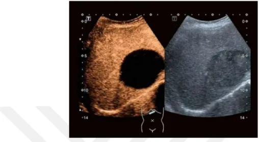
Figure 2.1: A comparison of an US image before and after the addition of the contrast agents.
before the US scans, and they provide considerable contrast in the images after the injection. Figure 2.1 shows two US images, the left image is taken before the addition of the contrast agents. Compared to that, a clear enhancement in the contrast can be seen in the picture on the right which was taken after the injection of the contrast agents.
Initially, the microbubbles produced encapsulated air inside them. Later mi-crobubbles were developed containing fluorinated gas, such as perfluoropentane (PFP), perfluoropropane (PFR), and perfluorohexane (PFH) etc. These are sur-rounded by either a polymer, lipid or a protein [22]. The microbubbles have a high compressibility compared to the tissue surrounding the bubbles. This gives a rise to the acoustic impedance and the microbubbles oscillates, which in turn produces nonlinear acoustic radiations ad a medium range of US frequencies [23]. It is this phenomenon that provides a higher contrast in the region which has these microbubbles [24, 25].
Currently, many of these microbubbles are commercialized and being used all over the world. Albunex was one of the first commercialized microbubbles which contained an air core surrounded by albumin. Fluorinated gas encapsulated mi-crobubbles were developed and commercialized later. Optison and Definity were developed which encapsulated PFR and had a shell of protein and phospholipid,
respectively [26].
More work had been done on these contrast agents to lower the size of these microbubbles to nano width. It was observed that lowering the size of these mi-crobubbles lowers the contrast enhancing capabilities of the mimi-crobubbles. On the contrary, a new gateway was open where these could be used as therapy agents. Since then, US has been used as a diagnostic device as well as a thera-peutic device. Present day, therapy using ultrasound can be classified in to three categories. Ultrasound for drug delivery using ultrasound, ultrasound based th-ernostics, and therapy using mechanical ablation techniques [27].
2.2
Drug delivery using ultrasound
US has shown evidence to raise the thrombolytic efficacy of urokinase [28]. This phenomenon can occur due to localized cavitation or the weakening of the clot by the use of US. Also, this will increase the penetration of the drugs into the cells or the tumor [29, 30]. Furthermore, it has been shown that the presence of microbubbles enhances this phenomenon. When the US is exposed to the body in the presence of the microbubbles, they produces pores on the surface of the cells through which the drugs can penetrate easily [31, 32].
If the tumor is small, ultrasound is used to cause hyperthermia to the tumor specific region. With the use of high intensities ultrasound, the temperature of the tumor is elevated high enough to go through coagulation [33, 34]. By this, the tumor is damaged and can also be killed by minimal damage to the normal tissues surrounding the tumor [35, 36].
Another way to enhance the delivery of chemotherapeutic drugs is by using lower intensities of the ultrasound. This increases the temperature moderately and amplifies the cytotoxity of the chemotherapeutic drugs [12, 11]. US has been tested to enhance the drug delivery in a number of biological and clinical trials. Unger et al. performed experiments using microbubbles which encapsulated a
drug filled layer of soy bean oil and the drug paclitaxel [37]. He concluded that the microbubbles enhances the drug delivery and helps to produce pores on the cell surfaces for easier penetration of the drug. Guzmen et al. also conducted experiments to quantify the molecular uptake of celcein in prostate cancer, and also tested aortic in smooth muscle cells. He investigated the heterogeneity of the cavitation and factors of that enhances the cell uptake [38, 38]. Literature also shows that microbubbles have been used with Doxorubicin and colloidal particles for enhanced cell uptake [39, 40].
2.3
Ultrasound based theranostics
The term theranostics was recently formulated to specify the ongoing research and efforts going on which were focused on to bringing together the advantages of diagnostics and therapy to form a single agent [41]. Once the size of the microbubbles was lowered, they were known as nanoparticles. Development of these nanoparticles opened a new portal for the therapeutic paradigm. It joins together the imaging as well was therapeutic functions giving rise to a theranostic agent capable of diagnosis, drug delivery and monitoring of therapeutic response [41].
To date, many different types of theranostic agents have been produced with different surface chemistries. It is because of their surface chemistry that pharma-ceutical and chemotherapeutic drugs can be loaded inside the core. Nanoparti-cles with different surface chemistries are available ranging from iron oxide, gold, quantum dots, silica and carbon nano tubes.
Zhang et al. developed pH sensitive iron oxide particles which contained methotrexate, a chemotherapeutic drug, onto the surface [42]. These particles were tested in vivo which revealed these particles enters the cells and upon reach-ing in the lysosomes, the drug is released due to the pH difference and because of the presence of proteases. Similarly, Hwu et al. demonstrated the use of pa-clitaxel drug onto the surface of these iron oxide nanoparticles [43] Other than
these, there are many other examples present which shows the use of doxorubicin as well [44].
Apart from drug delivery, RNA and DNA can also be delivered to the cells. This technique is knows as gene delivery or gene therapy. It was in 1996, that Kim et al. used the US to transfer the plasmid DNA to a variety of cells [45]. Also, they performed an in vivo experiment where they used albumin based theranostic agents to load plasmid DNA and transferred them to rat knees [30]. There are more examples in the literature with more work on delivering plasmid DNA, and also delivering DNA to heart and lungs of mice and rats [46, 47, 48, 49, 50, 51, 52, 53, 14].
2.4
Therapy using mechanical ablation
tech-niques
Ablation literally means chipping off, or vaporizing a material. In biological terms, mechanical cell ablation is referred to a technique in which a tumor is ablated locally, using different instruments, with minimal damage to the sur-roundings [54]. With the advancements, the combination of ablation with imag-ing techniques like ultrasound makes it more focused and efficient. Image guided ablation has a huge potential, and is an adroit alternative for the treatment, especially when the patient cannot undergo a surgical treatment [55, 56, 57].
Another advantage of using the ablation technique is that after the ablation, the remaining debris in situ may work as tumor antigens to the immune system. This might turn into a process known as the Abscopal effect [58]. This mean that the ablated tumor debris will work as an in situ cancer vaccine which would stimulate systemic immune responses towards metastases present everywhere in the body [59]. There are different types of cell ablation techniques available like radiofrequency ablation (RFA), laser ablation (LA), high-intensity focused ultrasound (HIFU), and histotripsy. Each of these techniques will be discussed
one by one below.
2.4.1
Radiofrequency ablation
Radiofrequency ablation (RFA) is an image guided, minimally invasive treatment for cancer. A thin needle electrode is inserted in the body near the tumor and alternating current at about 400 MHz frequency is passed through it. This results in frictional agitation of the molecules and generates heat, knows as the joule effect [60, 61]. This process creates a focal heat on the tumor and kills the cells surrounding the electrode, with little heat transferred to tumor free areas [62, 63]. Figure 2.2 displays how the RFA works using a US on the left and, on the right, it shows an RFA needle electrode is shown. This technique has advantages like option for local treatment. Also, it can be used in supplement with chemotherapy to provide a better result. When used with chemotherapy, it also prolongs the time without toxicity in the body [64]. Image modalities such as US, MRI or CT can be used as a guide for RFA .However, RFA needs to be operated by a well-trained personnel preferably with a high quality device as well. Additionally, if the tumor size is larger, the efficiency of RFA decreases [65].
Figure 2.2: The working of the RFA (left). The needle electrode used in RFA (right).
2.4.2
Laser thermal ablation
Laser Thermal Ablation (LTA) has not been used as much as RFA but its results as good enough to be compared with RFA. Research has proven that LTA can be used to fully ablate a tumor which is eligible for thermal ablation [66]. In LTA, a high energy laser radiation is delivered through laser optic fibers to the tissue. Due to the absorption of laser, temperatures as high as 150◦C can be reached. This much high temperature can cause coagulative necrosis [67, 68]. Due to a higher penetration of light in the near infrared spectrum, Neodymium:Yttrium Aluminum Garnet (Nd:YAG) and diode lasers of wavelengths 1064 nm and 800-980 nm, respectively, are most commonly used.
Usually, a single optical fiber is used for ablation, which creates a thermal lesion of about 15 mm [66]. A beam splitting device can be used as well, creating two beams with a single source. Also, a multi-source device can be used, which allows four fiber optics to be used at the same time, this results in a higher ablation area [69, 70]. Although LTA produces good results, there is still a risk of damage to the surrounding area due to extensive working temperature of the laser [71, 72].
2.4.3
High intensity focused ultrasound ablation
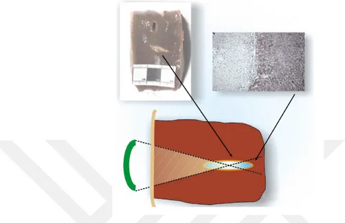
As everyone knows, today the application of US is not limited to diagnostics purposes only. They are also used to treat the diseases in multiple ways. High intensity focused ultrasound (HIFU) was developed in the 1940s but it didnt reach its pinnacle until recent developments. Now HIFU is one of the best noninvasive and extracorporeal technique. The best feature among many oth-ers is that there is no need for a lesion, or any penetration through the skin. HIFU operates from above the skin [73]. Figure 2.3 shows how the HIFU bean passes through the skin and focuses onto the tumor and induces tumor necrosis.
Figure 2.3: HIFU delivering a focused US beam to the tumor from outside the skin and a thin layer between dead and alive cells can be seen.
HIFU works on the same principle of a typical US. HIFU uses ultrasounds of higher intensities, 100-10,000 W/cm2, focused at a specific region using the special
probe. Figure 2.4 shows the transducer and the imaging probe used in a HIFU. These focused US increases the temperature of the tumor within 1 second. When the temperature of the tissue is rapidly increased above 60◦C for one second, it ruptures instantly via coagulation necrosis [73]. This cell death via thermal effect is the primary mechanism of HIFU.
Mechanical effect is the secondary mechanism of HIFU. It works by inducing cavitation, radiation force, and micro-streaming using acoustic pulses of very high intensities. When an ultrasound wave of high intensity propagates though the tissue, due to the compression and expansion of the tissue, a gas cavity is formed in the acoustic field, this is known as cavitation.
A cavitation can be classified in two types, stable and inertial cavitation [74]. A stable cavitation occurs when a low pressure acoustic wave passes through the tissue. Whereas, an inertial cavitation is induced using high pressure acoustic waves which causes the bubble to oscillate aggressively and it increases in size. After reaching the resonance size, the bubble collapses resulting in very high
pressure and temperature, 20,000 to 30,000 bars and 5000 K, respectively, in the microenvironment [74].
Although, HIFU is advancing in the field of treating cancer, and the results are very impressive, but it has a few limitations which needs to be addressed. Firstly, HIFU is incapable of predicting and controlling the formation of the lesion. Secondly, the cavitation might occur in the normal tissue as well. Finally, it has a poor tissue contrast with a limited field of view which makes it difficult to use [75].
Figure 2.4: An imaging probe and a transducer used for HIFU.
2.4.4
Histotripsy
Histotripsy is a recently developed technique which is completely non-invasive and extracorporeal [76]. It uses very short (15 —20 s), high intensity (500 MHz —1000 MHz) US pulses focused on the tissue. This produces a cloud of cavitation bubble in the tissue which oscillates and builds up the pressure. Once the negative pressure is raised above the critical threshold pressure of around 28 MPa, this bubble cloud bursts and ablates the tissue surrounding the cloud [76, 77].
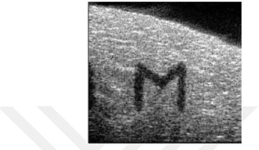
The histotripsy works on the mechanism of acoustic cavitation. The alteration in the US pressure form microbubbles in the human tissue. Once the energy level is high enough, the microbubble activities fragment and subdivide tissue, which results in cellular destruction. Histotripsy has some important advantageous
Figure 2.5: The letter ’M’ drawn on a tissue phantom using histotripsy. points when compared to non-invasive thermal therapy. First of all, microbub-bles are clearly visible through US imaging, so the operator can see the targeted volume. Additionally, the US imaging provides real-time feedback of the ener-getic microbubble activities and the cellular destruction, which allows for a more transparent treatment. Finally, this technique is very precise, and allows for the treatment to be performed in a controlled environment. Figure 2.5 shows an ex-ample of the precision of histotirpsy, as it shows a letter ’M’ made on the tissue phantom using the histotripsy. A clear depth can be seen where the phantom is ablated.
2.4.5
Nanodroplet mediated histotripsy
NMH is mediated by polymer nanodroplets which contains PFP inside them. It has been proven that using these PFP filled nanodroplets under high frequency ultrasounds vaporizes and form gas bubbles. Once these gas bubbles are formed, histotripsy uses these gas bubbles for cavitation which can be achieved at a sig-nificantly lower threshold pressure or 7 MPa [20]. This NMH approach takes advantage of the significantly reduced cavitation threshold of the nanodroplets localize allowing cavitation to be selectively generated only in regions where nan-odroplets [65]. NMH has the potential for selective ablation of tumors given the
small size (100 —400 nm) of the synthesized nanodroplets, which enables their diffusion across the leaky tumor vasculature and preferential accumulation in the tumor tissue [78]. Evidence was provided that this NMH can be used to create well-defined ablation similar to that obtained with histotripsy, but at significantly lower pressure, and has also indicated the potential to use NMH for simultaneous multifocal ablation [20].
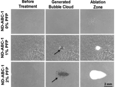
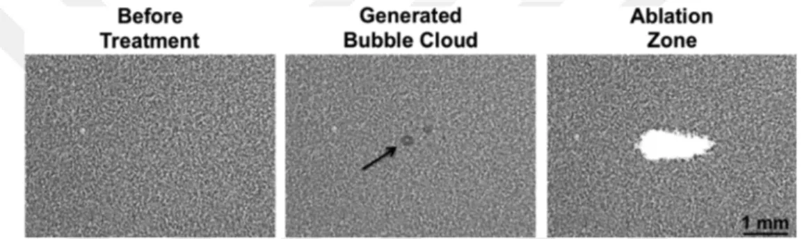
Durmaz et al. showed that because of the small size of the nanodroplets, the histotripsy threshold pressure, needed for cavitation, dropped significantly [76]. They claimed that encapsulating the nanodroplets with PFP will further decrease the cavitation threshold pressure making NMH totally non-invasive and safe for the normal tissues. The effect of adding PFP can be seen in Figure 2.6. It shows how a 2-cycle histotripsy pulse (20.7 MPa), with a pulse repetition of 10 MHz, generates a bubble cloud which leads to ablation. This ablation area increases with the increase in the percentage of PFP. In another experiment, they showed that same results can be achieved by the using the PFP encapsulated nanodroplets at 11.0 MPa. This result is shows in figure 2.7.
Figure 2.6: The effect of 2-cycle histotripsy pulse (20.7 MPa) with a pulse repe-tition frequency of 10 MHz on the nanodroplets with different PFP loading and embedded with RBCs in agarose gels.
Furthermore, it was also hypothesized that the small size of the nanodroplets would help them to accumulate inside the tumor through the EPR effect. Addi-tionally, targeting ligands can be attached on the surface of the nanodroplets and they can be directed towards a specific organ in the body. Moreover, these nan-odroplets also function as an US contrast agents. The encapsulated PFP inside them is, which is approved by the FDA. This allows the tumor to be easily seen through the US in real-time and also guides the histotripsy throughout treatment.
Figure 2.7: The effect of 2-cycle histotripsy pulse (11.0 MPa) with a pulse repe-tition frequency of 10 MHz on nanodroplets containing 1 —2 % PFP.
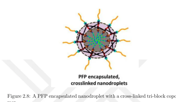
Although, the NMH is works in an excelling way, the synthesis of the polymer and fabrication of the nanodroplets is a complex part. The nanodroplets are composed of a tri—block copolymer which has to be synthesized in the lab and requires advanced skills and expertise in polymer chemistry [19]. Figure 2.8 shows a nanodroplet after complete synthesis of tri—block copolymer, PFP encapsula-tion and crosslinking. Once the tri-block copolymer is produced, it is then used to fabricate the nanodroplets encapsulating the PFP. Other than this, currently only this nanodroplets is available for NMH. There is no other type of droplets, or any other agent that can be used to provide the same results of lowering the threshold cavitation pressure of NMH.
Thus, there is an immediate need of new, innovative agents that can be used as histotripsy agents. We hypothesize that there is a simple and fast way to achieve new kind of histotripsy agents which are easy to produce and are cost effective. It uses BCD, a 7 sugar unit molecule, which has a cone like structure can be filled up with PFH to produce the same effect as the nanodroplets using a rather simple method. Both of these materials are approved by the FDA and
Figure 2.8: A PFP encapsulated nanodroplet with a cross-linked tri-block copoly-mer.
commercially available. The method used for the synthesis is relatively easier, economical, efficient, and more suitable for commercialization.
2.5
Host—guest inclusion complex
The origin of host-guest chemistry dates back in to 1987 when three scientists named Lehn, Cram, and Pedersen won the Nobel Prize on the discovery of the host-guest systems [79]. It gained much attention because this host-guest in-teraction utilized noncovalent inin-teractions, such as, hydrogen-bonding, van der Waals forces, π − π stacking interactions, and electrostatic interactions. During the past decade, numerous applications have been discovered in which host-guest interactions plays a vital role. Host-guest interactions contribute to the field of functional materials, electronic devices, catalysis, nanomedicine, and so on [80, 81, 82, 83].
The host-guest chemistry involves, the ’host’ molecule encapsulates the ’guest’ molecule through noncovalent interactions. Usually, the host has a macrocyclic
the field of biomedicine, cyclodextrins (CD), cucurbituril (CB), and calixarene (CA) are the most common host molecules used.
The ’guest’ molecule can be chosen from a lot of different options. Mostly, chemotherapeutic drugs are hydrophobic and encapsulated in the host molecules for better solubility. Gene delivery also benefits from host-guest interaction and DNA and RNA can also be delivered using the hosts. There are studies that showed promising results from these interactions. Jing et al. worked on the encapsulation of paclitaxel, a chemoterapeutic drug, in CD as a host[84]. Wheate et al. showed how CB can be used to lower the toxicity of anticancer drugs and the interaction with DNA [85]. It was also reported to have increased the solubility of albendazole by 2000-folds [86]. For CA, it has been reported to use the poorly soluble drug, mycophenolate mofetil, with CA and was made soluble [87]. Also, reports of using CA to increase the solubility of carvedilol and lamotrigine have been seen [88, 89].
Although work is being done for all the host molecules, but CD are one of the most famous host molecules. CDs are explained in detail below.
2.5.1
Cyclodextrin
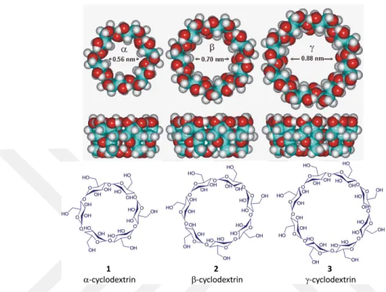
CDs, which are also known as cycloamyloses, belong to the family of cyclic oligosaccharides (sugar units) and they are bound together in a circular ring. They have unique physical properties, and their outer surface is hydrophilic but the internal cavity is hydrophobic [90]. CDs are mainly characterized in three forms based on the number of glucose monomer which ranges from six to eight. α-cyclodextrin (ACD, alpha cyclodextrin) contains six sugar units, β-cyclodextrin (BCD, beta cyclodextrin) consists seven sugar units whereas γ-cyclodextrin (GCD, gamma γ-cyclodextrin) accommodates eight sugar units in a circular shape. Each of these CD differ from each other in physical dimensions. Figure 2.9 gives a pictorial representation of the difference in the cavity dimen-sions of each of these CDs, also structural shape of the three CDs.
Figure 2.9: Comparison of the inner diameter of alpha, beta and gamma cy-clodextrins, also showing the structural shape of the cyclodextrins.
Even with the lowest solubility, BCD is the most famous of the three CDs and are the most thoroughly investigated of the CDs. Studies have shown that BCD has better dissociation characteristics than other CDs, when complexed with many drugs, including nonsteroidal anti-inflammatory drugs (NSAIDs) [91]. By complexing an NSAID with a BCD, a number of NSAIDs can be made soluble and their dissolution rates can be increased.
BCD is known for its use in the food industry, chemical industry and in phar-maceutical industry for drug delivery [92, 91]. In the food industry, BCD is used for the preparation of cholesterol free products. Oil, being hydrophobic, easily slides into the hydrophobic cavity of the BCD and these are later removed to pro-duce and oil free product. For chemical industries, BCD is used to make aerosols. The Table 2.1 lists different physical dimensions of each of the CDs.
The pharmaceutical industries takes the most advantage of the CDs because it is recognized as safe and is approved by the Food and Drug Administration
Table 2.1: A comparison of the physical properties of the CDs. CDs Molecular weight (g/mol) Outer dia (nm) Inner dia (nm) Solubility in water (g/kg) ACD 972 1.52 0.45 129.5 BCD 1134 1.66 0.60 18.4 GCD 1296 1.77 0.75 249.2
(FDA) [93]. Because of the hydrophobic cavity of the BCD, hydrophobic drugs can be inserted in the cavity and thus are soluble in water. BCD has the ability to undergo host-guest interactions resulting in the formation of inclusion com-plexes. In these inclusion complex, the hydrophobic guest particle goes inside the hydrophobic cavity of BCD. By this, the hydrophobic particle can be made soluble, and can be delivered to any part of the body.
Figure 2.10 shows the 3-D structure of BCD, also showing the hydrophobic cavity. Since, the hydrophobic cavity can interact with any hydrophobic material, we intent to react it with PFH to form an inclusion complex (IC). PFH, a type of perfluorocarbon, and forms an IS which is stable at room temperature and can be used as a histotripsy agent.
Figure 2.10: A 3D structure of BCD showing its hydrophobic cavity.
2.5.2
Perfluorocarbons
Perfluorocarbons (PFC) belongs to the family of organofluorine and has the for-mula CxFy. PFCs are made up of stabe C-F bonds and are chemically inert, therefore, they do not metabolize in the body. They can be removed from body by simple inhalation [94, 95]. There can be many types of PFC, such as perfluoroalka-nes, perfluoroalkeperfluoroalka-nes, and perfluoroalkynes etc. They are used in many different
industries. PFCs are used as refrigerants, solvents, fluoropolymers, gas/oxygen carriers, and also in the field of medicine as anesthetics and liquid ventilation [96, 97]. Recently, PFCs have gained much popularity due to its potential as the use of contrast agents in medical devices like MRI, CT, and US [98].
PFH being liquid at room temperature, with a boiling pint of 56 ◦C, has advantage to be used at room temperature. Moreover, having a small linear chain is suitable for reactions like host-guest interactions forming inclusion complex. Figure 2.11 shows the linear structure of PFH and also the molecular formula of PFH.
Currently, because of its ability to dissolve oxygen in high amounts, it is used in the liquid ventilation systems in case of lung diseases or lung failure [99, 100]. In the late 80s and early 90s, PFH began to gain popularity as a contrast agent for US and other medical devices [101, 102]. Since then there has been immense research on PFH being used as contrast agents for US and it has been very successful.
As histotripsy uses high frequency US waves, the use of PFH as inclusion complex is very effective. Due to its lower boiling point, PFH vaporizes at lower pressures and forms a bubble cloud which leads to cavitation. This was also confirmed by Vlaisavljevich et al. that PFH works better in histotripsy rather than PFP [76]. Furthermore, it also acts as a contrast agent for the US. This helps in the real-time imaging and also to locate the tumor more easily. Once in the US, it is seen that all the inclusion complexes containing PFH are attached or accumulate into the tumor, and cavitation can be achieved leading to tumor ablation without any damage to the normal cells.
Figure 2.11: A 2D and 3D structure of PFH.
2.6
Hypothesis
We hypothesize that we can obtain an inclusion complex as histotripsy agent using host-guest interaction between BCD and PFH. We expect it to lower cavitation threshold of the histotripsy, by acting as a histotripsy agent, and by working as effective as nanodroplets, whereas its production will be more user friendly, economical, practical and faster than nanodroplets. BCD, being an FDA ap-proved product, with its hydrophilic outer surface and hydrophobic inner cavity can accommodate at least one hydrophobic PFH molecule forming inclusion com-plex. We expect them to have smaller size than nanodroplets that can penetrate deeper into tumor tissue. Since primary face of BCD still will be available for desired modification, this agent might have potential to be easily targeted to-wards specific cell line. This inclusion complex will also have an ability to form a self-assembly depending of the concentration in the dispersion that could be effect on the cavitation threshold.
Chapter 3
Experimental section
3.1
Materials
β-Cyclodextrin (BCD, Sigma-Aldrich, 97%), tetradecafluorohexane (PFH, Sigma-Aldrich, 99%), dimethyl carbonate anhydrous (DMC, Alfa Aesar, 99%), potassium carbonate anhydrous (K2CO3, Sigma-Aldrich, 99.99%), anisole
anhy-drous (Aldrich, 99.7%), N,N-dimethylformamide anhyanhy-drous (DMF, Sigma-Aldrich, 99.8%), dimethyl sulfoxide (DMSO, Merck 99%), acetone (Sigma-Aldrich, 99.5%), diethyl ether anhydrous (DEE, Sigma-(Sigma-Aldrich, 99%), sodium chloride anhydrous (NaCl, Wisent Bioproducts, 99%), Phosphate Buffer Solu-tion (PBS, Multicell, Wisent Inc.), ethanol (Sigma Aldrich, 99.8%), Dulbecco’s Modified Eagle’s Medium (DMEM, Multicell, Wisent Inc.), Fetal Bovine Serum Advanced, Heat Inactivated (FBS, Capricorn Scientific GmbH), L-Glutamine, 200 mM solution (29.23 mg/mL in 0.85% NaCl) (Multicell, Wisent Inc.), Penicillin streptomycin solution (Multicell, Wisent Inc.) Trypsin EDTA (0.25%trypsin) in HBSS, 1X (Multicell, Wisent Inc.) Trypan Blue solution, 0.4% (Sigma Aldrich).
3.2
Methods
3.2.1
Preparation of β—cyclodextrin and
perfluorohex-ane inclusion complex
The first step in the preparation of BCD—PFH inclusion complex (IC) was to optimize the experimental conditions in order to obtain an IC with the high-est efficiency. Therefore, 50 mg CD (4.4 X 10⁻² mmol) was mixed with 1 mL double distilled water (Direct—Q 3 Ultrapure Water System, LabRepCo) in 4 different vials. The solutions were stirred at 80◦C until complete dissolution of CD. Afterwards, the temperature was slowly cooled down to 45◦C. Once 45◦C was obtained, different molar ratios of PFH (1, 2, 20, and 50 folds) were added to different solutions. After stirring the solutions overnight at 45◦C, they were kept in a fridge at 4◦C for one hour followed by centrifuge (Thermo scientific, MicroCL 21R) at 5000 rpm, for 10 min. The supernatant was decanted, and the precipitated was dried under vacuum. The dried ICs were stored at 4◦C for further use and for characterizations.
3.2.2
Methylation of β—cyclodextrin
BCD was randomly methylated using the procedure described in Gan et al.[103] Briefly, 3 g CD (2.64 mmol) was dissolved in 60.0 mL DMF in a two-necked round bottom flask connected to a condenser on the top. After complete dissolution of BCD, 8.6 g of K2CO3 was added to the solution. 8.0 mL of DMC was added, as
well, drop-by-drop and stirred overnight at room temperature. Subsequently, the catalyst was removed by centrifuging the solution at 2000 rpm for 5 minutes. The solvent and surplus DMC were separated from the solution by vacuum distillation during which the residue turned into a syrup-like concentration. Methylated β—Cyclodextrin (MCD) was then precipitated from acetone, and rinsed three times using diethylether. MCD was then filtered, and dried under vacuum. The dried MCD was stored at 4◦C for further use and for characterizations.
3.2.3
Preparation of methylated β—cyclodextrin and
per-fluorohexane inclusion complex
For the preparation of MCD-PFH IC (MIC), 50 mg MCD was dissolved in 1 mL double distilled water at room temperature in three separate vials. MCD was readily dissolved in water. Different molar ratios of PFH (5, 10, 50 folds) were added to each vial, and the solutions were stirred overnight. After 24 hours, the solutions were centrifuged at 5000 rpm, for 10 min. The supernatant was decanted, and the precipitate was dried under vacuum. The dried MIC was stored at 4◦C for further use and characterizations.
3.2.4
Hemolysis
To study the hemolysis profile of the IC and MIC, 4 mL of a blood sample was collected from a volunteer. The blood was centrifuged (Thermo scientific, SL 16R) at 3500 rpm for 5 minutes. With a permanent marker, a line was marked at the point where red blood cells (RBCs) were separated from plasma, and also, at the maximum height of the plasma. The plasma was then removed, and 0.15M saline solution was added until the upper mark. The saline solution and RBCs were mixed gently followed by centrifuge again using the same protocol. This procedure was performed to rinse the RBCs. It was repeated three times. After the third rinsing of the RBCs, the saline solution was removed using centrifuge, and it was replaced with 100 mM phosphate buffer saline (PBS) until the same mark. This was the stock solution for the experiment. The working solution was prepared by 10% dilution of the stock solution. More specifically, 1 mL of the stock solution was taken in a 15 mL falcon tube, and 9 mL PBS was added and mixed gently. In a 2 mL Eppendorf tube, 200 L of the working solution was mixed with solutions of BCD, MCD, IC, and MIC, each with concentrations of 1.0 mg/mL, 0.5 mg/mL,and 0.1 mg/mL, all prepared in 1 X PBS. A volume of 1 mL was obtained in all the Eppendorf tubes. A positive control containing a solution of Triton X, and a negative control with only RBCs in PBS were also
of the compounds. Afterwards, each solution was prepared as a triplicate. The solutions were placed in a water bath at 37◦C for one hour. The solutions were then again centrifuged at 5000 rpm for 5 minutes. Both the intact and ruptured RBCs were accumulated at the bottom in the form of a pellet. The supernatant, on the other hand, contained the released hemoglobin from the ruptured RBCs. This supernatants absorbance was measured at 541 nm using a 96 well plate reader (SpectraMax i3). The percentage hemolysis was calculated accordingR to the following equation:
% Hemolysis = Absorbance of sample − absorbance of negative control
Absorbance of positive control ∗ 100% (3.1) where negative control contains only the RBCs with PBS, while positive control contains RBS with PBS and triton X.
3.2.5
Cell toxicity studies
3.2.5.1 General cell culture procedure
Toxicity of BCD, MCD, IC, and MIC was investigated as a function of concentra-tion using the MTS assay. The procedure was performed with full protocols of cell culture. 15 minutes prior to the start, all the liquids, including the medium and PBS, were placed in a water bath at 37◦C. Meanwhile, the fume hood (Thermo Fisher, Maxisafe 2020) was cleaned using 70% ethanol. All the liquids from the water bath were sprayed with 70% ethanol and transferred to the fume hood.
Frozen human embryonic kidney cells (HEK-293T) were taken, and quickly melted by hands. In a 15 mL falcon tube, 10 mL Dulbeccos modified eagle medium (DMEM) was added, as well as the melted cells. It was then centrifuged at 1000 rpm for 5 min. After centrifuge, the cells were in form of a small pellet at the bottom of the tube. The medium was removed slowly without disturbing the
cells, and 5 mL of the growth medium (DMEM + 10% FBS+10% L-Glutamine + 5% penicillin streptomycin solution) was added to the cells. A T25 cell culture flask was prepared. The name, cell type, date, and passage number were written on both sides of the flask. 5 mL of the same growth medium was added to this cell culture. The 5 mL solution of cells from the falcon vial was transferred to this T25 flask so the total volume was 10 mL. This flask was sealed with a cap, and then viewed under a microscope (Zeiss Primo vert) to see if the cells are transferred successfully or not. After confirming successful transfer, the flask was placed in an incubator (Thermo Scientific, Forma Steri-Cycle CO2 Incubator) at
37◦C, and 5% carbon dioxide (CO2).
After 48 hours, the medium of the cells was changed. PBS and growth medium were placed in a water bath at 37◦C prior to the use. The T25 flask was taken out of the incubator, and first inspected using a microscope. Most of the cells adhered to the surface of the flask. The cells moving with the fluid were dead cells, and therefore, they were removed.
To change the medium and remove the debris, the T25 flask was opened in the fume hood and tilted so that all the medium was collected at the bottom corner of the flask. The medium was slowly drawn out using a pipette. 1-2 mL PBS was added and slowly swiped over the surface of the flask and then again drawn out in the same manner. Then, 10 mL of new growth medium was added to the flask. The T25 flask was then sealed, and checked under the microscope. After confirmation of the presence of the cells, the flask was again placed in the incubator at the same temperature and CO2 level.
After 48-72 hours, the T25 flask was examined for cell growth under the mi-croscope. If the cells covered more than 85% of the surface, the cells had to be transferred to a bigger flask, the T75 flask. The transfer of the cells is called passage. To pass the cells to the T75 flask, the medium was removed from the T25 flask. 2-3 mL PBS was added, the surface was rinsed, and PBS was removed. After this, 1-2 mL trypsin EDTA enzyme was added to the flask. The T25 flask was then incubated for 2 min. This enzyme helps the cells detach from the surface
After the incubation time of 2 minutes, the flask was opened in the fume hood, and 8-9 mL of growth medium was added. The total volume in the flask was 10 mL including the trypsin EDTA. The surface of the flask was rinsed 3-4 times using the same medium inside the flask. Then, the medium was transferred from the T25 flask to a 15 mL falcon tube, and it was centrifuged at 1000 rpm, for 5 min. Meanwhile, the T75 flask was prepared. The name, cell line name, date, and passage number were written on its sides. The passage number was one plus the previous flasks passage number. Once the centrifuge was done, the medium was carefully removed from the tube, and 10 mL growth medium was added to the tube. The tube was rinsed from the bottom in order for the cells to start floating in the medium. Once the cells were floating, 1 mL of this medium with cells was transferred to the T75 flask. Furthermore, 19 mL of growth medium was added to the T75 flask to make up 20 mL of total volume. The T75 flask was sealed and observed under the microscope. The cells were visible, so the T75 flask was incubated at 37◦C.
After 48 hours, the medium was changed using the same procedure, and the cells were again incubated until they covered about 85% of the surface of T75 flask. The cells had to be seeded on a 96 well plate. The medium was removed, cells were rinsed in PBS, trypsin EDTA was added to take off the cells from the surface, and after adding the medium, the cells were centrifuged. After centrifuge, 1 mL of the cells was transferred to a new T75 flask with passage number with an increment of one.
From the remaining 9 mL of the cell solution, cells were to be seeded on the well plate. Before that, a Neubauer hemocytometer (Celeromics) was used for cell counting. 100 µL cell solution was taken and diluted 50% by addition of 100 L trypan blue solution. The trypan blue solution is a blue color dye that helps living cells be more visible, and hence, they are counted with ease. After mixing the solution of dye with cells, 10 µL of the solution was placed on the hemocytometer with the lid on top. Once the solution filled the area, the hemocytometer was placed under a microscope.
The cells were counted from the 4 corners of the hemocytometer, and an av-erage number of the cells was taken. The avav-erage was then multiplied by the dilution factor, 2 in this case, and by the surface area of one block of the hemocy-tometer (10,000). This gave the total number of cells in 1 mL. After calculations, 19.4 µL of the cell solution, with a density of 2.0 * 104 per well, was transferred to each well with addition of 280.6 µL growth medium. The well plate was incubated at 37◦C, and the cells were allowed to adhere to the wells.
3.2.5.2 Cell viability assay
After 24 hours, in a separate 96 well plate, FBS free growth medium was added with addition of BCD, MCD, IC, and MIC, each at concentrations of 1.0 mg/mL, 0.5 mg/mL, and 0.1 mg/mL. These samples were prepared in 1 X PBS at the above stated concentrations. Additionally, PFH concentrations of 0.1 µL, 0.12 µL, and 0.15 µL were added to a separate set of FBS free medium. This medium was selected because FBS contains different proteins, and thus, it might interact with the samples. The total volume of the medium and sample solution was adjusted to be 200 µL. Then, the medium from above the cells was replaced by the FBS free medium plus the sample solutions. The well plate was then incubated at 37◦C for 24 hours. Afterwards, the medium from the well plate was removed, and 100 µL medium was added with addition of 10 µL CellTiter 96 R Aqueous One Solution Cell Proliferation Assay (MTS assay). The well plate was then incubated at 37◦C for 2 hours. After incubation, the well plate was read using the well plate reader at an absorbance of 490 nm.
The percentage of cell viability was calculated according to the following equa-tion:
%Cell viability = Absorbance of sample − absorbance of negative control
Absorbance of positive control ∗100% (3.2) where negative control contains only the cells with medium, and positive control
contains cells with medium and dye.
3.2.6
Fluorescence probe method (Investigation of critical
micelle concentration)
To see if the MIC is forming self-assembly or not, a critical micelle concentration test was conducted. This was done using the fluorescence probe method followed by Alkayal et al. [104]. Briefly, a pyrene solution (1.0 X 10⁻⁴ mmol/mL) in acetone was prepared. 0.1 mL of this solution was added to different vials, and acetone was evaporated in the air. After complete evaporation, MIC solutions were prepared in PBS, at concentration of 1.0 mg/mL, 0.5 mg/mL, 0.1 mg/mL, 0.01 mg/mL, and 0.001 mg/mL each, were added to these vials. The total volume of each vial was adjusted to be 1.0 mL. These solutions were vortexed for 5 minutes, followed by 10 minutes of sonication. 200 µL of these solutions were transferred to 96 well plate. The emission spectrum was recorded using spectra max at ex = 337 nm, and the emission bandwidth was selected to be 5.0 nm.
3.2.7
Ablation of agarose tissue phantom using inclusion
complex as a histotripsy agent
To test the MIC histotripsy agent, agarose phantoms were chosen as a medium. This was because the agarose phantoms provided a well-controlled viscoelastic medium, which was very important for the study as the damage in the tissue is induced by the histotripsy highly depends on the mechanical properties of the tissue [20]. The phantom was made using 1% agarose w/v by slowly mixing agarose powder (Agarose type VII, Sigma Aldrich) into saline solution. The temperature of the solution was raised above 70◦C until the solution became completely transparent. The solution was then degassed using a partial pressure vacuum of 2.7 KPa for 30 minutes. The agarose solution was then cooled down to 37◦C.
To achieve phantoms containing MIC, and only PFH, as a positive control, the MIC and pFH was slowly added to different gel solutions at around 40◦C while the solution was still stirring and being colled down. The agarose mixture was then poured into rectangular molds make of polycarbonate, and placed at 4◦C. The solutions were allowed to solidify, and the tissue phantoms embedded with MIC (test), with PFH (positive control), and without MIC (negative control) were obtained.
Two different histotripsy probes were used to perform two different experiment. In the first experiment, a 500 kHz transducer was used, and single cycle per pulse was transmitted at voltages of 30 V and 40 V. 40 V voltage corresponded to the maximum pressure used for histotripsy, without any use of the agents. 30 V voltage corresponded to the pressure lower than that threshold pressure. If the MIC worked at lower voltages, that means that it is working properly just like the nanodroplets. The cavitation behavior of the MIC was observed using a high-speed optical camera (V210, Vision research, Wayne). After every pulse, the camera captured images of the focal zone in the phantom. The camera was focused using a macro-bellows lens (Tominon, Kyocera) which was attached to the optical window of the chamber to observe the focal region. A light source was used at the back of the phantom, continuously, to produce shadowgraphic images of the cavitation bubbles.
3.3
Characterization
3.3.1
Hydrogen nuclear magnetic resonance spectroscopy
Once the IC and MIC were synthesized, a hydrogen nuclear magnetic resonance spectroscopy (1H NMR) was performed for the confirmation of the complex for-mation. For this purpose, MCD, IC and MIC were observed through Agilent VNMRS 500 MHz nuclear magnetic resonance spectrometer. A chemical peak shift was compared between BCD and MIC for the methylation, and between
MCD and MIC for the formation of MIC.
3.3.2
Fluorine nuclear magnetic resonance spectroscopy
For further confirmation of the synthesis of the complex, Agilent VNMRS 500 MHz nuclear magnetic resonance spectrometer was used to carry out the Fluo-rine nuclear magnetic resonance spectroscopy (19F NMR). MIC, and PFH were inspected, and the results were compared.
3.3.3
Fourier transform infrared spectroscopy
A Fourier transform infrared spectroscopy (FTIR) was carried out using the Ag-ilent Cary 630 FTIR spectrometer. FTIR of CD, MCD, IC, MIC, and PFH were performed using a diamond crystal, with range of 4000 cm−1 to 400 cm−1, reso-lution of 4 cm−1, and 16 scans per minute. The peak shifts were observed and compared to each other to confirm the synthesis of the complex.
3.3.4
Thermal gravimetric analysis
To see and compare the weight loss profiles of BCD, MCD, and MIC, they were tested through thermal gravimetric analysis (TGA) using EXSTAR TG/DTA 7300. The weight loss was measured as a function of temperature. The heat flow was 10◦C/min, from 25◦C-700◦C, under nitrogen flow. The results were compared to see the degradation of each sample, and also the stability of each compound.
3.3.5
Gas chromatography
Gas chromatography (GC) was performed as a part of the quantitative analysis to calculate the efficiency of the complexes. In other words, the amount of PFH
entrapped in the CD or MCD was calculated. Agilent Technologies 7820A GC System was used to perform this analysis. To calculate the amount of PFH present, first a calibration curve was formed using DMF as the solvent, anisole as internal standard and PFH. Initially, PFH, DMF, and anisole were injected in the GC individually. This was done to see the time at which the peak of each of the substance comes out. As the amount of PFH was very small in the samples (in µL range), it was impossible to compare it with the peak of DMF, as 200 mL of DMF was used. Thus, there was a need for an internal standard, whose peak should not overlap with either DMF or PFH. Anisole was a perfect match for this condition.
For the preparation of the calibration curve, in a 200 mL volume of DMF, 2 µL of anisole was added. This was the standard solution for the calibration curve, and also for the samples to be tested. The PFH amount was varied for each run, starting from 0.1 µL to 1.0 µL. This range was chosen because it corresponds to the range of the amount of PFH in a sample of 10 mg which was to be tested. A 6-point calibration curve was constructed.
After collecting the data, the six points were plotted on a graph and a line of best fit was constructed. Furthermore, the equation of the line of best fit was used to calculate the amount of PFH present in the sample. Following is the equation of the calibration curve, which was used to find out the volume of PFH.
A = 1.0273(V ) + 0.0005 (3.3)
Where A is the area of the PFH curve with reference to anisole and V is the volume of the PFH.
10 mg of IC and MIC, each with different ratios of PFH, were tested, and the peak areas were compared with anisole. The areas were then matched with the calibration curve to find out the amount of PFH present. The percentage efficiency of the complex was calculated accordingly. Each run for calibration curve and sample was tested for a temperature range of 40◦C 200◦C, at a heating
rate of 40◦C/min, and the sample was kept at 200◦C for one minute.
When the volume was calculated from this equation, the percentage of PFH present in the complex was calculated according to the following equation.
%P F H = V
U ∗ 100% (3.4) Where V is the volume of PFH in the sample calculated from the previous equation, and U is the theoretical volume at 100% encapsulation of PFH in the CD and MCD. The efficiencies of IC and MIC were compared based on the results of GC calculated using the calibration curve.
3.3.6
Dynamic light scaterring
Malvern Zetasizer Nano ZSP was used to calculate the dynamic light scattering (DLS) measurements. Solutions of CD, MCD, IC, and MIC were prepared in 1 X PBS. Each measurement was calculated using 3 runs, and each run contained 12—16 repetitions. The sizes of CD, MCD, IC, and MIC were calculated as an average of the sum of all the values, and the results were compared.
3.3.7
Scanning electron microscopy
To see the textural changes on the samples and how they differ form each other at nano level, BCD, MCD IC, MIC were observed in a scanning electron microscope (SEM, Philips XL 30S FEG with EDAX detector). Images of different samples were taken. Also, freeze dried samples of MIC at concentration of 1.0 mg/mL and 0.1 mg/mL in water and PBS were observed to see whether there is self-assembly formation or aggregation at higher concentrations. The samples were also checked for % fluorine content using the Energy dispersive x-ray spectroscopy (EDAX) detector in the SEM.
Chapter 4
Results and discussions
A histotripsy agent consisting of BCD and PFH was aimed to be synthesized using a host-guest chemistry. PFH is a small hydrophobic chain of six carbon atoms, all containing fluorine atoms attached to them. Because of its highly hydrophobic behavior, it is challenging to deliver it to the human body. Thus, the host guest chemistry is used, which is a perfect solution for carrying such hydrophobic molecules into the body. BCD, which is comprised of seven sugar chains, has a cone like structure. The exclusive feature of BCD, apart from being biocompatible, is that it has a hydrophilic outer surface with a hydrophobic inner cavity. BCD acts as a host and offers a place for hydrophobic guest molecules to fill the cavity. In our case, PFH acts as the guest molecule and fills the cavity of the BCD hence forming an IC. Figure 4.1 one depicts a visualization of the process of host-guest chemistry and shows the filled cavity.