The article was published by ACG Publications www.acgpubs.org/RNP © March-April 2018, EISSN:1307-6167
DOI: http://doi.org/10.25135/rnp.15.17.05.100
Rec. Nat. Prod. 12:2 (2018) 128-141
Evaluation of the in vitro Anti-inflammatory Activity of
Nerium oleander L. Flower Extracts and Activity-Guided Isolation
of the Active Constituents
İrem Atay Balkan
1 *, Ahmet C. Gören
2, Hasan Kırmızıbekmez
3and
Erdem Yeşilada
31Istanbul Medipol University, School of Pharmacy, Department of Pharmacognosy, 34810, Beykoz,
Istanbul, Türkiye
2TUBITAK National Metrology Institute, Chemistry Group Laboratories, 41470, Gebze, Kocaeli,
Türkiye
3Yeditepe University, Faculty of Pharmacy, Department of Pharmacognosy, 34755, Ataşehir, Istanbul,
Türkiye
(Received May 31, 2017; Revised August 11, 2017; Accepted August 24, 2017)
Abstract: The in vitro anti-inflammatory activity profile of the Nerium oleander flower EtOH extract/its subextracts (n-hexane, CH2Cl2, EtOAc, remaining H2O) were evaluated on LPS induced Raw 264.7
macrophages. The effects of the crude EtOH extract and its subextracts on nitric oxide (NO) production and cell viability were determined. The most active subextract was determined to be the EtOAc subextract without exerting any toxicity towards Raw 264.7 macrophages. This subextract significantly inhibited NO production of Raw 264.7 macrophages after LPS induction (62.56±1.91% at 200 µg/mL concentration). The levels of iNOS were reduced up to 67.50%. Moreover, this subextract slightly reduced the phosphorylation levels of MAP kinases (p-ERK, p-JNK, p-38). The highest inhibition was observed for ERK phosphorylation, which was inhibited by 20.53% at 200 µg/mL concentration. Through activity-guided fractionation procedures, kaempferol, kaempferol 3-O-β-glucopyranoside and chlorogenic acid were isolated as the main active components. The structures of the active compounds were determined by 2D-NMR techniques and HRMS analysis. All compounds significantly inhibited NO productions. Results of the present study supported the traditional use of N. oleander flowers to treat inflammatory complaints.
Keywords: Nerium oleander; nitric oxide; mitogen activated protein kinases; kaempferol; kaempferol 3-O-β-glucopyranoside; chlorogenic acid. © 2018 ACG Publications. All rights reserved.
1. Introduction
Inflammation is a beneficial defense mechanism against various stress factors. However, chronic inflammation contributes crucially to the pathogenesis of many diseases including inflammatory bowel diseases, asthma, arthiritis as well as neurodegenerative diseases (e.g. Alzheimer’s disease, multiple sclerosis) [1-6]. Such disorders are very common and constitute the main health problems of the worldwide society [7-10]. The complexity of the inflammatory reaction augments by the involvement of various pathways. These pathways include mediators like nitric oxide (NO), prostaglandins, leukotriens, platelet-activating factor, cytokines (IL-1, IL-6, TNFα) and various
enzymes including lipoxygenases, inducible nitric oxide synthase (iNOS), cyclooxygenases (COX-1 and COX-2), and phospholipases [11-13]. Among the mediators, NO which is primarily an essential molecule for physiological functions of the body, was found to be associated with inflammation, sepsis and carcinogenesis at higher concentrations [14]. In order to evaluate the activity of drug candidates against inflammation, inhibition of NO production through reduced levels of iNOS was reported to be an important measure [15].
The mitogen activated protein (MAP) kinases are classified among the family of serine/threonine protein kinases [16, 17]. Three major groups of MAP kinases (ERK, JNK and p38) regulate the synthesis of many inflammatory mediators at the level of translation and transcription [6, 18]. It has been shown that MAP kinases (ERK, JNK and p38) regulate the production of LPS induced iNOS. Moreover, MAP kinases are found to be essential for COX-2 gene expression [19-21]. In addition, MAP kinases regulate the expressions of inflammatory mediators such as TNFα, IL-1β and IL-6 [6].
N. oleander L. (Apocynaceae) or oleander is an evergreen ornamental shrub mainly distributed in the Mediterranean region [22]. Cardenolides, triterpenes, phytosterols, pregnanes and flavonoids were isolated from the leaves and steroidal glycosides were isolated from the roots of the plant [23-34]. Various pharmacological activities have been reported for the leaves and roots of N.oleander such as cytotoxic, anticancer, anti-inflammatory, antibacterial, antioxidant, skin degenerative and central nervous system stimulant activities [27, 35-37].
Almost all the chemical and pharmacological investigations have been carried out so far focused particularly on the leaves of oleander, while its flowers remained to be unexplored, except a number of neuroprotective, hepatoprotective and antioxidant studies [38-44]. In Turkish folk medicine, oleander flowers are soaked in olive oil or alcohol and the macerate is applied topically to the aching extremities against rheumatic pain as a home remedy [45]. In our previous study, the ethanol (EtOH) extract of the flowers was shown to exert significant in vivo anti-inflammatory activity [46]. The present study aims to elucidate the activity mechanism and to isolate potential anti-inflammatory metabolites through the in vitro activity-guided fractionation techniques. For that purpose, effects of the crude EtOH extract and the subextracts [n-hexane, dichloromethane (CH2Cl2),
ethyl acetate (EtOAc) and remaining water (R-H2O)] were evaluated on LPS-induced NO production
of Raw 264.7 cells. The EtOAc subextract which was the most potent subextract was further evaluated for its effects on iNOS and COX-2 protein levels. The effects on the phosphorylation levels of MAP kinases (ERK1/2, JNK and p38 kinases) were also targeted.
2. Materials and Methods 2.1. General
The column chromatography (CC) was achieved by using Sephadex LH-20 (Lipophilic Sephadex, 25–100 µm, Sigma-Aldrich, USA), and Polyamide (Sigma-Aldrich, USA) as stationary phases. Kieselgel 60 F254 (Merck) precoated plates were used for thin layer chromatography. Lipopolysachharides (LPS), dimethylsulfoxide (DMSO), HRP-conjugated goat mouse and anti-rabbit IgG and Griess reagent were purchased from Sigma-Aldrich (St. Louis, MO). Fetal bovine serum (FBS), Dulbecco’s modified Eagle’s medium (DMEM), penicillin-streptomycine was obtained from Invitrogen/Gibco (Grans Island, NY, USA). Antibodies for ERK1/2, ERK, phospho-p38, phospho-p38, phospho-JNK, JNK were purchased from Cell Signaling Technology (Beverly, MA, USA). Antibodies for iNOS, COX-2 and β-actin were purchased from Santa Cruz (CA, USA). ECL reagent obtained from Amersham (Buckhinghamshire, UK). WST-1 reagent was from Roch Applied Science (Mannheim, Germany).
2.2. Cells and Cell Culture
The Raw 264.7 macrophages (ATCC TIB-71) were grown in DMEM which was supplemented with 10% FBS, 4 mM L-glutamin, 100 IU/mL penicillin and 100 µg/mL streptomycin at 37 °C in a humidified atmosphere containing 5% CO2.
2.3. WST-1 Assay for Cell Viability
Extract/subextracts/fractions or compounds were evaluated for their non-toxic concentrations by WST-1 assay kit (Roche Applied Sciences) in agreement with the manufacturer’s instructions. Metabolically active cells reduces WST-1 which is a tetrazolium-based salt dye, to a formazan salt. The result can be directly quantified by spectrophotometric measurements. Raw 264.7 cells (22.500 cells/well) in 10% FBS-DMEM were seeded into 96-well plates and various concentrations of samples were added to the wells with or without 1 µg/mL LPS. After 24 hrs incubation at 37 °C, the supernatant was removed, WST-1 was directly added to the wells to a final concentration of 5% (v/v). After an additional 60 minutes of incubation at 37°C the absorbance was read between 420-480 nm (λmax 450 nm). DMSO was used to dissolve all test samples. The samples were then diluted with DMEM to the convenient concentrations. The DMSO concentrations in the culture medium did not exceed 0.1% (v/v).
2.4. Griess Assay
Raw 264.7 cells (22.500 cells/well) in 10% FBS-DMEM were seeded into 96-well plates. The extract/subextracts/fractions were added to cells and incubated for one hr, and then induced with 1 μg/mL LPS. After that the plates were incubated for additional 24 hr at 37°C, the nitrite accumulated in the medium was measured by Griess Assay. The cell culture medium (50 μL) was mixed with 50 μL of 1% (w/v) sulfanilamide prepared 5% (v/v) phosphoric acid. The reaction was incubated for 10 mins and then 50 μL of 0.1% (w/v) naphtylethylenediamine-HCl (Promega) was added. The mixture was incubated for another 10 mins at room temperature. The absorbance was read at 550 nm using a microplate reader. The nitrite amounts in the test samples were calculated from the NaNO2 serial
dilution standard curve.
2.5. Western Blotting
Cells were washed once with ice-cold PBS and the cellular proteins were extracted by RIPA lysis buffer composed of 1x TBS, 1% Nonidet P-40, 0.5 % sodium deoxycholate and 0.1 % SDS with 0.004 % sodium azide (Santa Cruz). The RIPA buffer was supplemented with 1mM phenylmethylsulfonyl fluoride 1 mM Na ortho-vanadate, 1% protease inhibitor cocktail (Sigma). The protein concentrations were calculated by using Lowry assay. Forty micrograms of cellular proteins were separated by 8% or %10 SDS-polyacrylamide gel electrophoresis. After that, the proteins were electroblotted onto a nitrocellulose membrane. The membranes were incubated with specific primary antibodies overnight at 4 °C with 5% skim milk as blocking solution. Blots were washed with Tween 20/Tris-buffered saline (TBST) for four times. Then the membranes were incubated for 2 hr at room temperature with a 1:1000 dilution of secondary antibodies. Eventually, blots were washed three times with TBST, and then visualised by ECL reagent (Amersham Life Science).
2.6. Plant Material
The flowers of N. oleander were collected from Kayışdağı-İstanbul (Turkey) in June, 2010. The identification of the plant material was achieved by Prof. Dr. Erdem Yesilada (one of the authors). A voucher specimen is deposited at the Herbarium of Yeditepe University (YEF 10009).
2.7. Extraction and Isolation
The fresh flowers of N. oleander (2.5 kg) were macerated with EtOH (700 mL) at room temperature for 50 days. The macerate was filtered through filter paper. The organic phase was evaporated to dryness under reduced pressure. As a result, crude EtOH extract was obtained (105.71 g, yield: 4.24%). EtOH extract was dispersed in 100 mL of MeOH in H2O (90%, v/v) and partitioned
against n-hexane (4 × 200 mL). The n-hexane phases were combined. Subsequently, n-hexane was evaporated under reduced pressure in order to obtain n-hexane subextract (3 g, yield: 6%). After removing the MeOH from the remaining extract, the volume of the solution was adjusted to 100 mL with distilled H2O and partition continued with CH2Cl2 (4 × 200 mL) and then EtOAc (4 × 200 mL),
respectively. Each organic phase was evaporated to dryness under reduced pressure to yield CH2Cl2 (2
g, yield: 4%), EtOAc (1.1 g, yield: 2,2%) subextracts while remaining-H2O subextract was lyophilized
(30.5 g, yield: 60,1%).
The EtOAc subextract (1.3 g) was submitted to a polyamide (20 g) column eluting with H2O
(200 mL) and mixtures of MeOH in H2O (10-100% in steps of 10%, each 100 mL) in order to obtain
four fractions, Fr. A-D. Fr. B (90 mg) was applied to a Sephadex (20 g) column using MeOH and chlorogenic acid (CA, 4 mg) was obtained. Fr. C (50 mg) was further subjected to a SiO2 (8 g) column
using mixtures of CH2Cl2-MeOH (90:10, 85:15, 70:30, 60:40 and 50:50; each 100 mL) to yield
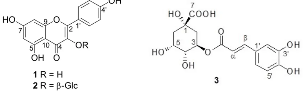
kaempferol 3-O-β-glucopyranoside (KG, 6 mg). The Fr. D (51 mg) was applied to a Sephadex (20 g) column using CH2Cl2-MeOH (1:1) mixture to yield kaempferol (KF, 22 mg). The chemical structures
of these compounds were given in Figure 1.
2.8. Structure Elucidation of the Compounds
The elucidation of the structures of the pure compunds were achieved by Nuclear Magnetic Resonance (1H and 13C NMR) and Mass Spectral (MS) techniques. NMR spectra were recorded on a
Varian Mercury-Mx spectrometer (USA) at 600 MHz for 1H NMR and at 150 MHz for 13C NMR, with
CD3OD as solvents. UV and IR spectra were recorded on an HP Aglient 8453 (USA) UV and
PerkinElmer-2000 FT-IR spectrometers respectively (USA).
2.9. Statistical Analysis
The data were recorded as mean ± standard error of the mean (SEM) of triplicate experiments. The data were analyzed by PASW Statistics. The significance of the difference between means were determined by Mann Whitney U test and p-values less than 0.05 were considered statistically significant. O OH HO O OH 1 R = H 2 R = -Glc OR 2 4 5 7 9 10 1' 4'
Figure 1. Chemical structures of the active compounds (1-3).
3. Results and Discussion
3.1. Activity-guided Fractionation of N. oleander EtOH Extract 3.1.1. Effects of EtOH Extract and Subextracts on Cell Viability
Non-toxic concentrations of the extract/subextracts on Raw 264.7 macrophage cells were determined by WST-1 assay. When EtOH extract was applied at 100 µg/mL, the viability of Raw 264.7 macrophage cells was 85.00±3.15% comparing to only media treated control group (Figure 2). The n-hexane, EtOAc and R-H2O subextracts were applied at 100 µg/mL concentrations and the
viability of Raw 264.7 macrophage cells were determined to be 109.67±2.48%, 103.75±3.54%, and 77.81±2.76, respectively. The CH2Cl2 subextract was applied at 50 µg/mL and the cell viability was
89.29±2.08.
3.1.2. Effects of EtOH Extract and Subextracts on NO Production
The NO levels of LPS-induced cells were significanly increased (14.64 ±0.32 µM) comparing to the untreated cells (0.14±0.14 µM). The EtOH extract of the flowers slightly decreased the NO levels (12.79 ±0.45 µM) at 100 µg/mL concentration. However, NO production was significantly inhibited by treatment of Raw 264.7 macrophage cells with EtOAc (7.68±0.55 µM) and n-hexane (8.98±0.53 µM) subextracts at 100 µg/mL concentrations. The CH2Cl2 subextract was applied to cells
at 50 µg/mL concentration and significantly decreased (8.40±0.63 µM) the NO production. As shown in Figure 2, the remaning aqueous extract did not decrease but slightly increased NO production of the cells (16.33±3.83 µM).Consequently, the most active subextract was found to be EtOAc subextract which inhibited 46.58% of NO production without showing toxicity towards Raw 264.7 macrophage cells at 100 μg/mL concentration. For that reason, we selected this subextract for further in vitro anti-inflammatory activity evaluation and activity guided fractionation procedures.
(A) (B) 0 20 40 60 80 100 120 C el l v ia bi li ty % LPS (1μg/mL) * * * * NO sbxs. 0 5 10 15 20 N O ( μ M ) LPS (1 μg/mL) ** ** * ** ** ##
Figure 2. The effects of EtOH (100 µg/mL) extract and its subextracts [n-hexane (100 µg/mL), CH2Cl2 (50 µg/mL), EtOAc (100 µg/mL), R-H2O (100 µg/mL)] on the viability (A) and NO
productions (B) of LPS induced Raw 264.7 cells. Cells were treated with the indicated concentrations of the extracts with LPS (1 µg/mL) for 24 hrs. L-NIL was applied at 10 µM. Error bars represent the mean ± SEM for three experiments. Values of * p ≤ 0.05, ** p ≤ 0.01 and *** p ≤ 0.001 were considered statistically significant.
3.2. In vitro Anti-inflammatory Effect of N. oleander EtOAc Subextract 3.2.1. Effects of EtOAc Subextract on Cell Viability and NO Productions
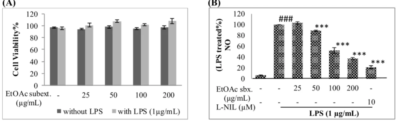
Raw 264.7 macrophage cells were treated with the EtOAc subextract at 25, 50, 100 and 200 µg/mL concentrations with or without LPS (1 µg/mL) for 24 hrs. The cell viability was not significantly affected (Figure 3). When EtOAc subextract was applied to cells at 50, 100 and 200 µg/mL concentrations, NO production of the cells were inhibited by 11.23±6.62%, 47.58±5.31% and 62.56±1.91%, respectively, while at a lower concentration (25 µg/mL) the inhibition rate was not significant.
(A) (B) 0 20 40 60 80 100 120 - 25 50 100 200 C el l V ia bi li ty % EtOAc subext. (µg/mL) without LPS with LPS (1µg/mL) 0 20 40 60 80 100 120 - - 25 50 100 200 -- - - 10 (L P S tr ea te d% ) N O LPS (1 µg/mL) EtOAc sbx. (μg/mL) L-NIL (µM) *** *** *** *** ###
Figure 3. The effects of N. oleander EtOAc extract on cell viability (A) and NO production (B) of LPS induced Raw 264.7 cells. Raw 264.7 macrophage cells were treated with the indicated concentrations of N. oleander EtOAc extract with or without LPS for 24 hours. Cell viablity was determined using WST-1 reagent (A). Raw 264.7 cells were treated with the indicated concentrations of N. oleander EtOAc extract and LPS (1 µg/mL) for 24 hrs. Media were collected and NO concentrations were determined by Griess Assay (B). Error bars represent the mean ± SEM for three experiments. Values of * p ≤ 0.05, ** p ≤ 0.01 and *** p ≤ 0.001 were considered statistically significant. (A) EtOAc sbx. (µg/mL) - - 25 100 200 β-actin iNOS LPS (1 µg/mL) EtOAc sbx. (µg/mL) - - 25 100 200 β-actin COX-2 LPS (1 µg/mL) (B) 0 20 40 60 80 100 120 - - 25 100 200 R el at iv e in te ns it y (i N O S /β -a ct in ) LPS (1 µg/mL) ## ** ** EtOAc sbx. (μg/mL) 0 20 40 60 80 100 120 - - 25 100 200 R el at iv e in te ns it y (C O X -2 /β -a ct in ) LPS (1 µg/mL) ## EtOAc sbx. (μg/mL)
Figure 4. Effects of N. oleander EtOAc subextract on iNOS and COX-2 protein levels of LPS induced Raw 264.7 macrophages. Raw 264.7 macrophage cells were pretreated for 1 hr with the indicated concentrations of N. oleander EtOAc subextract and then induced with LPS for a total of 24 hrs. After that cells were lysed and iNOS and COX-2 proteins were detected with Western Blot using specific antibodies. Equal loading was verified by β-actin antibody. A representative image of three independent experiments is shown (A). Relative band intensities measured by Image J. (B). The data are means ± SEM of three independent experiments. * p ≤ 0.05 ** p ≤ 0.01 *** p ≤ 0.001 compared to LPS treated group # p ≤ 0.05 ## p ≤ 0.01### p ≤ 0.001 compared to non-induced group.
3.2.2. Effects of EtOAc Subextract on iNOS and COX-2 Levels
In order to determine whether inhibition of NO release by EtOAc subextract was associated with the reduced levels of iNOS, protein levels were determined by Western Blot. Raw 264.7 macrophages were pretreated with 25, 100 and 200 µg/mL of EtOAc subextract for one hr and then treated with LPS for 24 hrs. As shown in Figure 3, the EtOAC subextract significantly decreased iNOS levels in a concentration-dependent manner. The iNOS levels reduced by 34.10% and 67.50% at 100 and 200 µg/mL concentrations, respectively, comparing to only LPS-treated (1 µg/mL) control group. Consistent with the effects on NO production, no inhibition was observed with 25 µg/mL of EtOAc subextract. On the other hand, EtOAc subextract slightly decreased (13.69%) COX-2 enzyme levels only at 200 µg/mL concentration (Figure 4).
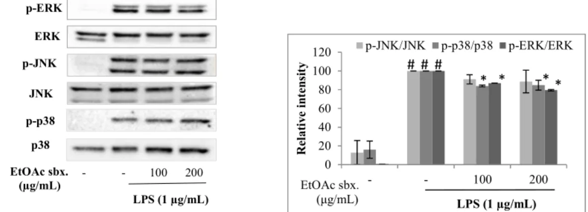
3.2.3. Effects of EtOAc Subextract on LPS-induced MAP Kinase Levels
Effect of the EtOAc subextract on the phosphorylation levels of MAP kinases were also investigated in order to explain its activity mechanism. Raw 264.7 macrophages were pretreated with 100 and 200 µg/mL EtOAc subextract and then treated with 1 µg/mL LPS for 15 min. Principally this stimulation would expected to increase ERK, JNK and p38 phosphorylations significantly (Figure 5). However, pretreatment of cells with EtOAc subextract slightly decreased the phosphorylation levels of MAP kinases. The highest inhibition was observed for ERK, which was 12.87% and 20.53% at 100 and 200 µg/mL concentrations, respectively. JNK phosphorylation was inhibited by 8.88% and 11.25% at the same concentrations. While the p38 phosphorylation was not affected by the increase in concentration (15.95% and 15.11% inhibitions at 100 and 200 µg/mL concentrations).
EtOAc sbx. (µg/mL) - - 100 200 p-p38 p-ERK p-JNK LPS (1 µg/mL) ERK JNK p38 0 20 40 60 80 100 120 - - 100 200 R el at iv e in te ns it y LPS (1 µg/mL) p-JNK/JNK p-p38/p38 p-ERK/ERK EtOAc sbx. (μg/mL) * * * # # # *
Figure 5. Effects of N. oleander EtOAc subextract on phosphorylation levels of MAP kinases on LPS induced Raw 264.7 macrophages. Raw 264.7 macrophage cells were pretreated for 2 hrs with the indicated concentrations of EtOAc subextract and then induced with LPS for 15 mins. Cells were lysed and MAP kinases (p-ERK, p-JNK, p-p38) proteins were detected with Western Blot using specific antibodies. Data was normalized to the corresponding total proteins (ERK, JNK, p38). A representative image of three independent experiments is shown (A). Relative band intensities measured by Image J (B). The data are means ± SEM of three independent experiments. * p ≤ 0.05 ** p ≤ 0.01 *** p ≤ 0.001compared to LPS treated group # p ≤ 0.05 ## p ≤ 0.01### p ≤ 0.001 compared to non-induced group.
3.3. Activity-guided Fractionation of N. oleander EtOAc Subextract and Isolation of Its Active Components
3.3.1. Effects of Fractions on Cell Viability
The EtOAc subextract was first fractionated by open column chromatography using polyamide as the stationary phase into four fractions: Fr. A, Fr. B, Fr. C, Fr. D. To determine the non-toxic concentrations, Fr. A, Fr. B and Fr. C were applied to cells at 100 µg/mL concentration and the cell viabilities were measured to be 95.99±2.61%, 83.29±1.08%, 90.64±3.37%. Fr D was applied to cells at 50 µg/mL and the cell viablity was determined to be 100.30±0.12% (Figure 6).
3.3.2. Effects of Fractions on NO Production
As shown in Figure 6, NO levels of the LPS treated cells (14,24 ±0,41 µM) were significantly increased comparing to untreated cells (0,36±0,23µM). The most active fractions were found to be Fr B (2.83±0.36 µM) and Fr C (6.94±0.22 µM). Fr D also moderately inhibited NO production of the cells (8.57±0.71 µM). Fr A slightly inhibited NO production (10.78±1.15) hence it was excluded from further studies. Consequently, through activity guided fractionation of the EtOAc subextract, kaempferol-3-O-β-D-glucoside (KG) was isolated from Fr.B, chlorogenic acid (CA) and kaempferol (KF) were isolated from Fr. C and Fr. D, respectively. The structure elucidation of these molecules were summarized at Section 3.5.
(A) (B) 0 20 40 60 80 100 120 - Fr A Fr B Fr C Fr D C el l v ia bi li ty % LPS (1μg/mL) * * * EtOAc Fr. 0 5 10 15 20 - - FrA FrB FrC FrD L-NIL N O ( μ M ) LPS (1μg/mL) * * * * * ### EtOAc Fr.
Figure 6. The effects of the fractions [Fr A (100 µg/mL), Fr B (100 µg/mL), Fr C (100 µg/mL), Fr D (100 µg/mL)] of the EtOAc subextract on cell viability (A) and NO production (B) of LPS induced Raw 264.7 cells. Raw 264.7 cells were treated with the indicated concentrations of fractions with LPS for 24 hrs. Cell viablity was determined by using WST-1 reagent (A). Raw 264.7 cells were treated with the fractions and LPS (1 µg/mL) for 24 hrs. L-NIL was applied at 10 µM. Media were collected and NO concentrations were determined by Griess Assay (B). Error bars represent the mean ± SEM for three experiments. Values of * p ≤ 0.05, ** p ≤ 0.01 and *** p ≤ 0.001 were considered statistically significant.
3.4. Effects of Active Compounds on NO Production
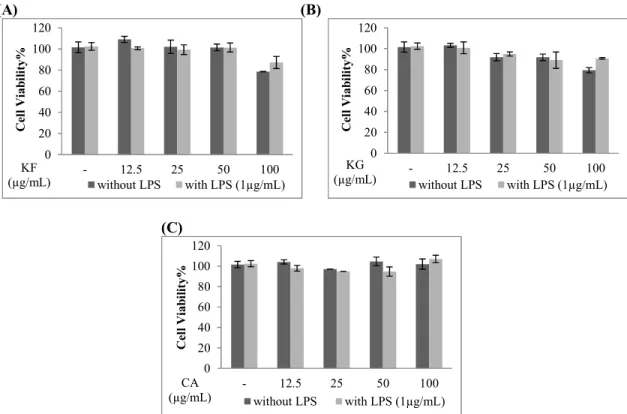
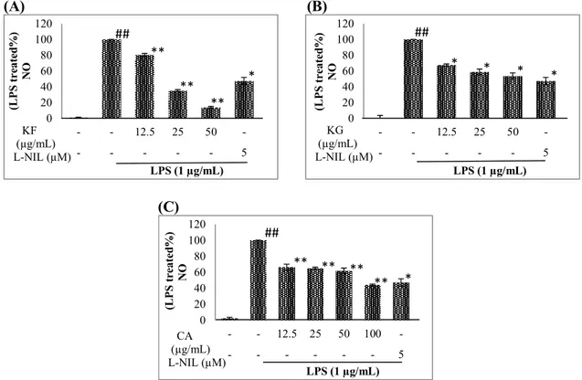
All compounds significantly reduced the levels of NO, but the most active compound was found to be KF. This molecule did not induce any significant decrease on the viability of Raw 264.7 macrophages up to 50 μg/mL concentration. KF inhibited NO production of the cells by 19.51%, 65.10%, 86.19% at 12.5, 25 and 50 µg/mL concentrations. That effect was higher than that of L-NIL (5 µM) which is a known NO inhibitor (52.77%). The viability of Raw 264.7 macrophages was not significantly affected when CA and KG was applied at 12.5, 25, 50 and 100 µg/mL concentrations. CA inhibited 33.74%, 35.01%, 38.12%, 55.96% of NO productions at these concentrations. KG inhibited 32.57%, 41.27 and 46.40% of NO at 12.5, 25 and 50 µg/mL concentrations. The inhibitory effects of CA and KG were also found to be comparable to L-NIL (Figure 7).
(A) (B) 0 20 40 60 80 100 120 - 12.5 25 50 100 C el l V ia bi lit y% KF (µg/mL) without LPS with LPS (1µg/mL) 0 20 40 60 80 100 120 - 12.5 25 50 100 C el l V ia bi lit y% KG (µg/mL) without LPS with LPS (1µg/mL) (C) 0 20 40 60 80 100 120 - 12.5 25 50 100 C el l V ia bi lit y% CA (µg/mL) without LPS with LPS (1µg/mL)
Figure 7. The effects of kaempferol (KF), kaempferol 3-O-β-glucopyranoside (KG) and chlorogenic acid (CA) on cell viability of Raw 264.7 cells. Raw 264.7 cells were treated with the indicated concentrations of KF (A), KG (B) and CA (C) with or without LPS for 24 hours. Cell viablity was determined by using WST-1 reagent. Error bars represent the mean ± SEM for three experiments. Values of * p ≤ 0.05, ** p ≤ 0.01 and *** p ≤ 0.001 were considered statistically significant.
3.5. Structure Elucidation of the Active Components
The structures of the isolates were elucidated on the basis of their NMR (1H- and 13C-NMR)
and HR-MS analysis and by comparison of their spectroscopic data with those published earlier. Kaempferol (1): (KF) was obtained as a yellow amorphous powder; C15H10O6, UV (CHCl3)
λmax:268, 369, IR (KBr) υmax cm−1: 3349, 1655, 1605, 1359, 1283, 1209, 1180, 1067, HR-MS: m/z = 287.0538 (calculated: 287.0555), 1H NMR (600 MHz, CD 3OD) δH: 8.08 (d, J = 8.6 Hz, H-2/6), 6.90 (d, J = 8.6 Hz, H-3/5), 6.39 (d, J = 1.8, H-8), 6.17 (d, J = 1.8, H-6) and 13C NMR (150 MHz, CD3OD) δC: 176.0 4), 164.0 7), 160.1 5), 159.0 4), 156.0 9), 153.5 2), 135.0 (C-3), 130.0 (C-2/6), 122.0 (C-1), 114.0 (C-3/5), 103.0 (C-10), 98.0 (C-6), 93.0 (C-8). These data were confirmed by the previous data [47].
Kaempferol-3-O-β-glucopyranoside (2): (KG) was also obtained as a yellow amorphous powder; C21H20O11, UV (MeOH) λmax: 266, 354,IR (KBr) υmax cm−1: 3365, 1686, 1594, 1403, 1265,
1121, HR-ESI-MS: m/z = 471.0920 [M+Na]+ (calculated: 471.0903), 1H NMR (600 MHz, CD 3OD) δH: 8.05 (d, J = 8.8 Hz, H-2/6) , 6.90 (d, J = 8.8 Hz, H-3/5), 6.36 (d, J = 2.0 Hz, H-8), 6.17 (d, J = 2.0 Hz, H-6), 5.21 (d, J = 7.7 Hz, H-1), 3.67 (dd, J = 2.3, 11.7 Hz, H-6a), 3.53 (dd, J = 5.3, 11.7 Hz, H-6b) 3.44 (dd, J = 7.7, 8.8 Hz, H-2), 3.40 (t, J = 8.3 Hz H-3), 3.30 (m, H-4), 3.19 (m, H-5), and 13C NMR (150 MHz, CDCl 3) δC: 176.0 (C-4), 166.5 (C-7), 162.0 (C-5), 160.3 (C-4), 157.2 (C-9), 153.5 (C-2), 133.0 (C-3), 130.3 (C-2/6), 120.4 (C-1), 114.0 (C-3/C-5), 103.8 (C-10), 103.0 (C-1), 98.0 (C-6), 93.0 (C-8), 77.5 (C-5), 76.8 (C-3), 74.5 (C-2), 70.0 (C-4), 60.8 (C-6). These data was found to be in accordance with the previous published data [47].
Chlorogenic acid (3): (CA) was obtained as a yellow amorphous powder; C16H18O9, UV
377.0981 [M+Na]+, (calculated: 377.0849), 1H NMR (600 MHz, CD
3OD) δH: 7.57 (d, J = 15.8 Hz,
H-), 7.05 (d, J = 2,3 Hz, H-2), 6.96 (dd, J = 2.4, 8.2 Hz, H-6), 6.77 (d, J = 8.2 Hz, H-5), 6.28 (d, J = 15.8 Hz, H-α), 5.20 (m, H-3), 4.12 (m, H-5), 3.70 (dd, H-4), 2.15 (m, H-2a), 2.01 (m, H-6a), 2.09 (m, H-6b), 1.95 (m, H-2b), and 13C NMR (150 MHz, CD
3OD) δC: 180.0 (C-7), 168.0 (C=O, caf.), 148.0
(C-4), 145.9 (C-3), 145.9 (C-), 126.5 (C-1), 121.9 (C-6), 115.0 (C-5), 114.0 (C-2), 113.9 (C-α), 76.5 (C-1), 73.9 (C-4), 71.9 (C-3), 71.2 (C-5), 39.2 (C-6), 38.0 (C-2). These data was consistent with the literature [48]. (A) (B) 0 20 40 60 80 100 120 - - 12.5 25 50 -- - - 5 (L P S tr ea te d% ) N O LPS (1 µg/mL) KF (µg/mL) L-NIL (µM) ** ** ** ## * 0 20 40 60 80 100 120 - - 12.5 25 50 -- - - 5 (L P S tr ea te d% ) N O LPS (1 µg/mL) KG (µg/mL) L-NIL (µM) * * * ## * (C) 0 20 40 60 80 100 120 - - 12.5 25 50 100 -- - - 5 (L P S t re at ed % ) N O LPS (1 µg/mL) CA (µg/mL) L-NIL (µM) ** ** ** ** ## *
Figure 8. The effects of kaempferol (KF), kaempferol 3-O-β-glucopyranoside (KG) and chlorogenic acid (CA) on NO production (B) of LPS induced Raw 264.7 cells. Raw 264.7 cells were treated with the indicated concentrations of KF (A), KG (B) and CA (C) with LPS (1 µg/mL) for 24 hrs. Media were collected and NO concentrations were determined by Griess Assay (B). Error bars represent the mean ± SEM for three experiments. Values of * p ≤ 0.05, ** p ≤ 0.01 and *** p ≤ 0.001 were considered statistically significant.
N. oleander has long been known for its toxic effects on oral use yet there are no toxicity or death report through the topical application or by corporal touch with the plant [49]. Consequently, there is only a limited number of utilization records in Turkish folk medicine mainly comprising external applications. The oily or alcohol extract of fresh flowers is applied to the aching joints against rheumatic pain or pounded flowers are directly wrapped externally on the affected extremities [45, 50].
A literature survey revealed that there are only a few reports regarding the anti-inflammatory activity of N. oleander. Anti-inflammatory activity of an Aloe vera-based extract of N. oleander leaves was investigated [35]. In another study, hydro-methanolic (70%) extract of the leaves was also shown to exert immunomodulatory activity by up-regulating signal molecules, i.e. interleukin-2 (IL-2), IL-10, interferon-gamma and down regulating IL-4, tumor necrosis factor-alpha (TNF-α), nitric oxide, cyclooxygenase-1 (COX-1) and COX-2 and PGE2 [51, 52]. On the other hand, in our previous study,
we investigated the anti-inflammatory and antinociceptive activities of fresh flowers. The EtOH extract was shown to exhibit potent in vivo antinociceptive activity as well as anti-inflammatory
activity against carrageenan-induced hind paw edema model in mice without inducing any gastric damage [46]. The activity was found to be as potent as reference drug, indomethacin.
In the present study, we aimed to discover the activity mechanism of EtOH extract of fresh flowers and to determine the active constituents in its active EtOAc subextract. For this purpose, firstly NO inhibitory activity of the EtOH extract and its subextracts (n-hexane, CH2Cl2, EtOAc,
R-H2O) were investigated. The crude EtOH extract did not significantly inhibit LPS induced NO
production of Raw 264.7 macrophages, but n-hexane, CH2Cl2 and EtOAc subextracts provided
significant inhibitions. This discrepancy might be due to the antagonistic effects of some of the components in the crude extract. Another explanation for this might be the elevation in relative concentrations of the active components in the subextract after partition. Hereby these results prompted us to focus on the EtOAc subextract which was the most active subextract without exerting toxicity towards Raw 264.7 macrophages.Despite exerting no inhibitory effect on COX-2 levels, a concentration dependent NO inhibitory activity of this extract was detected which is associated with the reduced levels of iNOS proteins. The subextract was also evaluated for its inhibitory effects on MAP kinases phosphorylation. However, the phosphorylation levels of MAP kinases were slighlty reduced by the treatment of this subextract. The highest effect was observed for ERK phosphorylation. Activity-guided fractionation of the EtOAc extract resulted in the isolation of one flavonol aglycone; kaempferol, one flavonol glycoside; kaempferol-3-O-β-glucopyranoside and one quinic acid derivative; chlorogenic acid. All of the compounds significantly inhibited NO productions in a concentration dependent manner.
The in vivo and in vitro anti-inflammatory activites of these three compounds were previously reported in several studies [53-56]. Chlorogenic acid was shown to decrease the levels of COX-2 and PGE2 and was also found to inhibit JNK, but not p38 and ERK (p44/42) phosphorylations on LPS
induced Raw 264.7 macrophages [57]. Besides chlorogenic acid was found to inhibit the mRNA expressions of COX-2 and iNOS, and also decreased the levels of pro-inflammatory (1β, TNFα, IL-6) cytokines [58]. Although it was reported that pretreatment of Raw 264.7 macrophage cells with chlorogenic acid up to 37.5 µg/mL concentration did not significantly affect iNOS and NO levels when the cells were treated with LPS for 10 to 12 hours, another study revealed that significant inhibitory effect is attainable at relatively lower concentrations such as 20 µM when cells were treated for 24 hrs [57, 58]. In the present study, CA exerted significant NO inhibitory activity at higher concentrations (100 µg/mL) without inducing notable toxicity to cells.
The NO inhibitory effects of kaempferol was also demonstrated on various in vitro models with cell lines such as Raw 264.7, J774 macrophages and Chang liver cells, while kaempferol-3-O-β-glucoside was shown to reduce LPS-induced production of NO in J774A.1 cells [59-61]. Moreover, kaempferol and kaempferol-3-O-β-glucoside have been shown to display significant inhibitory activities on various signaling molecules (TNFα, PGE2, COX-2, iNOS, NF-κB) which are known to
involve in inflammatory process [59-61]. Accordingly, these previously conducted bioactivity studies on these secondary metabolites are in line with the results obtained in the present study. The current findings will shed light on the chemical composition and bioactive components of the oleander flowers.
The results of the present study have clearly proved the traditional use of the oily or alcohol extracts of N. oleander flowers to treat inflammatory diseases. The EtOAc subextract obtained from the EtOH extract of the oleander flowers was found to exert significant in vitro anti-inflammatory activity. The active components in this subextract were determined to be kaempferol, kaempferol-3-O-β-D-glucoside and chlorogenic acid. Although these compounds were previously reported to be the anti-inflammatory components from several other plant remedies, their existence in the oleander flowers was reported for the first time here which would also contribute to the chemical profiling of the flowers. Apart from the EtOAc subextract, the n-hexane and CH2Cl2 subextracts also deserve to be
deeply investigated in order to discover potential bioactive components.
Acknowledgments
This research did not receive any specific grant from funding agencies in the public commercial, or not-for-profit sectors.
ORCID
İrem Atay Balkan: 0000-0002-7921-8621
Ahmet C Goren:: 0000-0002-5470-130X
Hasan Kırmızıbekmez:0000-0002-6118-8225
Erdem Yeşilada: 0000-0002-1348-6033
References
[1] L.M. Coussens and Z. Werb, (2002). Inflammation and cancer, Nature 420, 860-867.
[2] M. Feldmann and R.N. Maini (2003). TNF defined as a therapeutic target for rheumatoid arthritis and other autoimmune diseases, Nat. Med. 9, 1245-1250.
[3] C. Gabay (2006). Interleukin-6 and chronic inflammation, Arthritis Res. Ther. 6, 1-6.
[4] R. Gautam and S.M. Jachak (2009). Recent developments in anti-inflammatory natural products, Med. Res. Rev. 29,767-820.
[5] R.A. Heller, M. Schena, A. Chai, D. Shalon, T. Bedilion, J. Gilmore, D.E. Woolley and R.W. Davis (1997). Discovery and analysis of inflammatory disease-related genes using cDNA microarrays, Proc. Natl. Acad. Sci. U. S. A. 94, 2150-2155.
[6] B. Kaminska (2005). MAPK signalling pathways as molecular targets for anti-inflammatory therapy-from molecular mechanisms to therapeutic benefits, Biochim. Biophys. Acta. 1754, 253-62.
[7] A. Gibofsky (2012). Overview of epidemiology, pathophysiology, and diagnosis of rheumatoid arthritis, Am. J. Manag. Care. 18, S295-302.
[8] A.E. M’Koma (2013). Inflammatory bowel disease: An expanding global health problem, Clin. Med. Insights Gastroenterol. 6, 33-47.
[9] C. Reitz, C. Brayne and R. Mayeux (2011). Epidemiology of Alzheimer disease, Nat. Rev. Neurol. 7, 137-152.
[10] P.Subbarao, P.J. Mandhane and M.R. Sears (2009). Asthma: epidemiology, etiology and risk factors, Can. Med. Assoc. J. 181, E181-190.
[11] F. Kuralay and Z. Çavdar (2006). Enflamatuvar mediyatörlere toplu bakış, Genel Tıp Derg. 16, 143-152.
[12] A. Tincani, L.Andreoli, C. Bazzani, D. Bosiso and S. Sozzani (2007). Inflammatory molecules: A target for treatment of systemic autoimmune diseases, Autoimmun. Rev. 7, 1-7.
[13] E. Yesilada, O. Ustün, E. Sezik, Y. Takaishi, Y. Ono and G. Honda (1997). Inhibitory effects of Turkish folk remedies on inflammatory cytokines: Interleukin-1alpha, interleukin-1beta and tumor necrosis factor alpha, J. Ethnopharmacol. 58, 59-73.
[14] S.H. Tsai, S.Y. Lin-Shiau and J.K. Lin (1999). Suppression of nitric oxide synthase and the down-regulation of the activation of NFκB in macrophages by resveratrol, Br. J. Pharmacol. 126, 673-680. [15] Y.C. Chen, S.C. Shen, W.R. Lee, W.C. Hou, L.L. Yang and T.J.F. Lee (2001). Inhibition of nitric oxide
synthase inhibitors and lipopolysaccharide induced inducible NOS and cyclooxygenase-2 gene expressions by rutin, quercetin, and quercetin pentaacetate in RAW 264.7 macrophages, J. Cell. Biochem. 82, 537-548.
[16] G.M. Cooper and E.R. Hausman (2008). The cell: A molecular approach. 5th Edition. ASM Press. Washington, D.C., U.S.A.
[17] J. Gupta and A.R. Nebreda (2015). Roles of p38α mitogen-activated protein kinase in mouse models of inflammatory diseases and cancer, FEBS J. 282, 1841-1857.
[18] G.L. Johnson and R. Lapadat (2002). Mitogen-activated protein kinase pathways mediated by ERK, JNK, and p38 protein kinases, Science 298, 1911–1912.
[19] C.C. Chen and J.K. Wang (1999). p38 but Not p44/42 Mitogen activated protein kinase is required for nitric oxide synthase induction mediated by lipopolysaccharide in RAW 264.7 macrophages, Mol. Pharmacol. 55, 481-488.
[20] D. Hwang, B.C. Jang, G. Yu and M. Boudreau (1997). Expression of mitogen-inducible cyclooxygenase induced by lipopolysaccharide, Biochem. Pharmacol. 54, 87-96.
[21] H.G. Kim, D.H. Yoon, W.H. Lee, S.K. Han, B. Shrestha, C.H. Kim, M.H. Lim, W. Chang, S. Lim, S. Choi, W.O. Song, J.M. Sung, K.C. Hwang and T.W. Kim (2007). Phellinus linteus inhibits inflammatory mediators by suppressing redox based NF-kappaB and MAPKs activation in lipopolysaccharide induced RAW 264.7 macrophage, J. Ethnopharmacol. 114, 307-15.
[22] T. Baytop (1999). Treatment with Plants in Turkey, Past and Today [Türkiye’de Bitkilerle Tedavi Geçmişte ve Bugün]. 2. ed. Nobel Tıp Kitabevi, İstanbul (in Turkish).
[23] F. Abe and T. Yamauchi (1992). Cardenolide triosides of oleander leaves, Phytochemistry. 31, 2459-2463.
[24] F. Abe and T. Yamauchi (1992). Pregnanes from oleander leaves, Phytochemistry 31, 2819-2820. [25] S. Begum, R. Sultana and B.S. Siddiqui (1997). Triterpenoids from the leaves of Nerium oleander,
Phytochemistry. 44, 329-332.
[26] S. Begum, B.S. Siddiqui, R. Sultana, A. Zia and A. Suria (1998). Bio-active cardenolides from the leaves of Nerium oleander, Phytochemistry 50, 435-438.
[27] L.J. Rashan, K. Franke, M.M. Khine, G. Kelter, H.H. Fiebig, J. Neumann and L.A. Wessjohann (2011). Characterization of the anticancer properties of monoglycosidic cardenolides isolated from Nerium oleander and Streptocaulon tomentosum, J. Ethnopharmacol. 134, 781-8.
[28] S. Siddiqui, F. Hafeez, S. Begum and B.S. Siddiqui (1986). Isolation and structure of two cardiac glycosides from the leaves of Nerium oleander, Phytochemistry 26, 237-241.
[29] A. Siddiqui, S. Begum, F. Hafeez and B.S. Siddiqui (1989). Two triterpenes from the leaves of Nerium oleander, Phytochemistry 28, 1187-1191.
[30] B.S. Siddiqui, S. Begum, S. Siddiqui and W. Lichter (1995). Two cytotoxic pentacyclic triterpenoids from Nerium oleander, Phytochemistry 39, 171-174.
[31] B.S. Siddiqui, N. Khatoon, S. Begum, A.D. Farooq, K.,Qamar, H.A. Bhatti and S.K. Ali (2012). Flavonoid and cardenolide glycosides and a pentacyclic triterpene from the leaves of Nerium oleander and evaluation of cytotoxicity, Phytochemistry 77, 238-244.
[32] T. Yamauchi, N. Takata and T. Mimura (1975). Cardiac glycosides of the leaves of Nerium odorum, Phytochemistry 14,1379-1382.
[33] T. Yamauchi, M. Takahashi and F. Abe, (1976). Cardiac glycosides of the root bark of Nerium odorum, Phytochemistry 15, 1275-1278.
[34] R. Hanada, F. Abe and T. Yamauchi (1992). Steroid glycosides from the roots of Nerium odorum, Phytochemistry 31, 3183-3187.
[35] K.F. Benson, R.A. Newman and G.S. Jensen (2015). Antioxidant, anti-inflammatory, anti-apoptotic, and skin regenerative properties of an Aloe vera-based extract of Nerium oleander leaves (nae-8(®)), Clin. Cosmet. Investig. Dermatol. 8, 239-48.
[36] M.M. Huq, A. Jabbar, M.A. Rashid and C.M. Hasan (1999). A novel antibacterial and cardiac steroid from the roots of Nerium oleander, Fitoterapia. 70, 5-9.
[37] A. Zia, B.S. Siddiqui, S. Begum, S. Siddiqui and A. Suria (1995). Studies on the constituents of the leaves of Nerium oleander on behavior pattern in mice, J. Ethnopharmacol. 49, 33-39.
[38] M.S. Yu, S.W. Lai, K.F. Lin, J.N. Fang, W.H.Yuen and R.C. Chang (2004). Characterization of polysaccharides from the flowers of Nerium indicum and their neuroprotective effects, Int. J. Mol. Med. 14, 917-924.
[39] M.S. Yu, A.Y.Y. Wong, K.F.So, J.N. Fang, W.H. Yuen and R.C.C. Chang (2007). New polysaccharide from Nerium indicum protects neurons via stress kinase signaling pathway, Brain Res., 1153, 221-230. [40] K.G. Singhal and G.D. Gupta, (2012). Hepatoprotective and antioxidant activity of methanolic extract
of flowers of Nerium oleander against CCl4 induced liver injury in rats, Asian Pac. J. Trop. Med. 5,
677-685.
[41] K.G. Singhal and G.D. Gupta (2012). Neuroprotective appraisal of methanolic extract of flowers of Nerium oleander in a non classical rat model of Alzheimer disease, The Nat. Prod. J. 2, 235-245. [42] V. Gayathri, S. Ananthi, C. Chandronitha, G. Ramakrishnan, R. Lakshmisundaram and H.R. Vasanthi
(2011). Cardioprotective effect of Nerium oleander flower against isoproterenol-induced myocardial oxidative stress in experimental rats, J. Cardiovasc. Pharmacol. Ther. 16, 96-104.
[43] M. Mohadjerani (2012). Antioxidant activity and total phenolic content of Nerium oleander L. grown in North of Iran, IJPR. 11,1121-1126.
[44] D.S. Mohale, A.S. Tripathi, A.V.Shrirao, A.G. Jawarkar and A.V. Chandewar (2016). Evaluation of antioxidant effect of Nerium indicum in anxious rats. Ind. J.Pharmacol. 48, 430-433.
[45] E. Yesilada, (2002). Biodiversity in Turkish folk medicine. Kluwer Academic/Plenum Publishers. London, U.K.
[46] N. Erdemoglu, E. Küpeli and E. Yesilada (2003). Anti-inflammatory and antinociceptive activity assessment of plants used as remedy in Turkish folk medicine, J. Ethnopharmacol. 89, 123-129. [47] P.K. Agrawal (1989). Carbon-13NMR of Flavonoids. Elsevier Scientific Publishing Co. New York. [48] I. Saracoğlu, Ü.S. Harput and I. Çalış (2002). Phenolic constituents from Phlomis lycia, Turk. J. Chem.
26, 133-142.
[49] C. Kiran and D.N. Prasad (2014). A review on: Nerium oleander Linn. (Kaner), Int. J. Pharmacogn. Phytochem. Res. 6, 593-597.
[50] Y. Güzel, M. Güzelşemme and M. Miski (2015). Ethnobotany of medicinal plants used in Antakya: A multicultural district in Hatay Province of Turkey, J. Ethnopharmacol. 174, 118-152.
[51] P. Dey and T.K. Chaudhuri (2015). Anti-inflammatory activity of Nerium indicum by inhibition of prostaglandin E2 in murine splenic lymphocytes, Ind. J. Pharmacol. 47, 447-50.
[52] P. Dey and T.K. Chaudhuri (2016). Immunomodulatory activity of Nerium indicum through inhibition of nitric oxide and cyclooxygenase activity and modulation of TH1/TH2 cytokine balance in murine splenic lymphocytes, Cytotechnology 68, 749-761.
[53] M.D.D Santos, M.C. Almeida, N.P. Lopes and G.E. de Souza (2006). Evaluation of the anti-inflammatory, analgesic and antipyretic activities of the natural polyphenol chlorogenic acid, Biol. Pharm. Bull. 29, 2236-2240.
[54] L. Kong, C. Luo, X. Li, Y. Zhou and H. He (2013). The anti-inflammatory effect of kaempferol on early atherosclerosis in high cholesterol fed rabbits, Lipids Health Dis. 12, 115.
[55] M.Y.A. Mahat, N.M. Kulkarni, S.L. Vishwakarma, F.R. Khan, B.S. Thippeswamy, V. Hebballi, A.A. Adhyapak, V.S. Benade, S.M. Ashfaque, S. Tubachi and B.M. Patil (2010). Modulation of the cyclooxygenase pathway via inhibition of nitric oxide production contributes to the anti-inflammatory activity of kaempferol, Eur. J. Pharmacol. 642, 169-176.
[56] Y. Wang, P. Chen, C. Tang, Y. Wang, Y. Li and H. Zhang (2014). Antinociceptive and anti-inflammatory activities of extract and two isolated flavonoids of Carthamus tinctorius L, J. Ethnopharmacol. 151, 944-950.
[57] J. Shan, J. Fu, Z. Zhao, X. Kong, H. Huang, L. Luo and Z. Yin (2009). Chlorogenic acid inhibits lipopolysaccharide induced cyclooxygenase-2 expression in RAW264.7 cells through suppressing NF-κB and JNK/AP-1 activation, Int. Immunopharmacol. 9, 1042-1048.
[58] S.J. Hwang, Y.W. Kim, Y. Park, H.J. Lee and K.W. Kim (2014). Anti-inflammatory effects of chlorogenic acid in lipopolysaccharide-stimulated RAW 264.7 cells, Inflamm. Res. 63, 81-90.
[59] M. Hamalainen, R. Nieminen, P. Vuorela, M. Heinonen and E. Moilanen (2007). Anti-inflammatory effects of flavonoids: genistein, kaempferol, quercetin, and daidzein inhibit stat-1 and NF-κB activations, whereas flavone, isorhamnetin, naringenin, and pelargonidin inhibit only NF-κB activation along with their ınhibitory effect on iNOS expression and NO production in activated macrophages, Mediators Inflamm. 45673, doi: 10.1155/2007/45673. [60] M.S. Kim and S.H. Kim (2011). Inhibitory effect of astragalin on expression of
lipopolysaccharideinduced inflammatory mediators through NF-κB in macrophages, Arch. Pharm. Res. 34, 2101-2107.
[61] N.X. Nhiem, B.H. Tai, T.H. Quang, P.V. Kiem, C.V. Minh, N.H. Nam, J.H. Kim, L.R. Im, Y.M. Lee and Y.H. Kim (2011). A new ursane-type triterpenoid glycoside from Centella asiatica leaves modulates the production of nitric oxide and secretion of TNFα in activated RAW 264.7 cells, Bioorg. Med. Chem. Lett. 21, 1777-1781.
![Figure 2. The effects of EtOH (100 µg/mL) extract and its subextracts [n-hexane (100 µg/mL), CH 2 Cl 2 (50 µg/mL), EtOAc (100 µg/mL), R-H 2 O (100 µg/mL)] on the viability (A) and NO productions (B) of LPS induced Raw 264.7 ce](https://thumb-eu.123doks.com/thumbv2/9libnet/5445568.104598/5.918.138.783.447.644/figure-effects-extract-subextracts-etoac-viability-productions-induced.webp)
![Figure 6. The effects of the fractions [Fr A (100 µg/mL), Fr B (100 µg/mL), Fr C (100 µg/mL), Fr D (100 µg/mL)] of the EtOAc subextract on cell viability (A) and NO production (B) of LPS induced Raw 264.7 cells](https://thumb-eu.123doks.com/thumbv2/9libnet/5445568.104598/8.918.137.770.513.705/figure-effects-fractions-etoac-subextract-viability-production-induced.webp)