Summary
Type 2 diabetes mellitus (DM) affects a large population worldwide. DM is often considered as a syndrome of disordered glucose metabolism. Current conventional drug therapies for DM are usually insufficient and medicinal herbs with antihyperglycemic activities are increasingly sought by diabetic patients and health care professionals. Nerium Oleander (N.O.) is known to be effective in lowering of postprandial blood glucose in DM patients as a folk remedy. In this study we aimed to evaluate effect of N.O. distillate in glucose uptake activity of hepatocytes and adipocytes. The human hepatoma cells Hep3B and mouse adipocyte 3T3-L1 cells were cultured. Depending on the groups, different concentrations insulin (1, 10, 20 IU/ml) and N.O. (0.1, 1, 10, 50 µg/ml) were added to medium for 48 h. Cellular toxicity and proliferation were evaluated by LDH secretion levels and MTT test. A metabolizable fluorescent derivative of glucose, 2-NBDG and FITC-insulin were used for glucose uptake and insulin binding activity. Insulin increased cellular proliferation and decreased LDH leakage and apoptosis in both cell types. Lower dosages of N.O. has no significant effect on apoptosis and cell number while at the highest dosages minimal cytotoxicity was seen mainly in adipocytes. Main effect of N.O. treatment was increased glucose uptake in Hep3B and 3T3-L1 cells (P<0.001). Our results showed that N.O. may be offered as new approaches to treatment of type 2 diabetes by modulating cellular glucose uptake.
Keywords: Nerium oleander, Hepatocytes, Adipocytes, Glucose uptake, Insulin binding, Type II diabetes mellitus
Nerium Oleander’in Hepatositler ve Adipositlerde İnsulin
Bağlanma ve Glukoz Alımını Arttırıcı Etkisi
Özet
Tip 2 diabet glukoz metabolizmasının bozukluğu şeklinde tanımlanan tüm dünyada oldukça yaygın bir hastalıktır. Mevcut tedavi yöntemleri yeterli gelmemekte, hastaların antihiperglisemik etkiye sahip tıbbi bitkilerle tedaviye ilgisi artmaktadır. Nerium Oleander (N.O.) diabetik hastalarda postprandial glukoz düzeyini düşürmek amacıyla yerel olarak kullanılmaktadır. Bu çalışmada N.O. ekstraktının karaciğer ve adiposit hücrelerinde glukoz alımına ve insulin bağlanmasına etkilerinin belirlenmesi amaçlanmıştır. Farklı dozlarda insulin (1-20 IU/ml) ve N.O. (0.1-50 µg/ml) 48 h uygulamasının İnsan hepatosit Hep3B ve fare adipositleri 3T3-L1 hücre dizileri üzerindeki etkileri değerlendirilmiştir. Bu amaçla hücre toksitesi LDH sekresyonu, hücre çoğalması MTT yöntemi ve hücre içine glukoz alımı/insulin bağlanması florimetrik yöntemle 2-NBDG ve FITC-insulin kullanılarak ölçülmüştür. Insulin uygulaması her iki hücrede de proliferasyonu arttırmış, LDH sekresyonunu azaltmıştır. Düşük dozlarda N.O. uygulamasının hücre sayısına etkisi görülmezken kullanılan üst dozlarda adipositlerde sitotoksik etkisi gözlenmiştir. N.O. nun adiposit ve hepatositlerde hücre içine glukoz alımını anlamlı arttırdığı gözlenmiştir (P<0.001). Çalışmamız sonuçları N.O. ekstraktının tip 2 diabette özellikle insulin ve glukoz kullanımını düzenleyici etkisi nedeniyle önemli yeni bir tedavi alternatifi olabileceğini göstermiştir.
Anahtar sözcükler: Nerium oleander, Hepatositler, Adipositler, Glukoz alımı, İnsulin bağlanması, Tip II Diabetes mellitus
Increased Glucose Uptake and Insulin Binding Activity of
Nerium Oleander in Hepatocytes and Adipocytes
[1]Nuray YAZIHAN * Ahmet Levent BAS ** Ezgi ERMIS ***
Sule DEMIRCI **** Kamil UNEY **
[1] * ** *** ****
This research was granted by The Scientific and Technological Research Council of Turkey (TUBITAK, Project No: 107 O 222) Department of Pathophysiology, Moleculer Biology R&D Unit, Faculty of Medicine, Ankara University, TR-06100 Ankara - TURKEY
Department of Pharmacology and Toxicology, Faculty of Veterinary Medicine, Selcuk University, TR-42003 Konya - TURKEY
Moleculer Biology R&D Unit, Faculty of Medicine, Ankara University, TR-06100 Ankara - TURKEY
Department of Physiology, Faculty of Veterinary Medicine, Mehmet Akif Ersoy University, TR-15030 Burdur - TURKEY
Makale Kodu (Article Code): KVFD-2012-7044
İletişim (Correspondence)
+90 312 3103010/372INTRODUCTION
Type 2 diabetes mellitus (DM) affects a large population worldwide 1. There have been many attempts to develop
the safe and effective methods of curing diabetes. Although very intensive research is being conducted in this field, current protocols still have only limited applications.
The general consensus on treatment of type 2 diabetes is that lifestyle management is at the forefront of therapy options. In addition to exercise, weight control, and medical nutrition therapy, oral glucose-lowering drugs and injections of insulin are the conventional therapies. Alternative treatments for diabetes have become increasingly popular the last several years, including medicinal herbs, nutritional supplementation and acupuncture. Plant derivatives with purported hypo-glycemic properties have been used in folk medicine and traditional healing systems around the world. Approximately 1200 plants are used worldwide for the empirical treatment of DM. However, only about 350 of them are documented to present hypoglycemic activity although only a small number of these have received scientific and medical evaluation to assess their efficacy2,3.
The World Health Organization Expert Committee on diabetes has recommended that traditional medicinal herbs be further investigated 1,4.
Some of the most common plants used traditionally for treatment of diabetes are Pterocarpus marsupium 5, Bitter
Melon6, Gymnema Sylvestre 7, Asian Ginseng 8, Soybean 9and
Cinnamon 10.
Nerium oleander (N.O.) is a member of Apocynaceae familia and grown in Mediterrian region. Nerium indicum is known to be effective in lowering of postprandial blood glucose in DM patients and both oleander and indicum sub- forms are now used as a folk remedy for type II diabetes in some regions of mediterrian region and Asia 11. The hot
water extract of the leaves of N.O. has been used as a remedy against Type II DM with subjective success but without corroborating laboratory data. First report related to the hypoglycemic activity of the plant is given by Bellakhdar et al.12and then reported by other authors 13,14.
DM is a complex group of metabolic disorders including hyperglycemia and impaired insulin secretion and/or insulin response. Current theories of DM include a defect in insulin-mediated glucose uptake in muscle and adipocytes, a dysfunction of the pancreatic β-cells, an impaired insulin action in liver and decreased peripheral (muscle) glucose utilization. Changes in glucose clearance, an index of efficiency of glucose removal from the circulation, by itself do not affect plasma glucose concentrations independent of changes in rates of glucose entry and exit15,16. In this
study we aimed to evaluate in vitro direct effect of N.O. in hepatocyte and adipocyte cells especially with the aspect of glucose uptake metabolism and cellular proliferation.
MATERIAL and METHODS
Cell lines, Chemicals and MaterialsHuman hepatoma cell line Hep3B and mouse preadipo-cyte cells 3T3-L1 were obtained from the American Type Culture Collection(ATCC). Hep3B cells were cultured in Roswell Park Memorial Institute-1640 (RPMI, PAA, Austria) and 3T3-L1 cells in Dubellco’s Modified Eagles Medium (DMEM, PAA, Austuria), supplemented with fetal calf serum (FCS), (PAA, Austuria), L-glutamine, streptomycin and penicillin (Sigma, MO, USA). Insulin (Sigma, MO, USA) was dissolved in water, sterilized by 0.22 m pore size cellulose acetate membrane filters, and added to cultures at the indicated time and concentrations. Cell counts were tested by 3-4,5-dimethylthiazol -2-yl-2,5 diphenyltetrazolium bromide (MTT, Sigma, MO, USA). Lactate Dehydrogenase (LDH) and glucose levels were measured with commercial kits using an automatic multi-analyzer (Roche; P800).
Obtaining Liyophilized Nerium oleander
Nerium oleander plant was collected among new shoots in March-September period from Mediterranean region of Turkey. After washing collected plant, fresh shoots were chopped, and adequate distilled water added. The mixture was heated in heat resistant container. After liquid started to evaporate, container lid was covered and vapor was separated to other clean glass container by causing it to come to contact with a surface cooled with cold water. N.O. distillate was liyophilized in small glass bottle (20 ml) by using liyophilizator. N.O. liyophilized distillate was dissolved in according to dosage in saline solution then sterilized by 0.22 m pore size cellulose acetate membrane filters, and added to cultures at the indicated time and concentrations.
Cell Culture and Experimental Protocol
The human hepatoma cell line Hep3B was cultured in RPMI-1640 medium and 3T3-L1 cells in DMEM, supplemented with 10% v/v fetal calf serum, 2 mmol/L L-glutamine, streptomycin (100 mg/mL) and penicillin (100 IU/mL) in a humidified atmosphere containing 5% CO2 at 37°C. One day before the experiments, cells were seeded on 96-well microtitre plates (Nunc, Denmark) at 2X10⁵ cells/mL.
Depending on the groups, different concentrations insulin (1, 10, 20 IU/ml) and N.O. (0.1, 1, 10, 50 µg/ml) were added to medium for 48 h.
LDH levels were evaluated from control and treated cells at the 48th h. MTT was measured at the 2nd, 24th and 48th h. After supernatants were removed cell surface was washed with sterile phosphate buffered saline (PBS) and cells were harvested with lysis solution and caspase-3 levels of groups were measured from cell lysates. LDH measurement was done from both of the supernatant and cell lysates.
Evaluation of Cellular Proliferation or Death
MTT, a colorimetric assay based upon the ability of living cells to reduce MTT into formazan, was used for evaluation of the effects of dose and time dependent effects of glucose, insulin and N.O. on cellular death or proliferation (24th, 48th h). Cell number % was calculated as ratio of cell number of effected group vs control group 100 at the determined hours.
Biochemical Determination of Cell Death
Hep3B cells were plated in 96 multiwell cell culture plates as 3X10⁵ cells/mL. LDH is normally present in the cytosol of hepatocytes. In response to cell damage LDH is released from the cells. Therefore, to determine cell death, we measured secreted and intracellular LDH levels and calculated % released LDH at the 48th h for each group. To do this, the medium was collected to measure enzyme activities. The adherent cells were lysed. Both medium and cell lysates were used for quantitative determination of LDH activity (IU/L) which was performed with an automatic multianalyzer (Roche, MN, USA) using commercial kit (Roche, MN, USA). Released enzyme fractions for each sample were calculated as the ratio of enzyme present in the medium vs the sum of the levels of same enzyme in the supernatant and in the cells.
Glucose Uptake and Insulin Binding Activity
A metabolizable fluorescent derivative of glucose, 2-(N-(7-Nitrobenz-2-oxa-1,3-diazol-4-yl)Amino)-2-Deoxyglucose (2-NBDG, Molecular Probes, Oregon, USA), was used. Follo- wing incubation at 37°C for 120 min with the dye, culture medium was removed and after washing the cells with Hanks’ balanced salt solution (HBSS) fluorescence was measured in Spectromax M2 fluorescence microplate reader (Perkin- Elmer Corp. Norwalk, CT, USA) set at an excitation wavelength of 470 nm and an emission wavelength of 550 nm. To deter- mine the effect of insulin or N.O. on glucose uptake, the cell suspension was dispensed in 96-well microtiter plates at 2500 cells/well (200 ml). After 24 h of incubation (37°C/ 5% CO2), all culture medium was removed from each well and replaced with 100 ml of culture medium with different concentrations of insulin and and 100 ml of 300 μM 2-NBDG diluted in the same medium to give a final concentration of 150 μM /well. In this case, cells were incubated simultaneously
with both insulin/N.O. and 2-NBDG for 120 min, and then the plates were centrifuged, the culture medium was removed and the cells were washed once with HBSS. Fluorescence was measured as previously described and results of triplicate experiments expressed as 2-NBDG concentrations 17.
Same procedure was repeated with 0.3 mM Fluorescein isothiocyanate (FITC) labeled Insulin. Fluorescence was measured at an excitation wavelength of 490 nm and an emission wavelength of 520 nm 18.
Statistical Analysis
Results of the experiments were analyzed by One Way ANOVA, followed by a multiple comparison test using SPSS 13.0. P<0.05 was accepted as statistically significant. Results were given as mean± SEM.
RESULTS
We characterized the concentration-dependent effect of N.O. and insulin on human hepatocyte cell line (Hep3B) and mouse adipocyte cell line (3T3-L1) as a function of time. N.O. treatment decreased adipocyte cell number (P<0.01) whereas insulin increased (P<0.01, Fig. 1). Minimal toxic effect is seen at the highest dose of N.O. (P<0.05) in Hep3B cells (Fig. 2).
Insulin treatment decreased LDH leakage, higher doses of N.O. treatment increased LDH leakage (P<0.01, Fig. 3).
Main effect of N.O. treatment was seen in glucose uptake to Hep3B and 3T3-L1 cells (P<0.001, Fig. 4, 5) and this effect is more prominent in liver cells and significantly different from control and insulin treated cells (P<0.001). N.O. treatment increased insulin binding capacity in both adipocytes and hepatocytes (P<0.001, Fig. 6).
DISCUSSION
Both experimental and epidemiologic studies showed that insulin resistance is not key factor only in diabetes also in cardiovascular diseases 19. Control of blood glucose levels
is a function and coordination of different organ systems such as liver, pancreas, muscle and fat. These organs and tissues
Fig 1. Cell number was determined at 24th and 48th h in adipocyte
cell line 3T3-L1. NO treatment caused significant cytotoxicity with dose and time dependent manner (P<0.01). The effect is most prominent in N.O. 10 μg/ml dose and at 24 th h (P<0.001). Insulin
treatment increased cell number at 48 th h in all concentrations
(P<0.01). Data are presented as mean±SEM (n=6)
Şekil 1. Adiposit hücre dizisinde (3T3-L1) hücre proliferasyonunun 24 ve 48. saatlerde değerlendirilmesi. N.O. tedavisi doz ve zaman bağımlı olarak sitotoksitik etki göstermiştir (P<0.01). Bu etki NO 10 μg/ml dozda ve 24. saatte en belirgindir (P<0.001). Insulin tedavisi 48. saatte hücre proliferasyonunu tüm dozlarda arttırmıştır (P<0.01). Sonuçlar ortalama±SEM olarak verilmiştir (n=6)
have major roles in the use and storage of nutrients in the form of glycogen and triglycerides. Type II DM is the most common disorder which is characterized by impaired insulin stimulated glucose uptake in muscle and adipose tissue20.
It is striking that peripheral glucose levels should be well
regulated by physiological control mechanisms, which, when deregulated, trigger early signs of the pathogenesis of obesity and diabetes, such as abnormal suppression of glucagon and loss of insulin secretion in the fed state and decreased peripheral tissue glucose uptake. These dysregulations become prominent before full impairment
Fig 2. Cell number was determined at 24th and 48th h by the MTT
assay. N.O. treatment has minimal cytotoxic effect in highest dosage in Hep3B cell line (P<0.05). Insulin treatment increased cell number in all concentrations (P<0.001). Data are presented as mean±SEM (n=6)
Şekil 2. Hepatosit hücre dizisinde 24 ve 48. saatlerde hücre sayımının MTT ile değerlendirilmesi. N.O. tedavisi en yüksek dozda hepatositlerde minimal toksik etki göstermiştir (P<0.05). Insulin tedavisi tüm dozlarda proliferative etki göstermiştir (P<0.001). Sonuçlar ortalama±SEM olarak verilmiştir (n=6)
Fig 4. Basal and insulin-stimulated NBDG-glucose uptake in cultured human hepatocytes. N.O. treatment increased glucose uptake in hepatocytes compared to control and insulin treated cells (P<0.001). Data are presented as mean±SEM
Şekil 4. Hepatositlerde bazal ve insulinle uyarılmış NBDG-glukoz alımı. N.O. tedavisi hepatositlerde hücre içine glukoz alımını kontrole ve insulin gore belirgin arttırmıştır (P<0.001). Sonuçlar ortalama±SEM olarak verilmiştir (n=6)
Fig 3. N.O. treatment increased LDH released to medium at 48th h.
Starting from the 1 IU/mL dosage insulin treatment decreased LDH release from hepatocytes at the 24th and 48th h (P<0.01). Data are
presented as mean±SEM
Şekil 3. N.O. tedavisi hepatositlerde LDH sekresyonunu 48. saatte artırmıştır. İnsulinin 1 IU/mL dozundan itibaren hepatositlerde LDH sekresyonunu 24 ve 48. saatlerde azaltmıştır (P<0.01). Sonuçlar ortalama±SEM olarak verilmiştir (n=6)
Fig 5. Basal and insulin-stimulated NBDG-glucose uptake in cultured adipocytes N.O. treatment increased glucose uptake in adipocytes starting from lower dosages but most prominent at 10 μg/ml dosage compared to control and insulin treated cells (P<0.001). Data are presented as mean±SEM
Şekil 5. Adipositlerde bazal ve insulinle uyarılmış glukoz alımı. N.O. tedavisi adipositlerde düşük dozlardan itibaren hücre içine glukoz alımını arttırmıştır, bu etki en belirgin 10 μg/ml dozdadır (P<0.001). Sonuçlar ortalama±SEM olarak verilmiştir (n=6)
of beta-cell secretion and insulin resistance appears, a characteristic of established type 2 DM 20,21. Also
dysregula-tion of extrapancreatic glucose sensors, especially glucose uptake, may be early events in the pathogenesis of obesity and type 2 diabetes 21. From the physiologic standpoint,
activation of glucose transport and glycogen synthase is linked to the insulin-signaling mechanism in many systems.
Glucose entry into the primary insulin target tissues (skeletal muscle, heart, adipose tissue, and liver) occurs by facilitated diffusion, mediated by a family of transport proteins. Glucose transporters (GLUT) mainly GLUT 1-4 mediates insulin stimulated glucose uptake by skeletal muscle, heart, and adipose tissues 21. Adipocyte metabolism starts to take
famous roles in recent studies. It is accepted now that altered
adipocyte metabolism, fat storage and distribution is very important in the pathogenesis of glucose intolerancein Type 2 DM 22. Chronicallyincreased glucose and plasma free
fatty acids FFA stimulates adipogenesis and further gluco- neogenesis leading to hepatic/muscleinsulin resistance. Dysfunctional fat cells produceexcessive amounts of insulin resistance-inducing, inflammatory,and atherosclerotic-provoking cytokines and fail to secretenormal amounts of insulin-sensitizing adipocytokines. Fat cells start to proliferate and become enlarged and more insulin resistant. Different theraphies are used to enhance adipocyte insulin sensitivity,reduce plasma FFA, and favorably influencethe production of adipocytokines 23. Thiazolidinediones are
insulin-sensitizing antidiabetic agents which are used in DM patients 24. Researchers are seeking new alternative
treatments to decrease intracellular concentrations of tri- glyceridemetabolites in muscle, liver, and ß-cells, contri-butingto improvements in muscle/hepatic insulin sensitivity and pancreatic and liverfunction in type 2 diabetics.
Alternative therapies with anti-hyperglycemic effects are increasingly sought by patients with diabetes. This comes as no surprise since alternative treatments have been most widely used in chronic diseases, which may be only partially alleviated by conventional treatment. Herbal medications are the most commonly used alternative therapy for blood sugar control; however, their safety and efficacy need to be further evaluated by well-designed, controlled clinical studies. N.O. is well known toxic plant especially with its chemotherapic potential. Yassin and Mwafy 25showed anti-
lipidemic effect without toxicity at the dose Haeba et al.26
determined with toxicological studies of N.O. leaflets. Similarly; Gayathri et al.27found that N.O. extract had an
antilipidemic effect in high fat diet fed rats.
Hepatotoxicity of N.O. extract is tested by MTT, LDH leak- age measurements in human hepatocytes and we did not find toxic effect or minimal toxicity is found with higher dosages. However, the action mechanism of N.O. is not known yet in the regulation of glucose metabolism. We found that N.O. increases glucose uptake in both cell type, especially in liver cells which is more important for glucose retrival from blood. Our data suggest that N.O. acts through increase in insulin binding and may effect glucose utilization in adipo-cytes, and hepatocytes.
In summary, we conclude that N.O. is a bioactive compo- nent in hepatocytes and adipocytes and able to regulate glucose metabolism and insulin sensitivity in cells. Further investigations are required to evaluate the underlying mechanisms of benefit of N.O. treatment.
A
cknowledgementsThe authors declare that there isno conflict of interest that would prejudice the impartialityof this scientific work. Also the research results were applied to Turkish Patent Institute (Application No: 2009/00312) and Patent Cooperation Treaty (Application No: PCT/TR2009/000013).
REFERENCES
1. WHO: Library Cataloguing-in-publication data, Diabetes Action Now.
An initiative of the World Health Organization and the International Diabetes Federation 2004.
2. Alarcon-Aguilar FJ, Roman-Ramos R, Flores-Saenz JL, Aguirre-Garcia F: Investigation on the hypoglycaemic effects of extracts of four Mexican
medicinal plants in normal and alloxan-diabetic mice. Phytother Res, 16 (4): 383-386, 2002.
3. Modak M, Dixit P, Londhe J, Ghaskadbi S, Paul A Devasagayam T:
Indian herbs and herbal drugs used for the treatment of diabetes. J Clin
Biochem Nutr, 40 (3): 163-173, 2007.
4. Dey L, Attele AS, Yuan CS: Alternative therapies for type 2 diabetes. Altern Med Rev, 7 (1): 45-58, 2002.
5. Dhanabal SP, Kokate CK, Ramanathan M, Kumar EP, Suresh B:
Hypoglycaemic activity of Pterocarpus marsupium Roxb. Phytother Res,20 (1): 4-8, 2006.
6. Leung L, Birtwhistle R, Kotecha J, Hannah S, Cuthbertson S:
Anti-diabetic and hypoglycaemic effects of Momordica charantia (bitter melon):
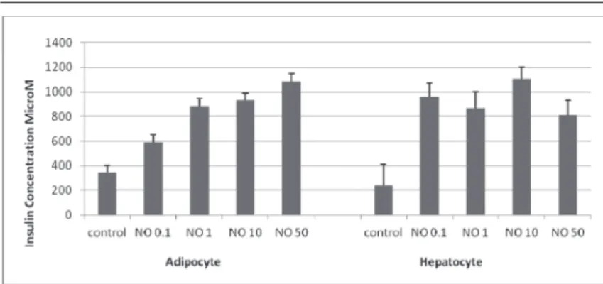
Fig 6. Basal and N.O.-stimulated FITC-Insulin binding activity in cultured adipocytes and hepatocytes. N.O. treatment increased insulin binding in adipocytes and hepatocytes as dose dependent manner (P<0.001). Data are presented as mean±SEM
Şekil 6. Hepatositlerde ve adipositlerde bazal ve N.O. ile uyarılmış insulin bağlanma kapasitesi. N.O. tedavisi adipositlerde ve hepato-sitlerde insulin bağlanmasını doz bağımlı olarak arttırmıştır (P<0.001). Sonuçlar ortalama±SEM olarak verilmiştir (n=6)
A mini review. Br J Nutr, 102 (12): 1703-1708, 2009.
7. Srivastava Y, Nigam SK, Bhatt HV, Verma Y, Prem AS: Hypoglycemic
and life-prolonging properties of Gymnema sylvestre leaf extract in diabetic rats. Isr J Med Sci, 21 (6): 540-542, 1985.
8. Banz WJ, Iqbal MJ, Bollaert M, Chickris N, James B, Higginbotham DA, Peterson R, Murphy L: Ginseng modifies the diabetic phenotype
and genes associated with diabetes in the male ZDF rat. Phytomedicinei, 14 (10): 681-689, 2007.
9. Kwon DY, Daily JW 3rd, Kim HJ, Park S: Antidiabetic effects of fermented
soybean products on type 2 diabetes. Nutr Res, 30 (1): 1-13, 2010.
10. Jitomir J, Willoughby DS: Cassia cinnamon for the attenuation of
glucose intolerance and insulin resistance resulting from sleep loss. J Med
Food, 12 (3): 467-472, 2009.
11. Ishikawa A, Yamashita H, Hiemori M, Inagaki E, Kimoto M, Okamoto M, Tsuji H, Memon AN, Mohammadio A, Natori Y: Characterization
of inhibitors of postprandial hyperglycemia from the leaves of Nerium indicum. J Nutr Sci Vitaminol (Tokyo), 53 (2): 166-173, 2007.
12. Bellakhdar J, Claisse R, Fleurentin J, Younos C: Repertory of standard
herbal drugs in the Moroccan pharmacopoeia. J Ethnopharmacol, 35, 123-143, 1991.
13. Bnouham M, Mekhfi H, Legssyer A, Ziyyat A: Ethnopharmacology
Forum Medicinal plants used in the treatment of diabetes in Morocco. Int
J Diabetes & Metabolism, 10, 33-50, 2002.
14. Tahraoui A, El-Hilaly J, Israili ZH, Lyoussi B: Ethnopharmacological
survey of plants used in the traditional treatment of hypertension and diabetes in south-eastern Morocco (Errachidia province). J Ethnopharmacol, 110 (1): 105-117, 2007.
15. Marshall S, Garvey WT, Traxinger RR: New insights into the metabolic
regulation of insulin action and insulin resistance: Role of glucose and amino acids. FASEB J, 5 (15): 3031-3036, 1991.
16. Khan AH, Pessin JE: Insulin regulation of glucose uptake: a complex
interplay of intracellular signalling pathways. Diabetologia, 45 (11): 1475-1483, 2002.
17. Leira F, Louzao MC, Vieites JM, Botana LM, Vieytes MR: Fluorescent
microplate cell assay to measure uptake and metabolism of glucose in normal human lung fibroblasts. Toxicol In Vitro, 16 (3): 267-273, 2002.
18. Gok E Olgaz S: Binding of Fluorescein Isothiocyanate to Insulin: A
fluorimetric labeling study. J Fluorescence, 14 (2): 203-206, 2004.
19. Alberti KG, Eckel RH, Grundy SM, Zimmet PZ, Cleeman JI, Donato KA, Fruchart JC, James WP, Loria CM, Smith SC Jr: International Diabetes
Federation Task Force on Epidemiology and Prevention; Hational Heart, Lung, and Blood Institute; American Heart Association; World Heart Federation; International Atherosclerosis Society; International Association for the Study of Obesity. Harmonizing the metabolic syndrome: a joint interim statement of the International Diabetes Federation Task Force on Epidemiology and Prevention; National Heart, Lung, and Blood Institute; American Heart Association; World Heart Federation; International Atherosclerosis Society; and International Association for the Study of Obesity. Circulation. 16, 1640-1645, 2010.
20. Thorens B: Glucose sensing and the pathogenesis of obesity and
type 2 diabetes. Int J Obes (Lond). 6 (Suppl): S62-S71, 2008.
21. Gould GW, Holman GD: The glucose transporter family: Structure,
function and tissue-specific expression. Biochem J, 95 (Pt 2): 329-341, 1993.
22. De Ferranti S, Mozaffarian D: The perfect storm: Obesity, adipocyte
dysfunction, and metabolic consequences. Clin Chem,54 (6): 945-955, 2008.
23. Antuna-Puente B, Feve B, Fellahi S, Bastard JP: Adipokines: the missing
link between insulin resistance and obesity. Diabetes Metab, 34 (1): 2-11, 2008.
24. Samuel VT, Petersen KF, Shulman GI: Lipid-induced insulin resistance:
Unravelling the mechanism. Lancet, 375 (9733): 2267-2277, 2010.
25. Yassin MM, Mwafy SN: Protective potential of glimepiride and Nerium oleander extract on lipid profile, body growth rate, and renal function in
streptozotocin-induced diabetic rats. Turk J Biol, 31, 95-102, 2007.
26. Haeba MH, Mohamed AI, Mehdi AW Nair GA: Toxicity of Nerium oleander leaf extract in mice. J Environ Biol, 23, 231-237, 2002.
27. Gayathri V, Ananthi S, Chandronitha C, Sangeetha MK, Vasanthi HR: Hypolipidemic potential of flowers of Nerium oleander in high fat