CORRESPONDENCE
Asbestos-related Diseases in Turkey: Caused Not Only by Naturally Occurring Fibers but Also by
Industrial Exposures To the Editor:
Many publications have been devoted to the epidemiology of asbestos exposure in Turkey. However, they have all focused on environmental exposure to“naturally occurring asbestos” (a natural component of soils or rocks), ignoring potential industrial asbestos exposure (1–4). Yet many industrial activities involved occupational asbestos exposure during Turkey’s industrialization, including construction, shipbuilding, and automotive manufacturing. Between 1900 and 2003, 1.2 million tons of asbestos were used in Turkish industry (5). The use of asbestos was officially banned in 2010.
It is likely that many patients have been misclassified as having had environmental asbestos exposure instead of occupational exposure. This can explain why there have been, as far as we know, no recognized compensation claims with mesothelioma in Turkey. Increasing awareness of occupational causes for asbestos-related diseases will be a helpful starting point for medicolegal processes. Methods
We screened radiology reports of patients in a university hospital in Istanbul between 2006 and 2017 for the terms“asbestosis,” “plaque,” “pleural calcification,” and “mesothelioma” to detect patients with asbestos-related diseases. We then matched these cases with control subjects of the same sex and age (63 yr) randomly taken from a data pool of patients with reports of chest computed tomography without evidence of any pulmonary diseases during the same period. Two thoracic radiologists reevaluated all computed tomography images for evidence of asbestos-related diseases, as well as other pulmonary disorders. When disagreement occurred, a third radiologist made thefinal decision. To avoid misclassification, asbestosis was only considered if compatible parenchymal changes were accompanied by pleural plaques/calcifications. Similarly, only patients with bronchial carcinoma accompanied by pleural plaques/calcifications were included.
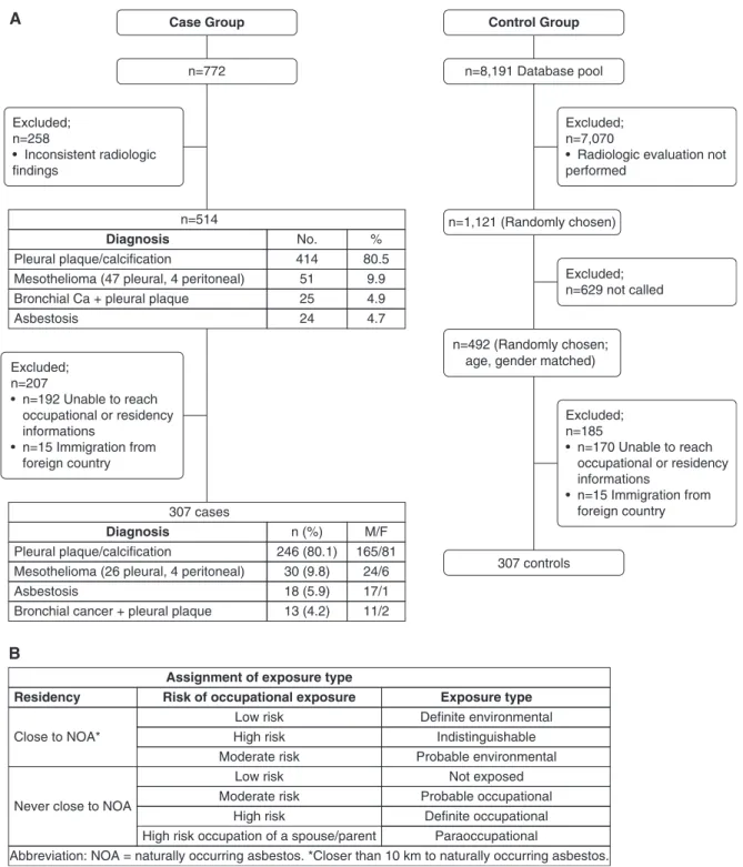
Cases and control subjects (or their relatives) were invited by telephone to participate in the study, and those having signed a consent form responded by telephone to a standardized questionnaire about their lifetime residential and occupational history, previous respiratory diseases, and the occupations of their spouses and parents. Interviewers were blinded to the case or control status of interviewees. Nonresponding control subjects were replaced by a randomly chosen eligible control subject (flow chart in
Figure 1A). This study was approved by the medical ethics board of Bezmialem Vakif University (#2017-12/179).
We classified occupational exposure to asbestos based on asbestosis proportionate mortality ratios (PMRs) published by National Institute for Occupational Safety and Health (6). We considered occupations with a PMR.3 as high, a PMR 1–3 as moderate, and a PMR,1 and all occupations with no recognizable sources of asbestos as lowest risk for asbestos exposure. On the basis of previous studies, we labeled patients born or having lived at least a year within 10 km of naturally occurring asbestos as having had environmental asbestos exposure (7, 8). We established categories of asbestos exposure based on a combination of probability of occupational asbestos exposure and proximity to environmental asbestos (Figure 1B). Results
Three hundred seven people were included in each of the case and control groups (Figure 1A). Among cases, 55% were considered as likely to have been exposed to naturally occurring asbestos compared with 9.8% among control subjects (P , 0.001). Definite occupational exposure was found in 15% of cases compared with 2% of control subjects; conversely, no known exposure was found in 11% of cases versus 66% of control subjects, and overlapping environmental and occupational asbestos exposure was found in 4% of cases against 0% for control subjects. No female cases or control subjects worked in occupations with a high risk of exposure.
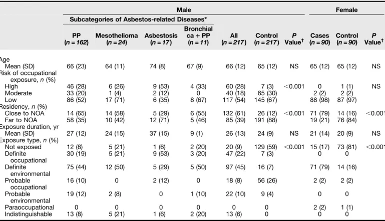
Table 1 presents the characteristics in both sexes. Briefly, in males, definite occupational asbestos exposure was more prevalent among cases (22%) than control subjects (3%). In the case group, 13 (6%) male patients had a high risk for both occupational exposure and environmental exposure.
Among males, the proportion of patients with occupations at high risk for asbestos exposure was significantly higher than in the control group. Similarly, for both sexes, the prevalence of patients with asbestos-related diseases who lived close to naturally occurring asbestos was significantly higher than in the control group. Within the case group, patients with high-, moderate-, or low-risk occupations had similar ages. In a multivariable logistic analysis including age, sex, and categories of environmental and occupational asbestos exposure, we found asbestos-related diseases independently associated (odds ratios with 95% confidence interval) with age (1.03 [1.01–1.05] for each year; P = 0.008), residency close to naturally occurring asbestos (24.4 [15.3–39.1]; P , 0.001) and high likelihood of occupational exposure (35.2 [14.5–85.2]; P , 0.001); male sex (1.24 [0.76–2.04]) and moderate likelihood of occupational exposure (1.20 [0.69–2.20]) were not significant.
The shipbuilding industry was the most common occupation with high risk of asbestos exposure (17%), whereas brake and clutch manufacturing was the second most common (16%).
Discussion
The abundance of naturally occurring asbestos in rural Turkey has created a feeling that all asbestos-related disease in the country is caused by this type of environmental exposure. The novelty of our study is that at least onefifth of the male patients with Author Contributions: M. Bayram provided conception and design,
acquisition of data, analysis and interpretation of data, and drafting the article and revising it critically for important intellectual content; D. ¨O. provided acquisition of data, analysis, and interpretation of data; E.H. and A.K. provided acquisition of data; E.M. provided acquisition of data and interpretation of radiological data; M. Bilgin provided interpretation of radiological data; F.O. provided acquisition of data and critical revision of the article; S‚.B. provided interpretation of radiological data; and M.E.A. provided acquisition of data and critical revision of the article.
Originally Published in Press as DOI: 10.1164/rccm.201810-1922LE on November 29, 2018
asbestos-related diseases diagnosed in Istanbul had been exposed to asbestos of industrial origin, and not of natural origin.
The main difficulty in this study was assigning the type of exposure. Although the proportion of patients at moderate risk for occupational exposure is important, we chose to focus on patients with definite occupational asbestos exposure, as this was the first study on occupational asbestos exposure in Turkey. The proportion
of occupational exposure in male patients with asbestos-related diseases may be up to 45% when including all the patients who worked in occupations with high or moderate risk for asbestos exposure.
Our data do not represent all individuals in Turkey. Although our hospital is in the largest city, internal migration to Istanbul is mainly from the eastern, northern, and central parts of Turkey. A Case Group n=772 Excluded; n=258 • Inconsistent radiologic findings Excluded; n=207 • n=192 Unable to reach occupational or residency informations • n=15 Immigration from foreign country 307 cases Diagnosis Pleural plaque/calcification n (%) 246 (80.1) 30 (9.8) 18 (5.9) 13 (4.2) Mesothelioma (26 pleural, 4 peritoneal)
Bronchial cancer + pleural plaque Asbestosis M/F 165/81 24/6 17/1 11/2 Asbestosis n=514 Diagnosis Pleural plaque/calcification No. 414 51 25 24 Mesothelioma (47 pleural, 4 peritoneal)
Bronchial Ca + pleural plaque
% 80.5 9.9 4.9 4.7 Control Group n=8,191 Database pool n=1,121 (Randomly chosen) 307 controls n=492 (Randomly chosen;
age, gender matched)
Excluded; n=7,070
• Radiologic evaluation not performed Excluded; n=629 not called Excluded; n=185 • n=170 Unable to reach occupational or residency informations • n=15 Immigration from foreign country B Residency Close to NOA*
Assignment of exposure type
Risk of occupational exposure Exposure type
Low risk Definite environmental Probable environmental Not exposed Probable occupational Definite occupational Paraoccupational Indistinguishable High risk Moderate risk Low risk High risk
High risk occupation of a spouse/parent
Abbreviation: NOA = naturally occurring asbestos. *Closer than 10 km to naturally occurring asbestos. Moderate risk
Never close to NOA
Figure 1. (A) Flow chart of the study. (B) Assignment of exposure type based on distance of residency from naturally occurring asbestos and occupational asbestos exposure risk. Ca = carcinoma.
Correspondence 657
A recently published study of the population at risk for environmental exposure in Turkey found that 1,789 (32%) of 5,617 of patients with mesothelioma were born in urban areas, and we suspect that these patients might have been exposed to occupational asbestos (4).
Ourfindings demonstrate that not only environmental but also occupational asbestos exposure is a relevant health problem in Turkey. Low awareness of occupational asbestos exposure risk, especially among physicians, discourages seeking compensation for occupational exposure. Occupational exposure should not be underestimated in countries that have high rates of asbestos-related diseases because of naturally occurring asbestos. Surveillance and prevention programs should examine occupational exposure as well as environmental asbestos exposure.n
Author disclosures are available with the text of this letter at www.atsjournals.org.
Acknowledgment: The authors thank the American Thoracic Society’s Methods in Epidemiologic, Clinical, and Operations Research Program, and especially Diana Buist for her help and guidance. They also thank Benoit Nemery for his valuable suggestions.
Mehmet Bayram, M.D.* Didem ¨Ozkan, M.D. Esat Hayat, M.D. Mehmet Bilgin, M.D. Elnur Mehdi, M.D. Bezmialem Vakif University Istanbul, Turkey
S¸ennur Bilgin, M.D. Medipol University Istanbul, Turkey
Muhammed Emin Akkoyunlu, M.D. Fatmanur Okyaltırık, M.D. Abdullah Kansu, M.D. Bezmialem Vakif University Istanbul, Turkey
ORCID IDs: 0000-0002-4933-4445 (M. Bayram); 0000-0003-2900-7794 (E.H.); 0000-0002-9465-4499 (F.O.); 0000-0001-8902-5498 (A.K.).
*Corresponding author (e-mail: [email protected]).
References
1. Bayram M, Bakan ND. Environmental exposure to asbestos: from geology to mesothelioma. Curr Opin Pulm Med 2014;20: 301–307.
Table 1. Comparison of Demographic Parameters in Cases and Control Subjects in Each Sex
Male Female
Subcategories of Asbestos-related Diseases* PP (n = 162) Mesothelioma (n = 24) Asbestosis (n = 17) Bronchial ca1 PP (n = 11) All (n = 217) Control (n = 217) ValueP † Cases (n = 90) Control (n = 90) ValueP † Age Mean (SD) 66 (23) 64 (11) 74 (8) 67 (9) 66 (12) 65 (12) NS 65 (12) 65 (12) NS Risk of occupational exposure, n (%) High 46 (28) 6 (26) 9 (53) 4 (33) 60 (28) 7 (3) ,0.001 0 1 (1) NS Moderate 33 (20) 1 (4) 2 (12) 0 40 (18) 65 (30) 2 (2) 2 (2) Low 86 (52) 17 (71) 6 (35) 8 (67) 117 (54) 145 (67) 88 (98) 87 (97) Residency, n (%) Close to NOA 14 (65) 14 (58) 5 (29) 6 (55) 132 (61) 26 (12) ,0.001 71 (79) 14 (16) ,0.001 Far to NOA 58 (35) 10 (42) 12 (71) 5 (46) 85 (39) 191 (88) 19 (21) 76 (84) Exposure duration, yr Mean (SD) 27 (12) 24 (15) 37 (15) 9 (1) 26 (13) 24 (9) NS 21 (14) 20 (9) NS Exposure type, n (%) Not exposed 12 (8) 5 (21) 1 (6) 2 (20) 20 (9) 129 (59) ,0.001 15 (17) 73 (81) ,0.001 Definite occupational 30 (19) 5 (21) 9 (53) 3 (20) 47 (22) 7 (3) 0 0 Definite environmental 75 (44) 12 (50) 5 (29) 5 (50) 97 (45) 16 (7) 71 (79) 14 (16) Probable occupational 16 (10) 0 2 (12) 0 18 (8) 56 (26) 2 (2) 2 (2) Probable environmental 19 (12) 2 (8) 0 1 (10) 22 (10) 9 (4) 0 0 Paraoccupational 0 0 0 0 0 0 2 (2) 1 (1) Indistinguishable 13 (8) 5 (21) 1 (6) 2 (20) 13 (6) 0 0 0
Definition of abbreviations: ca = carcinoma; NOA = naturally occurring asbestos; NS = not significant; PP = pleural plaques.
*See Figure 1A for type of asbestos-related diseases among women (not shown in table because no females had a high-risk occupation).
†P values were determined for all case and control groups.
CORRESPONDENCE
2. Dumortier P, Copl ¨u L, de Maertelaer V, Emri S, Baris I, De Vuyst P. Assessment of environmental asbestos exposure in Turkey by bronchoalveolar lavage. Am J Respir Crit Care Med 1998;158: 1815–1824.
3. C ¨opl ¨u L, Dumortier P, Demir AU, Selçuk ZT, Kalyoncu F, Kisacik G, et al. An epidemiological study in an Anatolian village in Turkey environmentally exposed to tremolite asbestos. J Environ Pathol Toxicol Oncol 1996;15:177–182.
4. Metintas¸ S, Batırel HF, Bayram H, Yılmaz ¨U, Karadağ M, Ak G, et al. Turkey national mesothelioma surveillance and environmental asbestos exposure control program. Int J Environ Res Public Health 2017;14:E1293.
5. Virta RL. Worldwide asbestos supply and consumption trends from 1900 through 2003. Reston, VA: US Geological Survey; 2006. pp. 1445–1463. 6. National Institute for Occupational Safety and Health. Work-related lung disease surveillance report 1999. Cincinnati, OH: National Institute for Occupational Safety and Health; 1999. DHHS [NIOSH] Publication No. 2000-105.
7. Bayram M, Dongel I, Bakan ND, Yalççn H, Cevit R, Dumortier P, et al. High risk of malignant mesothelioma and pleural plaques in subjects born close to ophiolites. Chest 2013;143:164– 171.
8. Baumann F, Maurizot P, Mangeas M, Ambrosi JP, Douwes J, Robineau B. Pleural mesothelioma in New Caledonia: associations with environmental risk factors. Environ Health Perspect 2011;119: 695–700.
Copyright © 2019 by the American Thoracic Society
Lectin Complement Pathway in Emphysema To the Editor:
The most common genetic cause of emphysema and chronic obstructive pulmonary disease (COPD) is AAT (alpha-1 antitrypsin) deficiency (AATD) (1). The risk of lung disease in AATD is associated with decreased levels of circulating AAT, a proteinase inhibitor with high affinity for neutrophil-derived serine proteases such as elastase (2, 3). Identification of additional proteases targeted by AAT may provide further insight into the pathogenesis of emphysema. Our work suggests that AAT targets a protease involved in complement activation, MASP-2 (MBL [mannose-binding lectin]-associated serine protease 2). Some of the results of these studies have been previously reported in the form of an abstract (4).
In contrast to other MASP-1 or -3 isoforms, MASP-2 alone is sufficient to initiate the lectin pathway within the complement system, a network of zymogen enzymes (C1–C9) activated by auto- or enzymatic cleavage (Figure 1A) (5). Whereas its main function is to form the membrane attack complex (C5–C9) that is critical for pathogen killing, the complement has been recently implicated in sterile inflammation and autoimmunity (5). Although the role of the lectin complement pathway in COPD pathogenesis is understudied, emerging reports point to complement activation (C4 and C3) in sera of patients with COPD and AATD (6, 7), and in lungs of mice in models of cigarette smoke (CS)-induced emphysema (8).
In this study, we investigated whether MASP-2 and the lectin pathway are activated in COPD and AATD plasma and lung tissue. Samples were obtained from subjects with COPD (n = 38, age = 646 10 yr, 21 men, FEV1% predicted = 456 27), AATD (n = 47)
off AAT augmentation therapy (AATD-off, n = 30, age = 67 6 9 yr, 18 men, FEV1% predicted = 736 32) or on augmentation therapy
(AATD-on, n = 17, age = 58 6 14 yr, 8 men, FEV1% predicted =
336 11), and healthy never-smokers (n = 23, age = 40 6 8.5 yr, 5 men). Subjects with active infections in the past 3 months were excluded. The subjects’ smoking status was recorded as active smoker, ex-smoker, never smoker, or unknown (the latter group was excluded from multivariate analyses). Statistical analyses were performed in R and Prism (GraphPad Software). Deidentified plasma samples were obtained from Leiden University (n = 8), Hannover University (n = 15), the Medical University of South Carolina (n = 24), and National Jewish Health (n = 62), and lung samples were obtained from the Lung Tissue Research Consortium and Hannover University. The institutional review board at National Jewish Health approved the study as exempt human subjects research.
Plasma MASP-2 levels in subjects with COPD (1,1356 670 ng/ml), AATD-off (1,1766 541 ng/ml), or AATD-on (1,085 6 428 ng/ml) were significantly increased compared with those in healthy volunteers (521.56 179 ng/ml) (Figure 1B). MASP-2 levels were not significantly different between males and females (1,326 6 1,153 ng/ml vs. 8876 568 ng/ml; P = 0.051, t test) or among never-smokers, ex-never-smokers, and active smokers with COPD (9836 590 ng/ml vs. 1,1546 653 ng/ml vs. 1,205 6 33 ng/ml, respectively; P = 0.53, Kruskal-Wallis test). The downstream product of MASP-2 proteolytic activity, the cleaved C4a fragment (expressed relative to total C4) was also elevated in COPD (2.336 1.1) and AATD-off (3.256 2.6), but not AATD-on (1.44 6 0.6), plasma compared with that from healthy individuals (0.966 0.7) (Figure 1C). Plasma MASP-2 tended to be positively associated with C4a levels (r = 0.24, P = 0.06). MASP-2 immunostaining in the airways and lung parenchyma was markedly increased in subjects with COPD of any severity, as defined by Global Initiative for Chronic Obstructive Lung Disease criteria (Figures 2A–2C). Interestingly, lower-molecular-weight MASP-2 isoforms (z65 and z50 kD), indicative of MASP-2 activation, were primarily present in COPD and AATD lungs, rather than in control lungs (Figure 2D).
Our data suggest that MASP-2 levels and activity are increased in subjects with COPD and AATD. The marked effect of AAT augmentation therapy on the abundance of C4a fragments generated, rather than on the MASP-2 levels, suggests that AAT inhibits MASP-2’s activity rather than its expression.
Our study has several limitations, including the small size of our groups and differences in demographics (with a predominance of young age and females in the control group), smoking status (fewer ex-smokers in the AATD-off group), and disease severity (milder COPD in the AATD-off group). Our statistical analyses did not include clinical covariates (e.g., treatment strategy and exacerbation frequency) and were not powered to investigate a relationship between MASP-2 and clinical markers of disease severity. For example, in multivariate linear regression models controlled for age, sex, AAT augmentation, and smoking status, MASP-2 was inversely associated with FEV1% predicted (b = 20.0056) and DLCO% predicted
(b = 20.0049), but it made a statistically insignificant contribution to the models (P = 0.06 and 0.16, respectively). Also, we did not evaluate Supported by a 2016 Laurell’s Award (European Respiratory Society and
Grifols, to K.A.S.) and European Research Council consolidator grant XHaLe (D.J.). This study used specimens and data provided by the Lung Tissue Research Consortium (NHLBI).
Originally Published in Press as DOI: 10.1164/rccm.201807-1380LE on December 17, 2018