IOS Press
Polymorphisms at the ligand binding site of
the vitamin D receptor gene and osteomalacia
1Duygu Gezen Ak
a, Hakkı Kahraman
b, Erdinc¸ Dursun
a, Belgin S ¨usleyici Duman
c, Nevin Erensoy
d,
Faruk Alag ¨ol
e, Refik Tanakol
eand Selma Yılmazer
a,∗aDepartment of Medical Biology, Cerrahpasa Faculty of Medicine, Istanbul University, Turkey
bDepartment of Internal Medicine, Faculty of Medicine, University of 19 Mayıs, Turkey
cDepartment of Medical Biology, Faculty of Medicine, University of Kadir Has, Turkey
dInstitute for Experimental Medicine, Istanbul University, Turkey
eDepartment of Endocrinology and Metabolism, Faculty of Medicine, Istanbul University, Turkey
Abstract. Vitamin D receptor (VDR) gene polymorphisms have been suggested as possible determinants of bone mineral density
(BMD) and calcium metabolism. In this study, our aim was to determine whether there is an association between VDR gene polymorphism and osteomalacia or not. We determined ApaI and TaqI polymorphisms in the vitamin D receptor gene in 24 patients with osteomalacia and 25 age-matched healthy controls. Serum calcium, phosphorus, ALP, PTH, 25OHD levels were also examined. We used PCR and RFLP methods to test for an association between osteomalacia and polymorphisms within, intron 8 and exon 9 of the VDR gene. When the control and patients were compared for their ApaI and TaqI genotypes there was no relationship between VDR gene allelic polymorphisms and osteomalacia. Whereas a nearly significant difference for A allele was found in the allellic distribution of the patients (p = 0.08). Also no association between biochemical data and VDR gene polymorphisms was observed.
Keywords: Vitamin D, osteomalacia, vitamin D receptor gene, ApaI, TaqI
1. Introduction
1.25-dihydroxyvitamin D3 (1.25 (OH)2 D3) is in-volved in biological actions such as calcium home-ostasis, bone mineralization, cell proliferation and cell differentiation [16,17,23]. Vitamin D deficiency usu-ally occurs in the lack of dietary intake, abnormal metabolism of vitamin D or inadequate synthesis of vitamin D in the skin. In addition, it may result from intestinal malabsorption, chronic renal disease or very rarely, from liver failure. Such kind of insufficiency of
1This study is supported by The Research Fund of Istanbul
University.
∗Corresponding author: Prof. Dr. Selma Yılmazer, Department of Medical Biology, Cerrahpasa Faculty of Medicine, Istanbul Uni-versity, Istanbul, Turkey. Tel.: +90 212 414 30 00/22032; +90 532 274 07 15; Fax: +90 212 414 30 42; E-mail: selmayilmazer@ mynet.com.
vitamin D causes mineralization defect of bone matrix rather than frank osteoporosis [21,26,28,31]. Without vitamin D, children develop rickets and adults exacer-bate their osteoporosis and develop osteomalacia [18]. The genomic actions of vitamin D3 are mediated through its nuclear receptor, the vitamin D receptor (VDR), which is a member of the nuclear hormone re-ceptor superfamily [16]. Genetic factors are consid-ered to be major determinants of bone mineral mass. Common polymorphisms in the 3’ – and 5’ – end re-gion of the VDR gene have been suggested as possible determinants of bone mineral mass and, hence, of the risk of osteoporosis. These polymorphisms are identi-fied by the restriction enzymes BsmI (BB, Bb, bb) or alternatively TaqI (TT, Tt, tt) and ApaI (AA, Aa, aa) [5, 26].
At least 22 unique loss of function mutations in the VDR gene have been reported. Single nucleotide changes producing amino acid substitutions in the
DNA-and ligand-binding domains are predominate mu-tations. These mutations cause hereditary vitamin D re-sistant rickets, a rare autosomal recessive disease result-ing from target organ resistance to 1.25(OH)2D3[9].
Osteomalacia is frequently seen in Turkey [2,4,24, 27]. The effects of genetic factors in pathogenesis of osteomalacia are not clear. Some individuals are more prone to have features of osteomalacia although they are living in the same habitat and having the same dietary. The only study about this subject could not be able to show any association between VDR gene BsmI polymorphism and osteomalacia [4]. We think that further investigation on the other polymorphisms of the VDR gene is required to determine the effect of these polymorphisms on osteomalacia. In the present study, the effect of VDR gene ApaI and TaqI polymorphisms on osteomalacia was investigated.
2. Subjects and methods
2.1. Subjects
Twenty-four patients (21 female, 3 male; age: 45.16
± 13.99 years.) who met the clinical (muscle
weak-ness of the lower extremities, walking difficulties, bone pain worsening by activity), biochemical (low or low-normal serum calcium and phosphorus, low serum 25 hydroxyvitamin D [OHD], increased alkaline phos-phatase and PTH levels), and/or radiological (pseud-ofractures) criteria of osteomalacia were included in this study. Patients are clinically diagnosed at Istanbul University, Faculty of Medicine, Department of En-docrinology and Metabolism, Bone Diseases Unit. Pa-tients with osteomalacia due to causes other than vita-min D depletion such as hypophosphatemia and renal osteodistrophy were excluded from the study. Twenty-five age matched healthy controls (22 female, 3 male; age: 41.08± 13.35 years.) had no history of diseases affecting bone metobolism. None of the subjects in the two groups were taking medicine affecting bone metabolism. Patients and healthy controls were not classified according to their calcium intake, which was determined to be above 800 mg per day, assessed by asking about 3-day calcium intake [6]. The study was approved by the Ethics Commitee of Research Fund of Istanbul University. Informed consents were obtained from all subjects.
2.2. Biochemical analysis
Fasting blood samples were collected in the same season (fall) for the measurement of calcium (Ca), phosphorus (P), alkaline phosphatase (ALP), parathy-roid hormone (PTH) and 25-hydroxyvitamin D (25OHD). Serum Ca, P and ALP were measured by Roche Diagnostics (Mannheim, Germany) Modular System. Serum intact PTH was measured with DSL-8000 Active Intact PTH IRMA kit from Diagnostic Systems Laboratories, Inc (Webster, Texas, USA), with intra-and interassay coefficients of variations of 2.8 and 3.6%, respectively. Serum 25OHD was measured by Diasorin 25OHD RIA kit (Stillwater, Minnesota, USA) with intra-and interassay coefficients of variations of 10.4 and 9.4%, respectively.
2.3. Genotype assignment
DNA was extracted from 10 ml of K3EDTA (Ethylenediaminetetraacetic acid) treated peripheral blood by the salting out method. A 740 base pair frag-ment which includes intron 8 and exon 9 of the vitamin D receptor gene in chromosome 12 was amplified by the polymerase chain reaction (PCR) with forward (5’-CAGAGCATGGACAGGGAGCAAG-3’) and reverse (5’-GCAACTCCTCATGGCTGAGGTCTCA-3’) [12] primers to detect Apa I and Taq I sites. PCR prod-ucts were generated in a 25µl reaction volume contain-ing 100 ng of genomic DNA, 1x PCR buffer, 1.8 mM MgCl2, 200µM of dNTP, 10 pmol/µl of each primer and 0.5U of Taq DNA polymerase. PCR was performed as follows: incubation for 5 min. at 94◦C, 10 cycles of incubation for 20 s at 94◦C, 40 s at 64◦C, and 1 min. at 72◦C, 25 cycles of incubation for 20 s at 94◦C, 40 s at 62◦C, and 1 min. at 72◦C, followed by an extension step of 6 min. at 72◦C. To determine the presence of Apa I and Taq I restriction sites, we performed Restric-tion Fragment Length Polymorphism (RFLP). Fiveµl PCR products were digested with 2µl of DNase, RNase free water and 2U of Apa I enzyme at 37◦C and 2U of Taq I enzyme at 66◦C, separately. Digestion prod-ucts were analyzed in 1.5% agarose gel stained with ethidium bromide (Applichem, Darmstadt, Germany). DNA fragments were visualized by ultraviolet illumi-nation and fragment size estimated by comparison to 50 bp ladder run on the same gel. The presence of ApaI restriction site causes spliting of the PCR product into two bands, 529 bp and 211 bp, respectively, des-ignated asa. If ApaI restriction site is not present in the corresponding sequence remained a 740 bp single
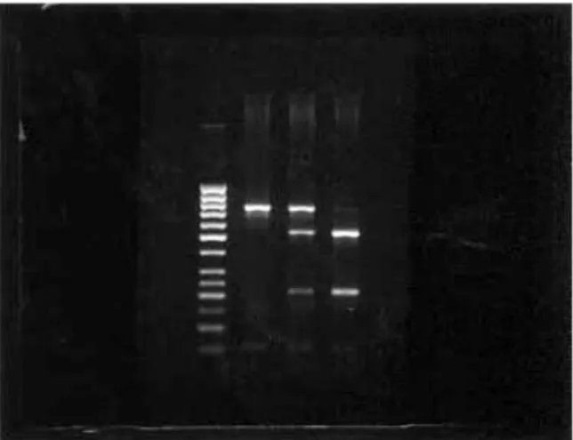
Fig. 1. RFLP results of ApaI enzyme. Lane 1, Gene RulerTM 50 bp DNA Ladder. Lane 2, AA genotype (740 bp, homozygous). Lane 3, Aa genotype (740 bp, 529 bp and 211 bp, heterozygous). Lane 4, aa genotype (529 bp and 211 bp, homozygous) (bp=base pairs).
Fig. 2. RFLP results of TaqI enzyme. Lane 1, Gene RulerTM 50 bp DNA Ladder. Lane 2, TT genotype (493 bp and 247 bp, homozygous). Lane 3, Tt genotype (493 bp, 291 bp, 247 bp and 202 bp, heterozygous). Lane 4, tt genotype (291 bp, 247 bp and 202 bp, homozygous).
band, designated as A (Fig. 1). The presence of TaqI restriction site causes spliting of the PCR product into three bands, 291 bp, 247 bp and 202 bp respectively, designated ast. If RFLP-associated TaqI restriction site is not found in the corresponding sequence were split into two bands, 493 bp and 247 bp respectively, designated asT (Fig. 2).
2.4. Statistics
Statistical analysis were performed by UNISTAT 5.0 software. To compare ALP and PTH levels the
nonparametric Mann Whitney U-Wilcoxon Rank Sum W test was used. Also to determine the distribution of Ca, P, 25OHD levels and age in groups, t- Test was used. The distribution of biochemical parameters and VDR genotypes by groups were determined by analysis of variance (two-way ANOVA).
3. Results
3.1. Biochemical parameters
Patients and healthy controls were age matched (45.16 ± 13.99 yrs.; 41.08 ± 13.35 years, respec-tivelyp = 0.306). Serum calcium (8.57 ± 0.99 mg/dl vs. 9.29 ± 0.53 mg/dl p = 0.004; reference range: 8.5–10.5 mg/dL), phosphorus (3.05± 0.71 mg/dl vs.
3.69± 0.45 mg/dl p = 0.001; reference range: 2.7–
4.5 mg/dL) and 25OHD (6.90± 5.04 ng/ml vs. 16.30 ± 12.15p = 0.0005; reference range: 15–56 ng/ml) lev-els were lower and serum ALP (329.15± 311.97 IU/L vs. 65.33± 15.53 IU/L p = 0.0004; reference range: 90–260 U/L) and PTH (225.41 ± 170.31 pg/ml vs. 40.54± 22.36 pg/dl p =< 0.001; reference range: 15– 65 pg/ml) were higher in the patient group than those in the control group (Table 1). These findings were statistically significant.
3.2. VDR alleles
After genetic analysis of the VDR gene, we found 50% genotype AA, 45.8% genotype Aa, 4.2% genotype aa and 33.3% genotype TT, 62.5% genotype Tt, 4.2% genotype tt for patients and 28% genotype AA, 56% genotype Aa, 16% genotype aa and 36% genotype TT, 52% genotype Tt, 12% genotype tt for healthy controls. When the control and patients were compared for their Apa I and Taq I genotypes, we observed that the geno-type distribution did not differ (p = 0.18; p = 0.55 respectively) whereas when the allellic distributions of the patients and controls compared for the ApaI poly-morphism a nearly significant difference was found for the A allele as it was slightly increased in the patients
(p = 0.08) (Table 2). Additionally no association
be-tween biochemical data and VDR gene polymorphisms was observed (Tables 3 and 4).
Table 1
Comparison of biochemical findings with vitamin D receptor (VDR) genotypes in osteoma-lacia and control groups
Parameters Patients Controls 2 way ANOVA
a b c aAge (years) 45.16± 13.99 41.08± 13.35 NS − − aCa (mg/dl) 8.57± 0.99 9.29± 0.53 0.004* NS NS aP (mg/dl) 3.05± 0.71 3.69± 0.45 0.001* NS NS bALP (U/l) 329.15± 311.97 65.33± 15.53 0.0004* NS NS a25OHD (ng/ml) 6.90± 5.04 16.30± 12.15 0.0005* NS NS bPTH (pg/ml) 225.41± 170.31 40.54± 22.36 <0.001* NS NS aMann-Whitney U;bt-test; *p < 0.05; NS, not significant; 2 way ANOVA, two way
analysis of variance; ALP, alkaline phosphatase, 25OHD, 25 hydroxyvitamin D; PTH, parathyroid hormone a, comparison of the two groups; b, Significance ofP value of ApaI genotype effect; c, Significance ofP value of TaqI genotype effect.
4. Discussion
It is suggested that VDR gene and the allelic varia-tions of the 3’ end region of this gene have an impor-tant role to determine the relation between vitamin D metabolism and the effects of genetic factors in bone formation [5,20,26].
In 1994 Morrison et al. suggested, in their study over healthy Caucasian twins, that there is a close associa-tion between VDR gene polymorphisms and BMD by over 75% [30] although many studies in several pop-ulations have failed to detect a significant association between bone mass and VDR gene alleles [1,7,15,22, 29]. It was suggested that, there is a relationship be-tween BB, tt, AA polymorphisms of VDR gene and the low BMD. It was found that twins with genotype bbT-Taa have 15% higher BMD than the twins with BbttAA genotype [22].
There were controversial results reported on the ef-fects of VDR genotypes on BMD. The studies which aim to explain this contradiction show that the relation-ship between BMD and VDR gene polymorphisms oc-curs in case of low calcium intake [11,20]. When pre-menopausal women with dietary high and low calcium intake were compared, it was found that the amount of calcium taken effects the BMD in individuals with Bb and BB genotypes, but does not effect individuals with bb genotypes [26]. In addition to this, parallel findings were obtained with the pre-adolescence girls with bb genotypes. Accordingly, there was a significant corre-lation between dietary calcium levels and BMD gain in Bb and probably BB but not in bb. It is suggested that calcium absorbtion decreases in low calcium intake due to of a possible functional error in vitamin D receptors of individuals with BB genotype [11]. Apa I and Taq I polymorphisms are also located in the ligand binding domain of VDR gene as BsmI polymorphism.
Table 2
Allellic distributions of VDR gene ApaI polymorphism
Allele A Allele a
Controls 28(0.56) 22(0.44)
Patients 35(0.73) 13(0.27)
p = 0.08. First numbers are the number of patients and controls.
The numbers in paranthesis are frequency.
The allelic variation of these polymorphic sites might cause a change in the affinity of VDR to its ligand [14, 20]. It was mentioned that calcium absorption is higher in bbTT genotype which is reported to be related with high BMD than in BBtt genotype and lower in BbttAA genotype which is related to low BMD than in bbTTaa and BbTtAa genotypes [13]. Calcium absorbtion in pre and postmenopausal women with the BAt haplotypes was found to be 11% and 37% lower, respectively when compared to that of women with the baT haplotype indicating that the effect of VDR gene variation on calcium absorption may also be modified by age or hormonal status [8,13]. However, a recent study reports that the Bat haplotype is associated with high bone density in normal subjects [10].
It is known that the vitamin D resistance is a con-sequence of mutations in VDR gene [17]. Although it is not clear, whether genetic factors have a role in the pathogenesis of osteomalacia or not, Kahraman et al. suggested that some VDR genotypes are more prone to osteomalacia [4]. VDR polymorphisms might have an effect on the osteomalacia, caused by the lack of vitamin D, as they effect the Ca+2 absorbtion. It is suggested that the Ca+2absorbtion might be decreased
by some polymorphisms in VDR gene, in low Ca+2
intake [11]. Since the major role of vitamin D and its receptor is to regulate the amount of Ca+2binding
pro-teins and the expression of Ca+2channels in cells it is possible that VDR polymorphisms effect the Ca+2
ab-sorbtion in low vitamin D intake, due to the alterations in the affinity of VDR to its ligand vitamin D [3,25].
Table 3
ApaI genotypes and biochemical findings in osteomalacia patients and controls∗
Parameters VDR Genotypes/Patients VDR Genotypes/Controls 2 way
AA Aa AA Aa ANOVA P value Age (years) 41.83± 14.76 49.00± 13.45 38.57± 11.41 41.69± 11.81 0.948 Ca (mg/dl) 8.55± 0.66 8.56± 1.33 9.27± 0.64 9.34± 0.47 0.594 P (mg/dl) 3.15± 0.70 2.94± 0.77 3.74± 0.33 3.66± 0.55 0.508 ALP (U/l) 276.27± 234.11 436.63± 396.53 61.83± 7.88 61.84± 9.13 0.202 25OHD (ng/ml) 8.45± 6.59 5.44± 2.24 13.41± 10.28 16.36± 9.47 0.16 PTH (pg/ml) 252.63± 199.47 218.64± 138.39 37.46± 26.44 35.94± 16.36 0.913 ∗“aa” genotype (n < 5) could not be analyzed with two way ANOVA.
Table 4
TaqI genotypes and biochemical findings in osteomalacia patients and controls∗
Parameters VDR Genotypes/Patients VDR Genotypes/Controls 2 way
TT Tt TT Tt ANOVA P value Age (years) 47.13± 13.56 44.33± 15.04 39.67± 15.07 41.58± 13.58 0.978 Ca (mg/dl) 8.26± 1.19 8.79± 0.86 9.47± 0.47 9.12± 0.43 0.248 P (mg/dl) 2.69± 0.67 3.17± 0.65 3.66± 0.43 3.75± 0.50 0.875 ALP (U/l) 431.63± 401.73 260.83± 229.53 65.86± 8.69 64.59± 19.86 0.429 25OHD (ng/ml) 5.73± 2.05 7.78± 6.32 14.16± 15.02 18.10± 9.35 0.792 PTH (pg/ml) 211.22± 162.85 230.66± 188.29 42.27± 26.02 43.19± 21.13 0.519 ∗“tt” genotype (n < 5) could not be analyzed with two way ANOVA.
In our study, the frequency of Apa 1 polymorphism was 50% for AA; 45.8% for Aa; 4.2% for aa in pa-tients and 28% for AA; 56% for Aa; 16% for aa in the healthy controls. The patients with AA genotype were 50% of the total patient number and when compared to the control group it had a higher ratio, however, this difference is not considered as statistically significant whereas allellic distribution of A allele was found to be slightly increased in the patients and became sta-tistically nearly significant. The frequency of Taq 1 polymorphisms were 33% for TT; 62.5% for Tt; 4.2% for tt in the patients, whereas 36% for TT; 52% for Tt; 12% for tt in the healthy controls. There was no sta-tistically significant difference between the genotype distribution of patient and control groups. Kahraman et al. have studied the relationship between osteomalacia and VDR gene Bsm 1 polymorphism and could not be able to determine an association [4]. The association between osteomalacia and VDR gene polymorphisms arise from the relative discussion of BMD and calcium absorbtion results of the other studies. These studies suggested that the AAtt genotype was related to low BMD and low calcium absorbtion [8,13,22]. Similarly, it can be suggested that in addition to vitamin D de-ficiency, also AAtt genotypes which may cause low affinity of VDR to vitamin D may affect the defective calcification of bone in osteomalacia.
In our study, the combined genotypes were consid-ered and it was found that the ratio of AATt genotypes in the patient and control groups were 50% and 12%,
respectively. Although these findings were not statis-tically significant, the results seem to be parallel with the other studies in terms of AA genotype which is suggested to be related with low BMD and low cal-cium absorbtion. On the other hand, according to our results, it is not consistent with those studies since tt genotype has been observed in 4.2% of the patients and 12% of the healthy controls. In our study, when the biochemichal analyses and the VDR genotypes of the patients were compared, the patients with Aa genotype had higher ALP and lower 25OHD values than patients with AA genotype, even not statistically significant. Likewise, the controls and the patients with TT geno-type had higher ALP and lower 25OHD values when compared with the controls and patients with Tt type. According to this, individuals with AaTT geno-type seemed more prone to osteomalacia. However, only 29.2% of osteomalacia group had this genotype. In this respect, a significant relation has not been found. Similarly, in a study carried out with postmenopausal Caucasian women it is reported that no relationship was observed between VDR genotypes and ALP, 25OHD, 1.25 (OH)2D3and PTH levels [13].
Consequently, in our study a probable but not con-vincing relationship has been found between the VDR gene polymorphisms and osteomalacia. This situation might have arised because of the insufficient number of samples. Although there is no significant differences between the distribution of the genotypes of the patients and the controls in our study, the higher ratio of the
TtAA and only AA genotypes of the patients and the finding that nearly significant difference of the A allele in the patients suggest that it should be investigated further by increasing the number of subjects.
Acknowledgments
This work was supported by the Research Fund of The Istanbul University. Project number: T-1221/01112001. We are also greateful for the help of ¨Omer Uysal, M.Sc., who carried out the statistical analysis of the study.
References
[1] B.D. Hughes, S.S. Harris and S. Finneran, Calcium absorption on high and low calcium intakes in relation to vitamin D receptor genotypes, J Clin Endocrinol Metab 80(12) (1995), 3657–3661.
[2] F. Alagol, Y. Shihadeh, H. Boztepe, R. Tanakol, S. Yarman, H. Azizlerli and O. Sandalci, Sunlight exposure and vitamin D deficiency in Turkish women, J Endocrinol Invest 23(3) (2000), 173–177.
[3] F. Bronner, Mechanisms of intestinal calcium absorption, J
Cell Biochem 88(2) (2003), 387–393.
[4] H. Kahraman, B. S¨usleyici-Duman, F. Alag¨ol, R. Tanakol and S. Yılmazer, Lack of association between vitamin D receptor gene polymorphism (Bsm I) and osteomalacia, J Bone Miner
Metab 22 (2004), 39–43.
[5] H.A.P. Pols, A.G. Uitterlinden and J.P.T.M. Van Leeuwen, How about vitamin D polymorphisms, Osteoporos Int 8 (1998), 20–23.
[6] Institute of Medicine. Dietary Reference Intakes for Calcium, Phosphorus, Magnesium, Vitamin D, and Fluoride. Washing-ton, DC: National Academy Press, 1997.
[7] J.E. Looney, H.K. Yoon, M. Fischer, S.M. Farley, J.R. Farley, J.E. Wergedal and D.J. Baylink, Lack of a high prevalence of the bb vitamin D receptor genotype in severely osteoporotic women, J Clin Endocrinol Metab 80(7) (1995), 2158–2162. [8] J.M. Wishart, M. Horowitz, A.G. Need, F. Scopacasa, H.A.
Morris, P.M. Clifton and B.E. Nordin, Relations between cal-cium intake, calcitriol, polymorphisms of the vitamin D recep-tor gene, and calcium absorption in premenopausal women,
Am J Clin Nutr 65 (1997), 798–802.
[9] J. Zmuda, J. Cauley and R. Ferrell, Vitamin D receptor gene variants and osteoporosis, Epidemiologic Reviews 22(2) (2000), 203–217.
[10] K. Douroudis, K. Tarassi, G. Ioannidis, F. Giannakopoulos, P. Moutsatsou, N. Thalassinos and C. Papasteriades, Associ-ation of vitamin D receptor gene polymorphisms with bone mineral density in postmenopausal women of Hellenic origin,
Maturitas 45 (2003), 191–197.
[11] K. Kohama, J. Uemasu, H. Kawasaki, E. Nanba and A. Toku-moto, Association between vitamin D receptor gene polymor-phisims and renal osteodystrophy in patients on maintenance hemodialysis, Yonago Acta Medica 43 (2000), 27–38.
[12] L. Fountas, P. Moutsatsou, I. Kastanis, N. Tamouridis, M. Tzanela, M. Anapliotou and C.E. Sekeris, The contribution of Vitamin D Receptor gene polymorphisms in osteoporosis and familial osteoporosis, Osteoporos Int 10 (1999), 392–398. [13] L. Gennari, L. Becherini, A. Falchetti, L. Masi, F. Massart and
M.L. Brandi, Genetics of osteoporosis: role of steroid hormon receptor gene polymorphisms, J Steroid Biochem Mol Biol 81 (2002), 11–24.
[14] L. Gennari, L. Becherini, L. Masi, S. Gonnelli, C. Cepol-laro, S. Martini, R. Mansani and M.L. Brandi, Vitamin D Re-ceptor Genotypes and Intestinal Calcium Absorption In Post-menopausal Women, Calcif Tissue Int 61 (1997), 460–463. [15] L.A. Houston, S.F.A. Grant, D.M. Reld and S.H. Ralston,
Vita-min D Receptor alleles, bone Vita-mineral density and osteoporotic fracture: studies in a UK population, Bone 17(3) (1995), 320. [16] L.L. Issa, G.M. Leong and J.A. Eisman, Molecular mechanism of vitamin D receptor action, Inflamm res 47 (1997), 451–475. [17] M. Thomas and M.B. Demay, Vitamin D deficiency and disor-ders of vitamin D metabolism, Endocrinol Metab Clin North
Am 29 (2000), 611–627.
[18] M.F. Holick, Noncalcemic actions of 1,25-dihydroxyvitamin D3and clinical applications, Bone 17(2) (1995), 107–111. [19] N.A.J. Morrison, C. Qi, A. Tokita, P.J. Kelly, L. Crofts, T.V.
Nguyen, P.N. Sambrook and J.A. Eisman, Prediction of bone density from vitamin D receptor alleles, Nature 367 (1994), 284–287.
[20] N.A. Morrison, R. Yeoman, P.J. Kelly and J.A. Eisman, Con-tribution of trans-acting factor alleles to normal physiologi-cal variability: vitamin D receptor gene polymorphisms and circulating osteocalcin, Proc Natl Acad Sci USA 89 (1992), 6665–6669.
[21] P. Chandrasoma and C.R. Taylor, Concise Pathology, 1st ed., Appleton and Lange, California, USA, 1991.
[22] Q. Cai, J.S. Chandler, R.H. Wasserman, R. Kumar and J.T. Penniston, Vitamin D and adaptation to dietary calcium and phosphate deficiencies increase intestinal plasma membrane calcium pump gene expression, Proc Natl Acad Sci USA 90 (1993), 1345–1349.
[23] R. Bouillon, G. Carmeliet, E. Daci, S. Segaert and A. Verstuyf, Vitamin D metabolism and action, Osteoporos Int 8 (1998), 13–19.
[24] R. Guzel, E. Kozanoglu, F. Guler-Uysal, S. Soyupak and T. Sarpel, Vitamin D status and bone mineral density of veiled and unveiled Turkish women, J Womens Health Gend Based
Med 10(8) (2001), 765–770.
[25] R.M. Evans, The steroid and thyroid hormone receptor super-family, Science 240 (1988), 889–895.
[26] S. Ferrari, J.P. Bonjour and R. Rizzoli, The vitamin D receptor gene and calcium metabolism, Trends Endocrinol Metab 9/7 (1998), 259–263.
[27] S. Gullu, M.F. Erdogan, A.R. Uysal, N. Baskal, A.N. Kamel and G. Erdogan, A potential risk for osteomalacia due to so-ciocultural lifestyle in Turkish women, Endocr J 45(5) (1998), 675–678.
[28] S. Kato, K. Sekine, T. Matsumoto and T. Yoshizawa, Molec-ular genetics of vitamin D receptor acting in bone, J Bone
Miner Metab 16 (1998), 65–71.
[29] S.K. Lim, Y.S. Park, J.M. Park, Y.D. E.J. Song, Lee, K.R. Kim, H.C. Lee and K.B. Huh, Lack of Association between vitamin D receptor genotypes and osteoporosis in Koreans, J
Clin Endocrinol Metab 80(12) (1995), 3677–3681.
[30] T.S. Hansen, B. Abrahamsen, F.L. Henriksen, A.P. Hermann, L.B. Jensen, M. Horder and J. Gram, Vitamin D receptor
alleles do not predict bone mineral density or bone loss in Danish premenopausal women, Bone 22(5) (1998), 571–575. [31] W.M. Kohrt, D.B. Snead, E. Slatopolsky and S.J. Birge,
Add-itive effects of weight-bearing exercise and estrogen on bone mineral density in older women, J Bone Miner Res 10(9) (1995), 1310–1322.
Submit your manuscripts at
http://www.hindawi.com
Stem Cells
International
Hindawi Publishing Corporation
http://www.hindawi.com Volume 2014
Hindawi Publishing Corporation
http://www.hindawi.com Volume 2014
MEDIATORS
INFLAMMATIONofHindawi Publishing Corporation
http://www.hindawi.com Volume 2014
Behavioural
Neurology
Endocrinology
International Journal of Hindawi Publishing Corporationhttp://www.hindawi.com Volume 2014
Hindawi Publishing Corporation
http://www.hindawi.com Volume 2014
Disease Markers
Hindawi Publishing Corporation
http://www.hindawi.com Volume 2014
BioMed
Research International
Oncology
Journal ofHindawi Publishing Corporation
http://www.hindawi.com Volume 2014
Hindawi Publishing Corporation
http://www.hindawi.com Volume 2014 Oxidative Medicine and Cellular Longevity Hindawi Publishing Corporation
http://www.hindawi.com Volume 2014
PPAR Research
The Scientific
World Journal
Hindawi Publishing Corporation
http://www.hindawi.com Volume 2014
Immunology Research
Hindawi Publishing Corporation
http://www.hindawi.com Volume 2014 Journal of
Obesity
Journal ofHindawi Publishing Corporation
http://www.hindawi.com Volume 2014
Hindawi Publishing Corporation
http://www.hindawi.com Volume 2014 Computational and Mathematical Methods in Medicine
Ophthalmology
Journal ofHindawi Publishing Corporation
http://www.hindawi.com Volume 2014
Diabetes Research
Journal ofHindawi Publishing Corporation
http://www.hindawi.com Volume 2014
Hindawi Publishing Corporation
http://www.hindawi.com Volume 2014
Research and Treatment
AIDS
Hindawi Publishing Corporation
http://www.hindawi.com Volume 2014
Gastroenterology Research and Practice
Hindawi Publishing Corporation
http://www.hindawi.com Volume 2014