Oligodendrogliomas comprise 5-9%of all primary intracranial gliomas (1). They mainly occur in the cerebral hemispheres. Intraventricular location as a primary site is rare (1). Although the histologic features of intraventricular and parenchymal oligodendrogliomas are similar, their imaging characteristics and presenting symptoms are quite different. In this paper we present computed tomography (CT) and magnetic resonance imaging (MRI) features of a purely intraventricular oligodendroglioma.
Case Report
A 54-year-old male patient presented with a two-month history of amnesia and seizures.
Physical and neurological examinations were normal. Routine laboratory investigations were unremarkable.
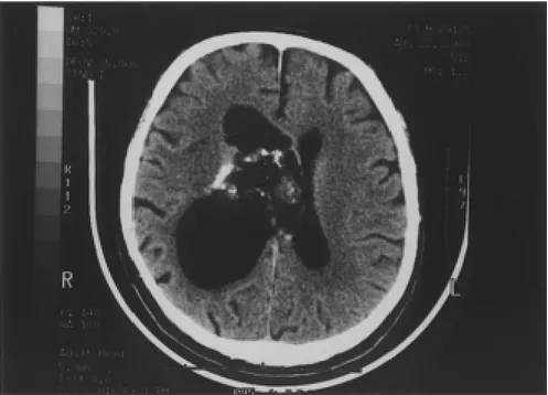
CT showed a partially calcified right ventricular mass, which was conforming to the shape of the ventricle. The mass was rather heterogeneous with cystic areas isodense with the cerebrospinal fluid and solid portions isodense with gray matter (Fig 1). The right lateral ventricle was dilated. MRI revealed a lobulated mass in the right lateral ventricle adjacent to the foramen of Monro and septum pellucidum, with displacement of the midline structures. The right lateral ventricle was dilated. There was no peritumoral edema. The cystic parts of the lesion Çetin Atasoy*, A.Tuba Karagülle**, İlhan Erden***, Serdar Akyar****
PRIMARY OLIGODENDROGLIOMA OF THE LATERAL
VENTRICLE: COMPUTED TOMOGRAPHY AND
MAGNETIC RESONANCE IMAGING FINDINGS
–––––––––––––––––––––––––
* M.D. Specialist in Radiology, Department of Radiology, Ankara University School of Medicine, 06100, Ankara-TURKEY **M.D. Resident in Radiology, Department of Radiology, Ankara University School of Medicine, 06100, Ankara-TURKEY ***M.D. Proffesor of Radiology, Department of Radiology, Ankara University School of Medicine, 06100, Ankara-TURKEY ****M.D. Proffesor of Radiology, Department of Radiology, Ankara University School of Medicine, 06100, Ankara-TURKEY
–––––––––––––––––––––––––––––––––––––––––––––––––––––––––––––––––––––––––––––––––––––––––––––––––––– Received: April 12, 2001 Accepted: May 28, 2001
SUMMARY
Oligodendrogliomas commonly arise from the cerebral hemispheres and may secondarily invade the adjacent ventricles. Primary intraventricular oligodendrogliomas are quite rare. In this report we describe computed tomographic and magnetic resonance imaging findings of a right lateral ventricular oligodendroglioma in a patient presenting with seizures and amnesia.
Key Words: Oligodendroglioma-Computed Tomography - Magnetic Resonance Imaging
ÖZET
Oligodenroglioma: CT ve MRI Görüntüleme
Oligodendrogliomlar genellikle serebral hemisferden k ken al r; nadiren ventrik lleri sekonder olarak invaze edebilir. Primer intraventrik ler oligodendrogliomlar olduk a enderdir. Bu olgu bildirisinde, unutkanl k ve n bet ge irme yak nmas ile ba vuran 54 ya ndaki erkek hastada saptad m z, primer intraventrik ler oligodendrogliomun Bilgisayarl tomografi ve Magnetik rezonans g r nt leme bulgular n sunduk.
Anahtar Kelimeler: Oligodendrogliom, Bilgisayarl Tomografi, Magnetik Rezonans G r nt leme
had a higher signal intensity than cerebrospinal fluid on T1-and proton density weighted images. The solid portions were iso-hyperintense than
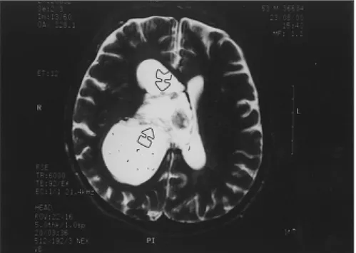
cortical gray matter on T1-weighted images and heterogeneously hyperintense compared with gray matter on T2-weighted images (Figs. 2,3).
Figure 1: Non enhanced axial CT section at the level of the lateral ventricle shows a
partially calcified mass near the foramen of Monro with cystic and solid areas and dilatation in the right lateral ventricle.
Figure 2: On non enhanced T1-weighted axial spin echo (SE) MR image, the cystic
areas of the lesion are hyperintense than cerebrospinal fluid and the solid portions are iso-hyperintense compared to cerebral gray matter.
Especially on T2-weighted images numerous hypointense septae rendered the lesion a multiloculated appearance (Fig. 3). The solid parts of the lesion enhanced remarkably
after the injection of gadolinium (Fig. 4). At surgery, the tumor was removed subtotally. Pathological examination revealed oligodendrog-lioma.
Figure 3: On the T2-weighted image, the cystic parts appear isointense with
cerebrospinal fluid, and the solid parts are slightly hyperintense than gray matter. Note several hypointense septa rendering the lesion a multiloculated appearance
(arrows)
Figure 4: Gadolinium enhanced T1-weighted image reveals marked enhancement in
Discussion
Oligodendrogliomas are composed of small oligodendrocytes of a rather uniform size (2). Nearly half of the lesions also contain astrocytes and spongioblasts (1,2). The majority occur in the cerebral hemispheres, particularly in the frontal lobes. Only a small percentage originates from within the ventricles. Although intraventricular oligodendrogliomas have the same histological fetaures as the parenchymal lesions, they have different clinical and imaging characteristics (3).
The intraventricular oligodendrogliomas are slightly more common in females (3), unlike the hemispheric variety, which shows a male predominance (4). Intraventricular oligodendrog-liomas frequently occur in the third and fourth decades of life. While it has previously been stated that intraventricular oligodendrogliomas present at an earlier age (5) several recent series showed no significant age difference between the parenchymal and intraventricular tumors (3-5). Although seizure is the most common presenting symptom of hemispheric oligodendrogliomas (4), the intraventricular variety commonly causes signs of increased intracranial pressure (3,5). The symptomatology consisting of seizures and amnesia and relatively older age of our patient differ from the typical presentation of an intraventricular oligodendroglioma.
Intraventricular oligodendrogliomas are most commonly found in the anterior parts of the lateral ventricles (3). There is only one case involving the occipital horn and atrium of the lateral ventricle (6). In our patient the mass was predominantly in the anterior part of the right lateral ventricle close to the foramen of Monro and adjacent to the septum pellucidum.
The matrix of an intraventricular oligodendroglioma tends to be hyperdense relative to the brain parenchyma (3), unlike parenchymal oligodendrogliomas, where the matrix is usually heterogeneous or hypodense (7). Low-density foci representing areas of necrosis and hyperdense areas caused by hemorrhage may be identified in both parenchymal and
intraventricular types. Nodular and plump calcifications are more prominent in the parenchymal type (3,5,8). Unless malignant features are present edema is not associated with either type (8). Contrast enhancement is mild or moderate in the intraventricular oligodendrogli-omas but is unusual in the parenchymal variety. Experience is limited on MR imaging of intraventricular oligodendrogliomas. MRI is superior over CT in determining the origin and extent of the lesions (3), which is crucial for planning of surgery. In our patient the intraventricular mass lesion had a heterogenous signal intensity on both T1-and T2-weighted images with cystic and solid portions and linear hypointense septal stuctures. The solid portions of the lesion enhanced remarkably on postgadolinium images.
Lateral ventricular masses that may be confused with intraventricular oligodendrogli-omas are subependymoligodendrogli-omas, astrocytoligodendrogli-omas, ependymomas, gangliogliomas, subependymal giant cell tumors, central neurocytomas and meningiomas.
Subependymomas occur in the same age group as intraventricular oligodendrogliomas but favor the fourth ventricle and usually have an extraventricular component (2). Rarely, however, a subependymoma involves a lateral ventricle, near the foramen of Monro, when the differential diagnosis from oligodendrogliomas is quite difficult. The matrix of a subependymoma is usually isodense with brain parenchyma and may demonstrate minimal enhancement after contrast administration (9).
Ependymomas are difficult to distinguish from subependymomas but they usually enhance after contrast administration and tend to calcify in an irregular manner (2). Some are partly cystic. Ependymomas are seen in a younger age group than subependymomas.
Like intraventricular oligodendrogliomas gangliogliomas also affect the young adults, however they are usually found in the anterior third ventricle rather than in the lateral ventricles.
Astrocytomas may be found in the lateral ventricles. Their typical location is in the frontal horns, more anterior than the intraventricular oligodendrogliomas. They usually enhance and have a hyperdense matrix (9).
Subependymal giant cell astrocytomas are typically seen in patients with tuberous sclerosis. They usually occur in the region of foramen of Monro and are accompanied by other cerebral findings of tuberous sclerosis (2), which are helpful in distinguishing them from intraventricular oligodendrogliomas.
Choroid plexus papillomas also show marked enhancement and usually occur in the trigones of the lateral ventricles, in contradistinction to oligodendrogliomas, which tend to locate more anteriorly near the foramina of Monro. Patient age is another important differential diagnostic feature in choroid plexus papillomas, which are typically seen in young children (10).
Central neurocytoma, a recently described variant of neuroblastoma, may occur in the lateral ventricles and be difficult to distinguish
from the oligodendroglioma on routine histopathological examinations. These lesions have a better prognosis than oligodendrogliomas and they usually enhance (2).
Intraventricular meningiomas tend to occur in the atria of the lateral ventricles. They are typically isointense with the cortical gray matter and show marked and homogeneous enhancement (2,9).
Epidermoid and dermoid tumors may also present as intraventricular masses. Epidermoids are frequently seen in the fourth ventricle. On MRI epidermoids are heterogenously isointens with cerebrospinal fluid and do not enhance after gadolinium administration. Dermoid tumors and teratomas can easily be identified by the presence of fat (9).
Due to their size and deep location, intraventricular oligodendrogliomas are not generally amenable to complete removal. Most cases are excised subtotally followed by CSF shunting to treat or prevent hydrocephalus.
1. Rubinstein LJ. Tumors of the central nervous system, second series, fascicle 6. Washington DC: AFIP, 1972: 85-103
2. Taveras JM. Intracranial Neoplasms. In: Taveras JM. Neuroradiology, 3rd ed, Baltimore Williams and Wilkins, 1996: 576-613. 3. Dolinskas CA, Simeone FA. CT characteristics
of intraventricular oligodendrogliomas. Am J Neuroradiol 1987; 8: 1077-1082.
4. Chin HW, Hazel JJ, Kim TH, et al. Oligodendrogliomas I. A clinical study of cerebral oligodendrogliomas. Cancer 1980; 45: 1458-1459.
5. Kikuchi K, Kowada M, Mineura K, et al. Primary oligodendroglioma of the lateral ventricle with computed tomographic and positron emission tomographic evaluations. Surg Neurol 1985; 23: 483-488.
6. Silver JA, Ganti SR, Hilal SK. Computed tomography of tumors involving the atria of the lateral ventricles. Radiology 1982; 145: 71-78. 7. Kazner E, Wende S, Grumme Th, et al. Computed tomography in intracranial tumors. Differential diagnosis and clinical aspects. Berlin: Springer Verlag, 1982: 42-44.
8. Gross CB, Masdeu JC, Maravilla KR, et al. Intracranial neoplasms of the adult. In: Gonzales CF, Grossman CF, Masdeu JC, eds. Head and spine imaging. New York: Wiley, 1985: 232-238.
9. Morrison G, Sobel DF, Kelley WM, et al. Intraventricular mass lesions. Radiology 1984; 153: 435-442.
10. Jelinek J, Smirniotopoulos JG, Parisi JE, Kanzer M. Lateral ventricular neoplasms of the brain: differential diagnosis based on clinical, CT and MR findings. Am J Neuroradiol 1990; 155: 365-372.