83
Ursodeoksikolik Asidin Streptozosin ile Diyabet Oluşturulmuş
Farelerde Kardiyomyopatideki Antiinflamatuar ve Anti-Fibrotik
Etkileri
Anti-Inflammatory and Anti-Fibrotic Effects of Ursodeoxycholic Acid in
Streptozocin-Induced Diabetic Rats
İsmail Polat Canbolat1, Gürkan Yiğittürk2, Oytun Erbaş3
1Demiroğlu Bilim Üniversitesi Tıp Fakültesi, Kardiyoloji Ana Bilim Dalı, İstanbul, Türkiye
2Muğla Sıtkı Koçma Üniversitesi Tıp Fakültesi, Histoloji ve Embriyoloji Ana Bilim Dalı, Muğla, Türkiye 3Demiroğlu Bilim Üniversitesi Tıp Fakültesi, Fizyoloji Ana Bilim Dalı, İstanbul, Türkiye
ÖZ
GİRİŞ ve AMAÇ: Diabetik kardiyomyopati, serbest yağ aside
oksidasyonu, mitokondrial disfonksiyon, oksidatif stress ve diffuz miyokardiyal fibrozise seconder olarak gelişmektedir. Bu deneysel çalışmada, ursodeoksikolik asidin streptozocin ile tetiklenmiş diabetik fare modelinde inflamatuar ve anti-fibrotik etkilerini araştırmayı hedefledik.
YÖNTEM ve GEREÇLER: Sprague Dawley albino 30 erişkin
fare 3 gruba ayrıldı: Grup-1: kontrol grubu (n=10); Grup-2 (n=10) diabetik fare grubu; Grup-3 (n=10) ursodeoksikolik asit verilen diabetic fare grubu. Histopatolojik ve biyokimyasal değerlendirmeler 4 hafta sonra kalp dokusundan yapıldı. Fibronektin ve TGF-β immunekspresyonu, TGF-β,
malondialdehid, pentraxin-3, pro-BNP ve troponin-T düzeyleri ölçüldü.
BULGULAR: Fibronektin immunekspresyonu, TGF-β,
pentraxin-3, troponin-t, pro-BNP ve malondialdehid düzeyleri diabetik farelerde control grubuna göre anlamlı olarak artmış saptandı. Ursodeoksikolik asidin inflamasyon belirteçlerini ve fibroz düzeyini anlamlı olarak azalttığı izlendi
TARTIŞMA ve SONUÇ: Bu deneysel çalışmada,
ursodeoksikolik asidin diabetik farelerde anti-inflammatuar ve anti-fibrotik etkilerini gösterdik. Diabetik hastalarda
ursodeoksikolik asidin ilaç olarak kullanımı klinik olarak fayda gösterebilir.
Anahtar Kelimeler: diabetik kardiyomyopati, ursodeoksikolik
asit, inflamasyon, kardiak fibroz
ABSTRACT
INTRODUCTION: Diabetic cardiomyopathy is a consequence
of free fatty acid oxidation, dysfunction in mitochondria, oxidative stress and diffuse myocardial fibrosis. We aimed to investigate the anti-inflammatory and anti-fibrotic effect of ursodeoxycholic acid in streptozocin-induced diabetic rat model.
METHODS: Male Sprague Dawley albino mature rats were
divided into 3 groups: Group 1 (n=10) control group; group 2 (n=10) diabetic rats group; group 3 (n=10): diabetic rats treated with ursodeoxycholic acid group. Diabetes mellitus model was established after injection of intraperitoneal streptozocin. Histopathological and biochemical examinations were done after 4 weeks from heart tissues. Immunoexpression levels of fibronectin and TGF-β were obtained.
Malondialdehyde levels were used to determine lipid peroxidation and pentraxin-3 levels were used to determine inflammation. Myocardial damage was also determined with troponin-T and pro-BNP levels.
RESULTS: Cardiac muscle cell thickness (hypertrophy),
TGF-β levels, fibronectin immunoexpression malondialdehyde, pentraxin-3, troponin-T and pro-BNP levels were increased significantly in groups 2 and 3 when compared to control group. Administration of ursodeoxycholic acid significantly reduced inflammation and fibrosis in group 3 compared to group 2.
DISCUSSION AND CONCLUSION: In this experimental
study, we demonstrated the anti-inflammatory and anti-fibrotic effects of UDCA on diabetic rats and it can be a good drug candidate for DM patients
Keywords: ursodeoxycholic acid, diabetic cardiomyopathy,
inflammation, cardiac fibrosis
İletişim / Correspondence: Dr. İsmail Polat Canbolat
Demiroğlu Bilim Üniversitesi Tıp Fakültesi, Kardiyoloji Ana Bilim Dalı, İstanbul, Türkiye E-mail: [email protected]
Başvuru Tarihi: 15.11.2019 Kabul Tarihi:17.02.2020
84 INTRODUCTION
Diabetes Mellitus (DM) is a persistent endocrinopathy with a global age-adjusted prevalence of 10% (1). Diabetic patients tend to establish twice-hold risk of heart failure compared with non-diabetic patients (2). Diabetes mellitus affects the heart mostly secondary to coronary atherosclerosis. Diabetic cardiomyopathy (DCM) is a type of heart failure associated with metabolic alterations secondary to diabetes after excluding atherosclerotic, hypertensive and structural heart disease (3). Hyperinsulinemia and insulin resistance exacerbate free fatty acid oxidation, dysfunction in mitochondria, oxidative stress and diffuse myocardial fibrosis (4-6).
Ursodeoxycholic acid (UDCA) is a widely used bile acid mostly for chronic cholestatic liver disease (7). The effect of UDCA is a result of reductions in cell apoptosis and resistance to oxidative stress (8,9). Administration of UDCA had been shown to reduce oxidative stress in aorta in a model of fructose induced metabolic syndrome in rats (10).
We aimed to investigate the beneficial effect of UDCA on the oxidative stress, vascular inflammation, myocardial damage and fibrosis in a diabetic rat model.
MATERIALS AND METHODS Animals
Totally 30 male Sprague Dawley albino mature rats were used. Animals were able to deliver food and water spontaneously. They were housed in steel cages and kept at room temperature (23 2 C) with light/dark (12/12 h) cycle. All the animal experiments performed in this study were done under the animal experiment guidelines.
Experimental design
Intraperitoneal (i.p.) injection of streptozocin (STZ) (Sigma-Aldrich, USA) was used to induce diabetes for 20 rats. Rats which were not injected STZ were selected as control group (n=10). Rats with higher than 250 mg/dl blood glucose level after 24 hours were confirmed as diabetic and included in this study. Then, 10 diabetic rats were randomly assigned as diabetes control group and 10 diabetic rats treated with ursodeoxycholic acid (UDCA) 250 mg/kg/day,
(Ursofalk) (Diabetes + UDCA) by oral way for 4 weeks as diabetic UDCA treatment group.
The animals were euthanized, and blood samples were collected by cardiac puncture for biochemical analysis and histopathological examination was performed after removal of the heart.
Histopathological examination of heart tissue
Rats were anesthetized with ketamin (80 mg/kg, i.p.) and xylazine (8 mg/kg, i.p.). Heart tissues were fixed with formalin. Hematoxylin and eosin (H&E) stained heart sections with 5 μm thickness were photographed.
Light microscopy was used to determine heart cell hypertrophy degree. The cardiac muscle fiber with the maximum cross section diameter was photographed. Image analysis software (Image- Pro Express 1.4.5, Media Cybernetics, Inc. USA) was used to measure muscle fiber. The analysis was performed after the average of 50 cardiac muscle cells were calculated for each rat.
Fibronectin immunoexpression
Endogenous peroxidase activity was eradicated with 30 min H2O2 (10%) and blocked with 10% normal goat serum (Invitrogen) for 1 hour at room temperature. Subsequently, sections were incubated in primary antibodies (Fibronectin, Bioss, Inc.; 1/100) for 24 h at 4 °C. The antibodies were detected with the Histostain-Plus Bulk kit (Bioss, Inc) and the final product was visualized with 3,3' diaminobenzidine (DAB). Brown cytoplasmic staining was scored as positive for immunoexpression. At least 10 fields of tissue section with 100 magnification was assessed and 100 cardiomyocytes per field was systematically scored.
Measurement of plasma TGF-β, Troponin T,
pro-BNP, pentraxin-3
A commercially available ELISA kit was used for measuring plasma TGF-β, troponinT, pro-BNP, pentraxin-3 levels.
Evaluation of lipid peroxidation
Plasma malondialdehyde (MDA) levels were measured to evaluate lipid peroxidation.
85
Statistical analysis
Descriptive analyses were presented as mean values ± standard derivation. Non-parametric parameters were analyzed with Mann-Whitney U test. The differences between groups was analyzed with Student’s-t test. Statistical significance was regarded as a p value equal or lower than 0.05. All analyses were performed using SPSS v.21.0 for Windows (SPSS, Inc., Chicago, Illinois, USA).
RESULTS
Histopathological changes
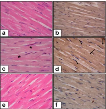
The histopathological changes in rats are demonstrated in Figure-1. Cardiac muscle cell thickness and immunoexpression of fibronectin were increased in diabetes group compared to the control group (p<0.01 for each). UDCA treatment in diabetic rats significantly decreased cardiac muscle cell thickness (p<0.05) and fibronectin immunoexpresion (p<0.001). The histopathological scores of the groups are shown in Table-1.
Oxidative stress, inflammation markers
Serum levels of MDA, TGF-, pentraxin-3 were significantly elevated in diabetic rats compared to the control group (p<0.001 for each). Administration of UDCA significantly decreased the level of MDA (p<0.001), TGF- (p<0.001) and pentraxin-3 (p<0.05) as shown in Table-1.
Myocardial damage markers
Serum levels of troponin-T and pro-BNP were significantly elevated in diabetic rats compared to the control group (p<0.001 for each). This elevation was significantly reduced by UDCA treatment (p<0.05 for pro-BNP and p<0.001 for troponin-T) as showed in Table-1.
Figure 1: Histopathological changes in streptozocin-induced diabetic
rats. a-Control Group, H & E stain x100 magnification. b- Control Group, Fibronectin expression x100 magnification. c- Diabetic Rats (Control group), H & E stain x100 magnification (* shown increased cardiac muscle cell thickness and arrow shown increased fibronectin expression). d- Diabetic Rats (Control group), Fibronectin expression x100 magnification. e- Diabetic Rats (UDCA treatment group), H & E stain x100 magnification. f- Diabetic Rats (UDCA treatment group), Fibronectin expression x100 magnification
Table-1: Cardiac muscle hypertropy,
immunoexpression, biomarker levels according to groups Normal Control (Group-1) Diabetic rat (control group) (Group-2) Diabetic rat (UDCA treatment) (Group-3) Cardiac muscle cell thickness (% of control) 100 125.3 ± 4.6 * 113.4 ± 4.1 # Immunoexpression Fibronectin percent (%) 13.2 ± 3.15 45.6 ± 8.3 * 15.1 ± 5.06 ## Blood glucose (mg/dl) 91.3 ± 8.8 367.1 ± 15.9 ** 408.1 ± 16.4 TGF-Beta (pg/ml) 10.2 ± 2.03 34.5 ± 4.4 ** 16.8 ± 4.08 ## MDA (nM) 80.1 ± 13.6 365.6 ± 15.3 ** 113.9 1± 6.4 ## Pro-BNP (pg/ml) 3.08 ± 0.68 16.7 ± 4.2 ** 8.6 ± 1.57 # Troponin T (pg/ml) 0.73 ± 0.1 3.4 ± 1.1 ** 1.6 ± 0.72 ## Pentraxin-3 (ng/ml) 1.38 ± 0.16 3.01 ± 0.25 ** 1.75 ± 0.33 #
* p<0.01, Group-2 compared to Group-1 ** p<0.001, Group-2 compared to Group-1 # p<0.05, Group-3 compared to Group-2 # # p<0.001, Group-3 compared to Group-2
86 DISCUSSION
In this experimental study, we demonstrated the
beneficial effects of UDCA on the end products of lipid peroxidation, vascular inflammation, myocardial damage and fibrosis.
Diabetes mellitus can affect heart from different mechanisms (11). Diabetic cardiomyopathy is a complication of DM related with impairment of microcirculation, abnormalities in subcellular components, alterations in lipid metabolism, maladaptive immune responses and can be diagnosed after ruling out coronary atherosclerosis, hypertension and structural heart disease (12). Inflammation is a contributor of DCM and different mechanisms of action had been shown in previous animal studies (13,14). Anti-inflammatory effects of UDCA had been shown in experimental models of acute liver injury, non-alcoholic liver disease (15,16). Pentraxin-3, a marker for vascular inflammation was significantly reduced in diabetic rats after UDCA administration in our study.
TGF- stimulates collagen production by coupling with angiotensin-1 receptor and promotes fibrosis in tissues (17). Pathil et al. demonstrated that fibrosis of liver was suppressed in hepatic stellate cells by blocking TGF-1/Smad2/3 signaling pathway after the administration of UDCA (18). The role of TGF- on pulmonary fibrosis was studied by Ko et al and demonstrated that TGF- alters mRNA to promote lung fibrosis (19). In our study, UDCA administration significantly reduced serum TGF- levels and immunoexpression of fibronectin leading to decreased cardiac muscle cell thickness and fibrosis. We demonstrated the reduced myocardial damage in diabetic rats treated with UDCA compared to diabetic control group via reduced troponin and pro-BNP levels which are cardiac biomarkers used for clinical assessment (20,21).
Oxidative stress had been described as a major contributor to DCM (22,23). Increased free fatty acid peroxidation is associated with overproduction of reactive oxygen species (24,25). Malondialdehyde (MDA) is a product of lipid peroxidation and reflects cellular damage (26). In an experimental model of cholestatic liver disease, UDCA administration was associated with decreased lipid peroxidation (27). In
our study, the use of UDCA lowered serum MDA levels reflecting lowered lipid peroxidation.
Ursodeoxycholic acid has been shown to protect heart muscles in different types of action. Gorelik et al. showed that UDCA protects cardiomyocytes from taurocholic acid’s arrhythmia risk by improving abnormal calcium dynamics (28). Hanafi et al. demonstrated the cardioprotective effect of UDCA from hypoxia by regulating ERK and Akt pathway (29). In our study, we demonstrated the cardioprotective role of UDCA in anti-inflammatory and anti-fibrotic ways.
Treatment with UDCA in heart transplant patients was retrospectively analyzed and UDCA was found to lower acute rejections, but the mechanism was not understood (30). von Haehling et al studied the beneficial effect of UDCA on inflammation in heart failure patients (31). In this study, UDCA had no beneficial effect on inflammatory cytokines, functional class or 6-min walk test, but UDCA was well tolerated in heart failure patients and improved endothelial functions. Treatment of UDCA may be more beneficial before overt heart failure occurs. In conclusion, in this experimental study, we demonstrated the anti-inflammatory and anti-fibrotic effects of UDCA on diabetic rats and it can be a good drug candidate in diabetic patients for protecting from DCM.
REFERENCES
1. International Diabetes Federation. Global Burden: Prevalence and Projections, 2015 and 2040. International Diabetes Federation; 2017. http://www.diabetesatlas.org/across-the-globe.html
2. Nichols GA, Gullion CM, Koro CE, Ephross SA,
Brown JB. The incidence of congestive heart failure in type 2 diabetes: an update. Diabetes Care 2004; 27: 1879–84.
3. Aneja A, Tang WH, Bansilal S, Garcia MJ,
Farkouh ME. Diabetic cardiomyopathy: insights into pathogenesis, diagnostic challenges, and therapeutic options. Am J Med 2008; 121: 748-57.
4. van de Weijer T, Schrauwen-Hinderling VB,
Schrauwen P. Lipotoxicity in type 2 diabetic cardiomyopathy. Cardio¬vasc Res 2011; 92: 10-8.
87
5. Aragno M, Mastrocola R, Medana C, Catalano
MG, Vercellinatto I, Danni O, et al. Oxidative stress-dependent impairment of cardiac-specific tran¬scription factors in experimental diabetes. Endocrinology 2006; 147: 5967-74.
6. Bugger H, Abel ED. Molecular mechanisms of
diabetic cardiomyopathy. Diabetologia 2014; 57: 660–71.
7. Makino I, Tanaka H. From a choleretic to an
immunomodulator: historical review of ursodeoxycholic acid as a medicament. Journal of gastroenterology and hepatology 1998; 13: 659–64.
8. Rodrigues CM, Fan G, Wong PY, Kren BT and
Steer CJ. Ursodeoxycholic acid may inhibit deoxycholic acidinduced apoptosis by modulating mitochondrial transmembrane potential and reactive oxygen species production. Mol Med 1998; 4: 165-78.
9. Lukivskaya O, Patsenker E and Buko VU.
Protective effect of ursodeoxycholic acid on liver mitochondrial function in rats with alloxan-induced diabetes: link with oxidative stress. Life Sci 2007; 80: 2397-402.
10. Mahmoud AAA, Elshazly SM.
Ursodeoxycholic Acid Ameliorates Fructose-Induced Metabolic Syndrome in Rats. PLoS ONE 2014; 9: e106993.
11. Miki T, Yuda S, Kouzu H, Miura T. Diabetic
cardiomyopa¬thy: pathophysiology and clinical features. Heart Fail Rev 2013; 18: 149-66.
12. Lee W, Kim J. Diabetic cardiomyopathy: where
we are and where we are going. Korean J Intern Med 2017; 32: 404-21.
13. Tschope C, Walther T, Escher F, Spillmann F,
Du J, Altman C, et al. Transgenic activation of the kallikrein-kinin system inhibits intramyocardial inflammation, endothelial dysfunction and oxidative stress in experimental diabetic cardiomyopathy. FASEB J. 2005; 19: 2057–9.
14. Westermann D, van Linthout S, Dhayat S,
Dhayat N, Escher F, Bücker-Gartner C, et al. Cardioprotective and anti-inflammatory effects of interleukin converting enzyme inhibition in
experimental diabetic cardiomyopathy. Diabetes. 2007; 56: 1834–41.
15. Pathil A, Warth A, Chamulitrat W, Stremmel
W. Comparison of different bile acid-phospholipid conjugates in acute hepatitis. Eur J Clin Invest. 2012; 42: 130-8.
16. Pathil A, Mueller J, Warth A, Chamulitrat W,
Stremmel W. Ursodeoxycholyl
lysophosphatidylethanolamide improves steatosis and inflammation in murine models of nonalcoholic fatty liver disease. Hepatology 2012; 55: 1369-78.
17. Chen K, Mehta JL, Li D, Joseph L, Joseph J.
Transforming growth factor beta receptor endoglin is expressed in cardiac fibroblasts and modulates profibrogenic actions of angiotensin II. Circ Res 2004; 95: 1167–73.
18. Pathil A, Mueller J, Ludwig JM, Wang J, Warth
A, Chamulitrat W, et al. Ursodeoxycholyl lysophosphatidylethanolamide attenuates hepatofibrogenesis by impairment of TGF-beta1/Smad2/3 signalling. Br J Pharmacol. 2014; 171: 5113-26.
19. Ko J, Mills T, Huang J, Chen NY, Mertens TCJ,
Collum SD, et al. Transforming growth factor beta 1 alters the 3’UTR of mRNA to promote lung fibrosis. J Biol Chem. 2019; 294: 15781-94.
20. Galsgaard J, Persson F, Hansen TW, Jorsal A,
Tarnow L, Parving HH, et al. Plasma high-sensitivity troponin T predicts end-stage renal disease and cardiovascular and all-cause mortality in patients with type 1 diabetes and diabetic nephropathy. Kidney Int 2017; 92: 1242-8.
21. Huelsmann M, Neuhold S, Strunk G, Moertl D,
Berger R, Prager R, et al. NT-proBNP has a high negative predictive value to rule-out short-term cardiovascular events in patients with diabetes mellitus. Eur Heart J 2008; 29: 2259-64.
22. Jay D, Hitomi H, Griendling KK. Oxidative
stress and diabetic cardivascular complications. Free Radic Biol Med. 2006; 40: 183-92.
23. Giacco F, Brownlee M. Oxidative stress and
88
24. van de Weijer T, Schrauwen-Hinderling VB,
Schrauwen P. Lipotoxicity in type 2 diabetic cardiomyopathy. Cardio¬vasc Res 2011; 92: 10-8.
25. Kanter M, Akpolat M, Aktas C. Protective
effects of the volatile oil of Nigella sativa seedsonβ-cell damage in streptozotocin-induced diabetic rats: a light and electron microscopic study. J MolHist 2009; 40: 379–85.
26. Cordis GA, Das DK, Riedel W.
High-performance liquid chromatographic peak identification of 2,4-dinitrophenylhydrazine derivatives of lipid peroxidation aldehydes by photodiode array detection. J Chromatogr A 1998; 798: 117-23.
27. Ljubuncic P, Tanne Z, Bomzon A.
Ursodeoxycholic acid suppresses extent of lipid peroxidation in diseased liver in experimental cholestatic liver disease. Dig Dis Sci 2009; 45: 1921– 8.
28. Gorelik J, Shevchuk AI, Diakonov I, de Swiet
M, Lab M, Korchev Y, et al. Dexamethasone and ursodeoxycholic acid protect against the arrhythmogenic effect of taurocholate in an in vitro study or rat cardiomyocytes. BJOG Int J Obstet Gynaecol 2003; 110: 424-9.
29. Hanafi NI, Mohamed AS, Md Noor J, Abdul
Hamid Hasani N, Siran T, Osman NJ, et al. Ursodeoxycholic acid upregulates ERK and Akt in the protection of cardiomyoctes against hypoxia. Genet Mol Res GMR 2016; 15.
30. Bahrle S, Szabo G, Stiehl A, Theilmann L, Tj
D, Zimmermann R, et al. Adjuvant treatment with ursodeoxycholic acid may reduce the incidence of acute cardiac allograft rejection. J Heart Lung Transplant 1998; 17: 592-8.
31. von Haehling S, Schefold JC, Jankowska EA,
Springer J, Vazir A, Kalra PR, et al. Ursodeoxycholic acid in patients with chronic heart failure: a double blind, randomized, placebo-controlled crossover trial. J Am Coll Cardiol 2012; 59: 585-92.