Abstract.
Background/Aim: This study was designed to
provide further evidence for the interactions between
hydrogen sulfide (H
2S) and nitric oxide (NO) in
ischemia/reperfusion (I/R) injury. Materials and Methods: Rat
hearts were studied with the Langendorff technique using the
H
2S donor sodium hydrosulfide (NaHS, 40 μM) and the
cystathionine gamma-lyase (CTH or CSE) inhibitor
DL-propargylglycine (PAG, 1 mM). NO synthase inhibitor
L-NG-nitroarginine methyl ester (L-NAME, 30 mg/kg, 7 days) was
administered before the isolation. The hearts were
homogenized for biochemical and molecular analysis.
Results: NaHS reversed I/R-induced cardiac performance
impairment, increased tissue nitric oxide production and
decreased tissue markers for cardiac injury, while L-NAME
inhibited these effects. The expression of CTH was increased
with PAG, which was suppressed by L-NAME. Conclusion:
H
2S and NO increase each other’s production suggesting
their interaction and cooperation in cardioprotection against
I/R injury.
Nitric oxide (NO), carbon monoxide (CO), and hydrogen
sulfide (H
2S), in the order of their discovery, are
gasotransmitters, a term that refers to a gaseous transmitter,
and was first coined by Wang (1). All are endogenously
produced small signaling molecules with low molecular
weight (NO, 30 Da; CO, 28 Da; H
2S, 34 Da). Because they
are small gaseous molecules, they reach easily their
intracellular targets to activate them, by diffusing freely across
the plasma membrane. They play a pivotal roles in the control
of many physiological functions, including regulation of
cardiovascular, nervous, gastrointestinal, excretory, immune,
and reproductive systems (2-5). Of these three gaseous
transmitters, H
2S that was first introduced as a metabolic
product in mammals by the American biochemist Vincent Du
Vigneaud, has gained much attention in recent years due to its
involvement in the above-mentioned physiological functions
(2, 6, 7). It is endogenously synthesized in most mammalian
tissues from L-cysteine and/or L-homocysteine by
cystathionine beta-synthase (CBS), cystathionine gamma-lyase
(CTH or CSE), and cysteine aminotransferase together with
3-mercaptopyruvate sulfurtransferase (2, 4, 8).
Heart failure, the major health issue in the world and the
leading cause of deaths, is a complicated disease caused by
This article is freely accessible online.
Correspondence to: Savas Ustunova, Ph.D., Department of
Physiology, School of Medicine, Bezmialem Vakif University, 34093 Istanbul, Turkey. Tel: +90 5335106592, e-mail: [email protected]
Key Words: Hydrogen sulfide, nitric oxide, isolated heart,
ischemia/reperfusion injury, oxidative damage.
Cardioprotection Against Ischemia/Reperfusion
Injury in Isolated Rat Heart
SAVAS USTUNOVA
1, SELCUK TAKIR
2, NADIM YILMAZER
3, HURI BULUT
4, DIDEM ALTINDIREK
5,
OZDEN HATIRNAZ NG
6, CIHAN DEMIRCI TANSEL
7, B. SONMEZ UYDES DOGAN
8,
UGUR OZBEK
9, ELIF ILKAY ARMUTAK
10and EBRU GUREL GUREVIN
71
Department of Physiology, School of Medicine, Bezmialem Vakif University, Istanbul, Turkey;
2Department of Medical Pharmacology, School of Medicine, Giresun University, Giresun, Turkey;
3Department of Biology, Faculty of Arts and Sciences, Namik Kemal University, Tekirdag, Turkey;
4Department of Medical Biochemistry, School of Medicine, Istinye University, Istanbul, Turkey;
5
Department of Genetics, Aziz Sancar Institute of Experimental Medicine, Istanbul University, Istanbul, Turkey;
6Department of Medical Biology, School of Medicine, Acibadem Mehmet Ali Aydinlar University, Istanbul, Turkey;
7
Department of Biology, Faculty of Science, Istanbul University, Istanbul, Turkey;
8Department of Pharmacology, Faculty of Pharmacy, Istanbul University, Istanbul, Turkey;
9
Department of Medical Genetics, School of Medicine, Acibadem Mehmet Ali Aydinlar University, Istanbul, Turkey;
10Department of Histology and Embryology, Faculty of Veterinary Medicine,
a variety of common stresses to the heart, such as
hypertension, diabetes, and myocardial infarction that is the
result of ischemic heart disease (9, 10). Therefore, novel
complementary compounds that are safe and effective
alternatives to conventional pharmacotherapy of heart failure
are needed. In recent years, a considerable number of studies
have revealed that H
2S plays important roles in alleviating
ischemia/reperfusion (I/R) injury (11), and that plasma sulfur
concentration is inversely proportional to the severity of
congestive heart failure (12). In addition, exogenous
administration of H
2S or cardiac-specific CTH overexpression
provides protection against acute myocardial I/R injury and
heart failure (10, 12, 13).
Since 1997, when the first experimental study by Hosoki,
et al. (14) revealed that endogenous H
2S may regulate smooth
muscle tone in synergy with NO, many studies have provided
strong and growing evidence that these two molecules could
modulate each other’s activities by altering the functions of
the related proteins (15-17). Kondo, et al. (18) has shown that
H
2S protected against heart failure via up-regulation of
endothelial nitric oxide synthase (eNOS) activity, while a new
thiol sensitive molecule resulted from the reaction of H
2S with
NO was found to regulate heart function (19). However, the
precise mechanisms of interactions between NO and H
2S that
affect heart failure, and whether H
2S modulates the biological
effects of NO are not entirely clear (8). Therefore, it is
urgently needed to deeply understand the underlying
mechanisms, so that novel strategies can be developed to
provide protection against heart failure (10, 17).
In view of these facts, the present study aimed to provide
further evidence for the effects of H
2S and NO, and their
interactions in I/R injury by employing the Langendorff
technique of isolated rat heart perfusion.
Materials and Methods
Animals. Forty-eight male Wistar albino rats weighing 250-300 g
were used. They were housed under 12/12 h day/night cycle and controlled room temperature (22±2˚C) and were allowed free access to food and water, and received humane care according to the criteria outlined in the ‘Guide for the Care and Use of Laboratory Animals (2011)’ prepared by the National Academy of Science and published by the National Institutes of Health. Animal experiments were reviewed and approved by the Animal Care and Use Committee of Istanbul University.
Isolated heart perfusion. All Langendorff isolated heart studies were
performed as previously described (20). Briefly, animals were anesthetized by intraperitoneal injection of 75 mg/kg pentobarbital sodium (Pental Sodyum, IE Ulagay, Istanbul, Turkey). Tracheotomy was performed, and mechanical ventilation (Small Animal Ventilator Model 683, Harvard Apparatus, Holliston, MA, USA) was initiated soon after surgical opening of the thorax. Heparin (150 IU) was administered from the abdominal vein, and before excision of the heart the aorta was cannulated in situ. The hearts were then Langendorff-perfused at 37˚C with Krebs-Henseleit buffer containing (mM) 118 NaCl, 0.5 EDTA, 4.7 KCl, 2.25 CaCl2, 1.2 MgSO4, 25
NaHCO3, 1.2 KH2PO4, and 11 glucose, 1 lactate, 0.5 glutamine, and
0.1 pyruvate, gassed with 95% O2- 5% CO2. End-diastolic pressure
was adjusted at 5-10 mmHg. The hearts were perfused by a mini pulse peristaltic pump (ML172B, ADInstruments, Sydney, Australia) at a constant flow with initial perfusion pressure of approximately 80 mmHg. After stabilization of pressure development during the first 20 min of Langendorff-perfusion, 6 groups of hearts, each composed of 8 animals, were studied (Figure 1). All hearts were subjected to 30 min ischemia and 60 min reperfusion by switching the peristaltic pump off and on. The ischemia/reperfusion (IR) group was just perfused with Krebs-Henseleit solution for 20 min, while the sodium hydrosulphide (NaHS) group was perfused with 40 μM NaHS (as H2S donor), and the DL-propargylglycine (PAG) group with 1 mM PAG (as CTH inhibitor) prior to ischemia. L-NG-Nitroarginine methyl ester (L-NAME), L-NAME+NaHS, and L-NAME+PAG groups additionally received 30 mg/kg L-NAME (as NOS inhibitor) intraperitoneally for 7 days before Langendorff studies.
Cardiodynamic analysis. Left ventricular pressure was recorded by
means of a balloon catheter placed inside the left ventricle and connected to a physiological pressure transducer (MLT844, ADInstruments) for assessment of contractile performance, while a second physiological pressure transducer was connected to the system in order to record the perfusion pressure via the data acquisition unit (PowerLab ML870B2, ADInstruments). The obtained data were analyzed with an appropriate software (LabChart 7; ADInstruments), and the records at certain time points [0thmin:
end of stabilization (baseline), 20thmin: end of drug administration
before ischemia; 55thmin: 5thmin of reperfusion, 60thmin: 10thmin
of reperfusion, 110thmin: end of experiment] were used for further
analyses of cardiac parameters, including end diastolic pressure (EDP), left ventricular developed pressure (LVDP), Max dP/dt (a specific index used to determine the ability of the heart to contract), and rate pressure product (RPP, an indirect index of myocardial oxygen consumption and cardiac function).
Biochemical analysis. At the end of the experiment, the hearts were
homogenized with a teflon piston homogenizer (Sartorius Potter S, Goettingen, Germany) in ice-cold PBS (pH 7.4) in a borosilicate Figure 1. Experimental design (: time points for cardiodynamic analyses).
glassware of 15 ml. The homogenate was centrifuged at 15.000×g at 4˚C for 20 min.
As tissue markers for cardiac injury, the levels of creatine kinase-MB (CK-kinase-MB) (Uscn Life Science Inc., Wuhan, PR China), a marker of myocardial injury, lactate dehydrogenase (LDH) (Elabscience, Wuhan, PR China), a marker of necrosis, and glutathione peroxidase (GPx) (Elabscience), an endogenous antioxidant enzyme, were measured in supernatant by ELISA. The production of NO and H2S was measured with a nitrate/nitrite assay
kit (Cayman Chemical, Ann Arbor, MI, USA), and a H2S ELISA kit
(Elabscience), respectively.
Gene expression analysis
Homogenization and RNA isolation. Total RNA was isolated
from cardiac tissue via TRIzol® Reagent (Invitrogen-Thermo
Fisher Scientific, Waltham, MA, USA) as described in the manufacturer’s protocol. Briefly, 0.1 g of tissue was thawed, and homogenized in 1 ml TRIzol® Reagent with a benchtop
homogenizer (MP Biomedicals LLC, CA, USA). Then, the resulting homogenate was incubated for 5 min at room temperature. After the addition of 200 μl chloroform to each sample, they were incubated for 2-3 min at room temperature, and centrifuged for 15 min 12.000×g at 4˚C, forming a lower red phenol-chloroform interphase and a colorless upper aqueous phase. The latter containing RNA was transferred to a new tube, and the former containing protein was stored at –20˚C for protein isolation. Precipitation of RNA was performed with the addition of 70% ethanol and incubation for 10 min at room temperature. The quality and quantity of RNA was measured by a spectrophotometer (Nanodrop ND-1000 spectrophotometer; Thermo Fisher Scientific, Waltham, MA, USA).
cDNA synthesis and real-time PCR. Random hexamers (pdN6)
(Roche Applied Science, Mannheim, Germany) and M-MLV reverse transcriptase (Life Technologies, Carlsbad, CA, USA) were employed to synthesize cDNA from 1 μg of total RNA. The obtained cDNA samples were kept at –20˚C. eNOS, inducible nitric oxide synthase (iNOS), and CTH gene expression analysis was performed using a LightCycler 480 instrument (Roche Applied Science, Penzberg, Upper Bavaria, Germany). Gene-specific primers and probes were designed using Universal ProbeLibrary reference gene assays for mouse or rat (Universal Probe Library Rat-ACTB Gene Assay; Cat. No. 05 046 203 001, Roche Applied Science, Penzberg, Upper Bavaria, Germany) (Table I). The housekeeping gene ACTB (β-actin) was used to standardize quantification in gene expression, by performing two-color real-time PCR. Based on the mathematical model described by Livak et al. (21), double delta Ct analysis was used to calculate relative gene expressions.
Statistical analysis. All data were statistically analyzed with
GraphPad Prism 6.0 (GraphPad Prism Software, San Diego, CA, USA), and presented as mean±standard error of mean (mean±SEM). A value of p<0.05 was considered as statistically significant. Cardiodynamic results were evaluated by two-way analysis of variance (ANOVA), while gene expression and biochemical results were analyzed by one-way ANOVA and followed by post-hoc Bonferroni's multiple comparison test.
Results
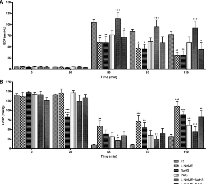
Cardiodynamic results. The end diastolic pressure values were
close to each other in all groups before ischemia, which
increased them significantly (p<0.001), implying diminished
cardiac contractility that is observed in heart failure.
Administration of L-NAME, NaHS or PAG induced a
statistically significant (p<0.01) decrease in EDP values.
Following ischemia, the highest EDP value was determined at
the 5
thmin of reperfusion in the L-NAME+NaHS group, while
the lowest value was in the groups of L-NAME and NaHS. At
the end of reperfusion, values were approximated to those of
the initial point in the groups of L-NAME and NaHS. It was
interesting that perfusion with PAG alone reversed this effect
and caused an increase in EDP values when compared to
perfusion with NaHS alone (Figure 2A). As to values of LVDP,
there was a significant decrease only in the NaHS group
compared to the IR (p<0.001) and L-NAME (p<0.001) groups.
The decrease observed due to ischemia at the 5
thmin of
reperfusion was found to be increased in the later stages of
reperfusion in all groups. The highest value was observed in
the L-NAME group, whereas the lowest value was in the IR
group (Figure 2B). Max dP/dt values were also similar with the
LVDP results (Figure 3A). Before ischemia, the initial RPP
values of all groups were in parallel with each other except for
the NaHS group. Following ischemia, the values in all groups
decreased until the first 5
thmin of reperfusion, and then
increased with the progression of reperfusion. The best
recovery occurred in L-NAME (p<0.001), NaHS (p<0.01), and
L-NAME+PAG (p<0.05) groups in descending order, whereas
the lowest recovery was in the IR (p<0.001) and
L-NAME+NaHS (p<0.001) groups (Figure 3B).
Biochemical results. CK-MB levels showed a significant
increase (p<0.01) in the L-NAME+PAG group compared to the
Cth ACACTTTCATGTCTGCATATTTCC TTTGTGGCAGAACACATACAAA 21IR group, while a significant decrease was observed in all other
groups (p<0.001) (Figure 4A). Apart from the L-NAME+PAG
group (p<0.001), all groups had decreased levels of LDH in
comparison with both the IR and L-NAME groups (Figure
4B). NaHS and PAG administration increased the levels of
GPx compared to both the IR and L-NAME groups (p<0.001).
In the L-NAME+NaHS group, the levels of this enzyme also
increased compared to the IR group, while they significantly
decreased in the L-NAME+PAG group (Figure 4C).
The levels of H
2S were increased by NaHS administration
in the tissue compared to both the IR and L-NAME groups
(p<0.001), but PAG administration caused a decrease
(p<0.001). However, H
2S increased in the NAME and
L-NAME+NaHS groups (p<0.001 for both), whereas it decreased
in the L-NAME+PAG group as in the PAG group (p<0.001)
(Figure 4D). NaHS administration led to an increase in NO
levels (p<0.001), but PAG administration did not cause a
significant change. Nitrate/nitrite levels significantly decreased
(p<0.001) in all L-NAME-administered groups compared to
the IR group. L-NAME+NaHS combination increased
nitrate/nitrite levels (p<0.05) compared to the L-NAME group,
whereas PAG had no effect (Figure 4E).
Figure 2. Cardiodynamic analysis. (A) The end diastolic pressure (EDP), B) Left ventricular developed pressure (LVDP) values of all groups.
*p<0.05, **p<0.01, ***p<0.001, statistical significance compared to the IR group; +p<0.05, ++p<0.01, +++p<0.001, statistical significance
Gene expression results
eNOS, iNOS, and CTH mRNA expression. Relative mRNA
expressions of genes were evaluated by RT-PCR analysis. The
expression levels of eNOS mRNA were similar in all groups
except for the NaHS-administered groups, in that NaHS
administration caused a decrease in eNOS mRNA expression,
while a further decrease was detected in the L-NAME+NaHS
group. However, this decrease was not statistically significant
(Figure 5A). L-NAME administration alone caused a
prominent increase in the expression of iNOS mRNA,
although it was not statistically significant. In contrast to the
increment with L-NAME alone, the administration of NaHS
and PAG with L-NAME resulted in a decrease (Figure 5B).
The expression profile of CTH was intriguingly different
among all groups; although PAG increased its expression,
L-NAME administration suppressed it significantly (p<0.01) in
the PAG group. PAG co-administered with L-NAME showed
a similar decrease with the L-NAME group (Figure 5C).
Discussion
A considerable number of studies have been performed to
examine the cardioprotective effects of H
2S and NO, and the
interactions between them in I/R injury, by employing
several in vitro and in vivo experimental models of cardiac
injury, including cultured cardiomyocytes, isolated perfused
Figure 3. Cardiodynamic analysis. A) The Max dP/dt, B) Rate pressure product (RPP) values of all groups. *p<0.05, **p<0.01, ***p<0.001,
hearts, and rodent and large animal (rabbit, dog, pig) models
(17, 22-26). However, to the best of our knowledge, no study
has examined the effect of NaHS, PAG and L-NAME on
isolated rat heart administered, and the present study is the
first to establish the role of these donors and inhibitors in
cardioprotection against I/R injury in rat heart.
The studies that aim to reveal the role of H
2S in
physiological functions are generally based on two strategies:
(i) inhibition of endogenous H
2S, and (ii) administration of
exogenous H
2S by employing NaHS as donor. Although the
latter strategy is found inconvenient because the large and
quick release of H
2S from NaHS may have detrimental
effects on the experimental animals, this may be negligible
since the resulting effects will be fairly short lasting (27).
Similarly, the NO synthase inhibitor L-NAME is
exogenously administered to the experimental animals in
studies investigating the biological functions of NO.
Accordingly, the experimental setup of our study is based on
these strategies.
In our study, the EDP values that were decreased by
ischemia, suggesting diminished cardiac contractility in heart
failure, were restored to those of initial levels at the end of
Figure 4. Biochemical analysis in heart tissue samples. A) CK-MB, B) LDH, C) GPx, D) H2S and E) Nitrate/nitrite levels of all groups. **p<0.01,
***p<0.001, statistical significance compared to the IR group; +p<0.05, ++p<0.01, +++p<0.001, statistical significance compared to the L-NAME
reperfusion in the L-NAME and NaHS groups, while a
significant decrease in Max dP/dt and LVDP values was
observed in the NaHS group. Concerning the RPP values, the
best recovery was observed with the L-NAME, NaHS, and
L-NAME+PAG. These results obviously show the role of
both H
2S and NO in cardioprotection against I/R injury in
rat heart, and are concordant with many studies. Johansen et
al. (28) have shown that exogenous H
2S decreased left
ventricular Max dP/dt in a concentration- and
time-dependent manner in rat heart. In a study performed in
isolated rat hearts, NaHS administration led to a significant
reduction in heart rate (HR) and LVDP, and this effect was
ascribed to the muscle relaxant role of H
2S, suggesting that
it has a similar effect on myocardium (29). Moreover,
postconditioning with H
2S improved the contractile and
diastolic functions of the heart subjected to I/R, as revealed
with improved HR, Max dP/dt, Min dP/dt, and LVDP, and
reduced left ventricular EDP in the left atrium after
reperfusion in the study in isolated rat hearts by Luan et al.
(24). We obtained similar results in our isolated heart study,
which suggests that a decrease in H
2S and NO levels due to
the inhibition of synthesizing enzymes might be responsible
for the impaired cardiac parameters in ischemia. It has been
reported that cardioprotection by H
2S occurs in I/R injury
through the inhibition of oxidation, increase in mitochondrial
biogenesis, restoration of mitochondrial dysfunction,
inhibition of heart cell apoptosis, reduction in the expression
of proinflammatory cytokines and iNOS, up-regulation of
eNOS, modulation of autophagy, and increase in
angiogenesis (30).
There are numerous studies demonstrating the role of NO
in protecting the heart against I/R injury, although some
results highlight a controversial role (31). NO plays a
positive role by being involved in the mechanisms of
protection triggered by cardiac adaptation and ischemic
preconditioning, on the other hand it has deleterious effects
on the normal heart subjected to I/R alone (32). It is accepted
that H
2S and NO cooperatively provide a cardioprotection,
although there are fewer studies on the interaction between
H
2S and NO in the cardiovascular system. In our study,
exogenous NaHS resulted in increased NO levels, and
combined L-NAME and NaHS caused an increase in
nitrate/nitrite levels, indicating that H
2S participates in NO
production. As it is known, NO and H
2S interact with each
Figure 5. RT-PCR analysis in heart tissue samples. A) eNOS B) iNOS and C) Cth mRNA expressions. All expression levels were measured relative
other’s synthesizing enzymes, and affect their generation, but
the precise mechanism remains unclear (17).
It has been shown that H
2S provides cardioprotection
against I/R injury by augmenting NO bioavailability via
activation of eNOS (33). Congestive heart failure in mice
was attenuated by eNOS overexpression, while eNOS
deficiency resulted in heart failure and congenital septal
defects during cardiac development due to increased
apoptotic cardiomyocyte death (34, 35). Based on the results
of the studies with L-NAME-induced hypertensive rats, Ji et
al. (36) and Zhong et al. (37) have suggested that the
eNOS/NO pathway was involved in the antihypertensive
effects of H
2S. As confirmed by the significantly increased
expression of iNOS mRNA and protein, iNOS is often
induced to produce higher NO in certain pathological
conditions involving I/R injury (38, 39). However,
exogenous NaHS administration suppressed iNOS activity
and reduced NO content in the plasma and myocardial tissue
to improve heart function in a metabolic syndrome model of
rats (38). In a mice study by Hua et al. (40), exogenous H
2S
provided protection against virus-induced myocardial injury
through the inhibition of myocardial iNOS mRNA and
protein expression. However, studies reporting conflicting
results are available in the current literature. For instance,
H
2S inhibited the activity of eNOS in rat and mouse aortic
rings (41), and both exogenous and endogenous H
2S reduced
NO generation and prevented eNOS activity and
transcription (42). Additionally, in the present study, a slight
non-significant increase of iNOS expression was observed
with L-NAME administration, while the administration of
NaHS and PAG in combination with L-NAME resulted in
decreased expression. Moreover, NaHS administration
caused a decrease in eNOS mRNA expression.
Our biochemical results also indicate that H
2S and NO are
required for cardioprotection since the levels of CK-MB and
LDH were increased in the L-NAME+PAG group in which
NO and H
2S were inhibited, while their levels were decreased
in the other groups. Our results are in agreement with those of
Yang et al. (38) who have found that exogenous NaHS
ameliorated cardiac hypertrophy and myocardial injury in
diabetic cardiomyopathy and reduced LDH and CK-MB
activities in rats. In addition, another study has shown that
CK-MB and LDH levels decreased following NaHS administration
(4). Reactive oxygen species (ROS) production is accelerated
and cellular antioxidants become depleted during myocardial
ischemia. H
2S is a cytochrome C oxidase inhibitor and
therefore inhibits respiration and thus can decrease the
production of ROS and preserve mitochondrial function at low
concentrations. In addition, it has been reported that,
glutathione peroxidase, an antioxidant enzyme, was increased
by NaHS application (4). The GPx, which functions in the
detoxification of hydrogen peroxide, increased in the NaHS
group of our study, as well as in other studies (4, 38). Thus,
H
2S may be involved in the activation of endogenous
antioxidant mechanism by elevating enzyme levels.
We also found that PAG increased expression of CTH,
whereas L-NAME suppressed it. In the PAG group, NO
levels were not changed despite increased CTH expression,
and this result is in conflict with studies stating that CTH
overexpression promotes NO production (10), and that mice
lacking CTH exhibit reduced NO levels (42). It is possible
that different H
2S/NO donors/inhibitors and amounts, and
different experimental models and parameters may result in
conflicting results. Therefore, there is need to examine further
the interactions between H
2S and NO in cardioprotection.
In conclusion the results of our study strengthen the
evidence that NaHS and L-NAME alone reverse I/R injury
induced cardiac performance impairments, while
co-administration adversely affected cardiodynamic values as
reflected by the biochemical results of tissue markers of
cardiac injury. It was also demonstrated that both H
2S and
NO increased each other’s production. We suggest that H
2S
and NO cooperated in cardioprotection against I/R injury in
isolated rat heart. However, there is no doubt that the precise
mechanisms underlying these interactions require further
studies.
Conflicts of Interest
The Authors declare that there are no conflicts of interest associated with this work.
Authors’ Contributions
S.U. conceived the work, designed and performed the experiments, analysed the data, and wrote the manuscript. S.T., H.B., D.A., O.H.N., C.D.T., S.U.D., U.O. and E.I.A. designed and performed the experiments and analysed the data. N.Y. and E.G.G. conceived the work, designed and performed the experiments, and critically reviewed and supervised the study.
Acknowledgements
This work was supported by Scientific Research Projects Coordination Unit of Istanbul University [grant numbers; 31508, 55736 and 47109].
The Authors would like to thank to Prof. Dr. Ismail Meral for providing language help and Aysu Kilic, MSc. for the technical support and writing assistance.
References
1 Wang R: Two’s company, three’sa crowd: Can H2S be the third
endogenous gaseous transmitter? FASEB J 16(13): 1792-1798, 2002. PMID: 12409322. DOI: 10.1096/fj.02-0211hyp
2 Bełtowski J: Hydrogen sulfide in pharmacology and medicine– an update. Pharmacol Rep 67(3): 647-658, 2015. PMID: 25933982. DOI: 10.1016/j.pharep.2015.01.005
4 Nicholson CK and Calvert JW: Hydrogen sulfide and ischemia– reperfusion injury. Pharmacol Res 62(4): 289-297, 2010. PMID: 20542117. DOI: 10.1016/j.phrs.2010.06.002
5 Shefa U, Yeo SG, Kim M-S, Song IO, Jung J, Jeong NY and Huh Y: Role of gasotransmitters in oxidative stresses, neuroinflammation, and neuronal repair. Biomed Res Int 2017, 2017. PMID: 28386548. DOI: 10.1155/2017/1689341
6 Kashfi K: The role of hydrogen sulfide in health and disease. Biochem Pharmacol 149: 1, 2018. PMID: 29501583. DOI: 10.1016/j.bcp.2018.02.030
7 Szabo C: A timeline of hydrogen sulfide (H2S) research: From
environmental toxin to biological mediator. Biochem Pharmacol
149: 5-19, 2018. PMID: 28947277. DOI: 10.1016/j.bcp.
2017.09.010
8 Jin S, Teng X, Xiao L, Xue H, Guo Q, Duan X, Chen Y and Wu Y: Hydrogen sulfide ameliorated l-name-induced hypertensive heart disease by the akt/enos/no pathway. Exp Biol Med
242(18): 1831-1841, 2017. PMID: 28971696. DOI: 10.1177/
1535370217732325
9 Mozaffarian D, Benjamin EJ, Go AS, Arnett DK, Blaha MJ, Cushman M, Das SR, De Ferranti S, Després JP and Fullerton HJ: Heart disease and stroke statistics-2016 update a report from the american heart association. Circulation 133(4): e38-e48, 2016. PMID: 26673558. DOI: 10.1161/CIR.0000000000000350 10 Wu D, Hu Q, Xiong Y, Zhu D, Mao Y and Zhu YZ: Novel H2
S-no hybrid molecule (zyz-803) promoted synergistic effects against heart failure. Redox Biol 15: 243-252, 2018. PMID: 29288927. DOI: 10.1016/j.redox.2017.11.020
11 Hu MZ, Zhou B, Mao HY, Sheng Q, Du B, Chen JL, Pang QF and Ji Y: Exogenous hydrogen sulfide postconditioning protects isolated rat hearts from ischemia/reperfusion injury through Sirt1/PGC-1α signaling pathway. Int Heart J 57(4): 477-482, 2016. PMID: 27357440. DOI: 10.1536/ihj.15-506
12 Kovačić D, Glavnik N, Marinšek M, Zagožen P, Rovan K, Goslar T, Marš T and Podbregar M: Total plasma sulfide in congestive heart failure. J Card Fail 18(7): 541-548, 2012. PMID: 22748487. DOI: 10.1016/j.cardfail.2012.04.011 13 Calvert JW, Elston M, Jha S, Gundewar S, Aragon JP,
Grinsfelder DB, Ramachandran A, Elrod JW and Lefer DJ: Hydrogen sulfide therapy attenuates ischemia-induced heart failure via nrf2 and nrf1 signaling. J Am Coll Cardiol 55: A20-E186, 2010. DOI: 10.1016/S0735-1097(10)60187-8
14 Hosoki R, Matsuki N and Kimura HJB The possible role of hydrogen sulfide as an endogenous smooth muscle relaxant in synergy with nitric oxide. Biochem Biophys Res Commun 237(3): 527-531, 1997. PMID: 9299397. DOI: 10.1006/bbrc.1997.6878 15 Faro MLL, Fox B, Whatmore JL, Winyard PG and Whiteman
M: Hydrogen sulfide and nitric oxide interactions in inflammation. Nitric oxide 41: 38-47, 2014. PMID: 24929214. DOI: 10.1016/j.niox.2014.05.014
16 Whiteman M and Moore PK: Hydrogen sulfide and the vasculature: A novel vasculoprotective entity and regulator of nitric oxide bioavailability? J Cell Mol Med 13(3): 488-507, 2009. PMID: 19374684. DOI: 10.1111/j.1582-4934.2009. 00645.x
S, Murohara T, Predmore BL, Gojon Sr G and Gojon Jr G: H2S protects against pressure overload–induced heart failure via upregulation of endothelial nitric oxide synthase. Circulation
127(10): 1116-1127, 2013. PMID: 23393010. DOI: 10.1161/
CIRCULATIONAHA.112.000855
19 Yong QC, Cheong JL, Hua F, Deng LW, Khoo YM, Lee HS, Perry A, Wood M, Whiteman M and Bian JS: Regulation of heart function by endogenous gaseous mediators—crosstalk between nitric oxide and hydrogen sulfide. Antioxid Redox Signal 14(11): 2081-2091, 2011. PMID: 21194352. DOI: 10.1089/ars.2010.3572
20 Gurel E, Ustunova S, Kapucu A, Yilmazer N, Eerbeek O, Nederlof R, Hollmann MW, Demirci-Tansel C and Zuurbier CJ: Hexokinase cellular trafficking in ischemia–reperfusion and ischemic preconditioning is altered in type i diabetic heart. Mol Biol Rep 40(7): 4153-4160, 2013. PMID: 23652994. DOI: 10.1007/s11033-013-2495-5
21 Livak KJ and Schmittgen TD: Analysis of relative gene expression data using real-time quantitative pcr and the 2− ΔΔCt
method. Methods 25(4): 402-408, 2001. PMID: 11846609. DOI: 10.1006/meth.2001.1262
22 Xie YH, Zhang N, Li LF, Zhang QZ, Xie LJ, Jiang H, Li LP, Hao N and Zhang JX: Hydrogen sulfide reduces regional myocardial ischemia injury through protection of mitochondrial function. Mol Med Rep 10(4): 1907-1914, 2014. PMID: 25198340. DOI: 10.3892/mmr.2014.2391
23 Nandi S, Ravindran S and Kurian GA: Role of endogenous hydrogen sulfide in cardiac mitochondrial preservation during ischemia reperfusion injury. Biomed Pharmacother 97: 271-279, 2018. PMID: 29091875. DOI: 10.1016/j.biopha.2017.10.118 24 Luan HF, Zhao ZB, Zhao QH, Zhu P, Xiu MY and Ji Y: Hydrogen
sulfide postconditioning protects isolated rat hearts against ischemia and reperfusion injury mediated by the jak2/stat3 survival pathway. Braz J Med Biol Res 45(10): 898-905, 2012. PMID: 22948409. DOI: 10.1590/s0100-879x2012007500090 25 Rossoni G, Sparatore A, Tazzari V, Manfredi B, Soldato PD and
Berti F: The hydrogen sulphide-releasing derivative of diclofenac protects against ischaemia–reperfusion injury in the isolated rabbit heart. Br J Pharmacol 153(1): 100-109, 2008. PMID: 17965734. DOI: 10.1038/sj.bjp.0707540
26 Szabó G, Veres G, Radovits T, Gerő D, Módis K, Miesel-Gröschel C, Horkay F, Karck M and Szabó C: Cardioprotective effects of hydrogen sulfide. Nitric Oxide 25(2): 201-210, 2011. PMID: 21094267. DOI: 10.1016/j.niox.2010.11.001
27 Caliendo G, Cirino G, Santagada V and Wallace JL: Synthesis and biological effects of hydrogen sulfide (H2S): Development
of H2S-releasing drugs as pharmaceuticals. J Med Chem 53(17): 6275-6286, 2010. PMID: 20462257. DOI: 10.1021/jm901638j 28 Johansen D, Ytrehus K and Baxter GF: Exogenous hydrogen
sulfide (H2S) protects against regional myocardial ischemia– reperfusion injury. Basic Res Cardiol 101(1): 53-60, 2006. PMID: 16328106. DOI: 10.1007/s00395-005-0569-9
29 Hussain A, Maddock H, Al-Rajaibi H and Carson RJ: Effects of hydrogen sulphide on the isolated perfused rat heart. Sultan Qaboos Univ Med J 11(2): 236, 2011. PMID: 21969896.
30 Zhang L, Wang Y, Li Y, Li L, Xu S, Feng X and Liu S: Hydrogen sulphide (H2S)-releasing compounds: Therapeutic
potential in cardiovascular diseases. Front Pharmacol 9: 1066, 2018. PMID: 30298008. DOI: 10.3389/fphar.2018.01066 31 Matejikova J, Kucharska J, Pancza D and Ravingerova T: The
effect of antioxidant treatment and nos inhibition on the incidence of ischemia-induced arrhythmias in the diabetic rat heart. Physiol Res 57(Suppl 2): S55-S60, 2008. PMID: 18373392.
32 Andelova E, Bartekova M, Pancza D, Styk J and Ravingerová T: The role of no in ischemia/reperfusion injury in isolated rat heart. Gen Physiol Biophys 24(4): 411, 2005. PMID: 16474186. 33 Shen Y, Shen Z, Luo S, Guo W and Zhu YZ: The cardioprotective effects of hydrogen sulfide in heart diseases: From molecular mechanisms to therapeutic potential. Oxid Med Cell Longev 2015: 925167, 2015. PMID: 26078822. DOI: 10.1155/2015/925167
34 Jones SP, Greer JJM, van Haperen R, Duncker DJ, de Crom R and Lefer DJ: Endothelial nitric oxide synthase overexpression attenuates congestive heart failure in mice. Proc Natl Acad Sci USA 100(8): 4891-4896, 2003. PMID: 12676984. DOI: 10.1073/ pnas.0837428100
35 Feng Q, Song W, Lu X, Hamilton JA, Lei M, Peng T and Yee S-P: Development of heart failure and congenital septal defects in mice lacking endothelial nitric oxide synthase. Circulation
106(7): 873-879, 2002. PMID: 12176963. DOI: 10.1161/
01.cir.0000024114.82981.ea
36 Ji W, Liu S, Dai J, Yang T, Jiang X, Duan X and Wu Y: Hydrogen sulfide defends against the cardiovascular risk of nw-nitro-l-argininemethyl ester-induced hypertension in rats via the nitric oxide/endothelial nitric oxide synthase pathway. Chin Med J (Engl) 127(21): 3751-3757, 2014. PMID: 25382331. DOI: 10.3760/cma.j.issn.0366-6999.20141573
37 Zhong G, Chen F, Cheng Y, Tang C and Du J: The role of hydrogen sulfide generation in the pathogenesis of hypertension in rats induced by inhibition of nitric oxide synthase. J Hypertens 21(10): 1879-1885, 2003. PMID: 14508194. DOI: 10.1097/00004872-200310000-00015
38 Yang R, Jia Q, Liu XF, Wang YY and Ma SF: Effects of hydrogen sulfide on inducible nitric oxide synthase activity and expression of cardiomyocytes in diabetic rats. Mol Med Rep
16(4): 5277-5284, 2017. PMID: 28849194. DOI: 10.3892/
mmr.2017.7247
39 Soliman H, Craig GP, Nagareddy P, Yuen VG, Lin G, Kumar U, McNeill JH and MacLeod KM: Role of inducible nitric oxide synthase in induction of rhoa expression in hearts from diabetic rats. Cardiovasc Res 79(2): 322-330, 2008. PMID: 18411229. DOI: 10.1093/cvr/cvn095
40 Hua W, Chen Q, Gong F, Xie C, Zhou S and Gao L: Cardioprotection of H2S by downregulating inos and
upregulating ho-1 expression in mice with cvb3-induced myocarditis. Life Sci 93(24): 949-954, 2013. PMID: 24140888. DOI: 10.1016/j.lfs.2013.10.007
41 Kubo S, Doe I, Kurokawa Y, Nishikawa H and Kawabata A: Direct inhibition of endothelial nitric oxide synthase by hydrogen sulfide: Contribution to dual modulation of vascular tension. Toxicology 232(1-2): 138-146, 2007. PMID: 17276573. DOI: 10.1016/j.tox.2006.12.023
42 King AL, Polhemus DJ, Bhushan S, Otsuka H, Kondo K, Nicholson CK, Bradley JM, Islam KN, Calvert JW and Tao YX: Hydrogen sulfide cytoprotective signaling is endothelial nitric oxide synthase-nitric oxide dependent. Proc Natl Acad Sci USA
111(8): 3182-3187, 2014. PMID: 24516168. DOI: 10.1073/pnas.
1321871111