TURCICA Acta Orthop Traumatol Turc 2008;42(2):97-105
The results of internal fixation of proximal humeral fractures with
the PHILOS locking plate
Proksimal humerus kırıklarında kilitli plak PHILOS ile internal tespit sonuçları
Mehmet Fatih KORKMAZ, Neslihan AKSU, Abdullah GOGUS, Mursel DEBRE,1Ayhan Nedim KARA, Zekeriya Ugur ISIKLAR Amaç: Humerus proksimal ve diyafiz kırıkları,
osteopo-rotik hastalarda sık görülen ve cerrahisi komplikasyon-larla seyreden sorunlu kırıklardır. Çalışmamızda AO/ ASIF grubu tarafından geliştirilen ve kilitli vidalarla sa-bit açılı stabilizasyon sağlayan yeni internal tespit siste-mi PHILOS (Proximal Humeral Internal Locking System) ile tedavi edilen hastaların sonuçları değerlendirildi. Çalışma planı: Çalışmaya, proksimal humerus kırığı nede-niyle PHILOS plak sistemiyle tedavi edilen 41 hasta alın-dı. Hastalar 65 yaş altı (grup A) ve 65 yaş veya üstü (grup B) olarak iki grupta değerlendirildi. Grup A’da 24 hasta (12 erkek, 12 kadın; ort. yaş 47, dağılım 24-64); grup B’de 17 hasta (4 erkek 13 kadın; ort. yaş 78, dağılım 67-90) vardı. Radyografik olarak tüm kırıklar AO/ASIF ölçütlerine göre sınıflandırıldı. Grup A ve B’de sırasıyla 10 hasta ve iki hasta-da deltopektoral girişim; 14 hasta ve 15 hastahasta-da deltoid split girişim uygulandı. Ortalama 15 ay (dağılım 6-28 ay) takip süresi sonunda her iki grubun fonksiyonel ve radyografik so-nuçları değerlendirildi.
Sonuçlar: Constant omuz skoru grup A’da ortalama 95.0 (dağılım 74-100), grup B’de 92.8 (dağılım 72-100) bulundu (p>0.05). Deltopektoral ve deltoid split girişim uygulanan olgularda altıncı aydan sonra Constant skoru ve fonksiyonel açıdan fark saptanmadı. Hiçbir hastada kaynamama veya implant yetersizliği gözlenmedi. Komplikasyonlar şunlardı: Vidanın eklem içine girmesi (n=1), tüberkülum majus frag-manında deplasman (n=1) ile birlikte plağın oblik yerleşimi (n=1), yetersiz redüksiyon (n=4) ve başın varus pozisyonun-da tespiti (n=3). Hiçbir olgupozisyonun-da avasküler nekroz görülmedi. Çıkarımlar: Kilitli vida plak sistemi, özellikle osteopo-rotik kırıkların tespitinde bugüne kadar kullanılan oste-osentez yöntemlerinden, erken harekete izin vermesi ve implant yetersizliği olmaması nedeniyle üstündür.
Anahtar sözcükler: Kemik plağı; kemik vidası; kırık tespiti,
internal/yöntem; humerus kırığı/cerrahi; omuz kırığı/cerrahi.
Objectives: Proximal and diaphyseal humeral fractures are common especially in the elderly, presenting as a challeng-ing problem due to their high complication rates followchalleng-ing surgical treatment. In this prospective study, we evaluated the results of patients treated with the PHILOS (Proximal Humeral Internal Locking System) locking plate, a new technique recently developed by the AO/ASIF.
Methods: Forty-one patients who were treated with the PHILOS plate for proximal humeral fractures were evaluated in two age groups. Group A included 24 patients (12 males, 12 females; mean age 47 years; range 24 to 64 years) younger than 65 years, and group B involved 17 patients (4 males, 13 females; mean age 78 years; range 67 to 90 years) at or above 65 years. Radiographically, all fractures were classified ac-cording to the AO/ASIF system. Surgery was performed with the deltopectoral approach in 10 and two patients, and with a deltoid split in 14 and 15 patients in group A and B, respec-tively. Functional and radiographic results were evaluated af-ter a mean follow-up of 15 months (range 6 to 28 months). Results: The mean Constant scores were 95.0 (range 74 to 100) and 92.8 (range 72 to 100) in group A and B, respec-tively (p>0.05). After six months of surgery, Constant scores and functional outcomes were similar in patients operated on with the deltopectoral approach or deltoid split. There was neither nonunion nor implant failure. Complications included intra-articular screw penetration (n=1), displacement of the greater tuberculum (n=1) with oblique placement of the plate (n=1), insufficient reduction (n=4), and varus displacement of the humeral head (n=3). No avascular necrosis was seen. Conclusion: Locking plate system is superior over other means of fixation methods, particularly in osteoporotic fractures, because it allows early rehabilitation and does not result in implant failure.
Key words: Bone plates; bone screws; fracture fixation, internal/
methods; humeral fractures/surgery; shoulder fractures/surgery.
Correspondence / Yazışma adresi: Mehmet Fatih Korkmaz, M.D. T.R. Istanbul Bilim University, The Department of Orthopedics and Traumatology
Büyükdere Caddesi No:120 34394 Esentepe/Sisli
Submitted / Başburu tarihi: 26.11.2007 Accepted / Kabul tarihi: 03.03.2008
©2008 Türk Ortopedi ve Travmatoloji Derneği / ©2008 Turkish Association of Orthopaedics and Traumatology
Istanbul Bilim University, Department of Orthopaedics and Traumatology; 1Florence Nightingale Hospital,
98 Acta Orthop Traumatol Turc
Proximal fractures of the humerus constitute 5-8% of all humeral fractures[1,2] Although more than 80% of these heal without surgical intervention, the rates of nonunion vary between %1 and 23% in displaced and nonimpacted fractures of the surgical neck. [1,3-5] These fractures are displaced in 20% of the cases and may coexist with other injuries.[6,7] The aim of treatment in proximal humeral fractures is to attain a painless and simultaneously functional shoulder. This result depends on the age, medical condition, bone quality and expec-tations of the patient as well as a good evaluation of the current fixation techniques. Loosening or failure of the implant and nonunion are possible complicati-ons of surgery in humeral fractures. There is still no treatment that can be the golden standart in these frac-tures.[8] Shoulder arthroplasty for proximal humerus fracture is effective in the elimination of pain, however the functional results are limited.[9,10,11,12] Especially in comminuted fractures of the humerus that involve the traculae the functional results are not good. The factors which negatively affect the functional results after frac-ture have been studied. In comminuted osteoporotic fractures where the trabeculae are also fractured, the placement of the prosthesis in the appropriate height and position, establishment of trabecular stability and repair of the rotator cuff may be difficult. These dif-ficulties negatively affect the functional success of the prosthesis.[11,12] In order to decrease the high complica-tion rates of proximal humeral fractures, the AO/ASIF group developed the PHILOS (The Proximal Humeral Internal Locking System) plate (Synthes, Stratec Me-dical ltd, Mezzovico Switzerland), an internal fixation system that enables angled stabilization with multiple interlocking screws. In this study, the results of 41 pa-tients with proximal humeral fractures who were tre-ated with internal fixation using PHILOS plate in our hospital between September 2005 and July 2007 are analyzed. Functional evaluation was carried out using Constant shoulder scale. The functional results were investigated in this plate, which early results are being reported in the literature.
Material and method
Between September 2005 and December 2007, a total of 64 patients underwent surgical treatment with PHILOS plate system. Forty one patients who had lon-ger than 6 months follow up and who fulfilled the inc-lusion criteria were taken into the study.
Inclusion criteria: 1. Closed proximal humerus
frac-ture (AO/ASIF bifocal, unifocal, intraarticular). 2. Frac-tures not treated with conservative means (inadequate position, osteoporotic fracture, patients who did not ac-cept conservativie treatment) 3. Patients older than 18 years. Exclusion criteria: 1.Pathologic fractures 2. Pati-ents with primary or metastatic tumors (one patient who had enchondroma was not included ) 3. Fractures with nonunion. In four patients older that 65, there were as-sociated fractures in the contralateral femur diaphysis, ipsilateral femur diaphysis fracture, ipsilateral intraar-ticular humerus distal edge fracture and distal radius fracture, and ipsilateral distal radius fracture. The pati-ents (n=41) were separated into two groups, below age 65 (Group A), and above age 65 (Group B). In group A, there were 24 patients (12 males, 12 females; mean age 45.66, range 24-64). In group B, there were 15 patients (4 males, and 13 females; mean age: 77.73, range 67-90). In order to completely analyse the fractue type AP and transthoracic lateral imaging was used, and CT scans were used only in selected cases. Using X-rays, all frac-tures were classified according to AO/AIF classification ( Müler et al, 1990) (Table 1). Computer tomography was used only in selected cases to evaluate the extensi-on to the articular surface and to evaluate the amount of major tuberculum displacement in comminuted frac-tures. All operations were controlled using fluoroscopy. After a mean follow up period of 14.68 (6-28) months the functional and radiologic results of both groups were assessed. In the surgical treatment of proximal humeral fractures, most surgeons prefer the deltopec-toral approach due to their education and habits. The deltoid splitting approach is a good choice especially in comminuted fractures or where the trabecular frag-ments are displaced. We used both approaches. The pa-tients were positioned in the beach chair position. Ten patients in Group A underwent deltopectoral approach and 14 underwent deltoid split approach. Two patients in Group B underwent deltopectoral approach and 15 underwent deltoid split approach. In proximal femur
Table 1. The classification of fractures according to AO/ ASIF.
Below age 65 Above age 65
(Group A) (Group B) 1.1 A.1 – – 1.1 A.2 7 5 1.1 A.3 1 3 1.1 B.1 9 7 1.1 B.2 2 1 1.1 C.1 4 –
fractures, after the fracture site was exposed, reducti-on was enabled with a K wire under fluoroscopy and with ethibond sutures passed through the rotator cuff tendons. The PHILOS plate was position lateral to the bicipital groove and distal to the major tuberculum, and the correct position was checked with fluoroscopy. The tubercular fragments and rotator cuff tendons were fixa-ted using sutures passing from these structures and the plate. Finally, fracture reduction and screw length were assessed with fluroscopy. Preoperative and postoperati-ve images of our cases are shown in Figures 1,2,3.
In 5 patients who underwent deltoid split approach, the axillary nerve was seen to be compressed between fracture fragments and was released. None of the pati-ents had hypoesthesia in the axillary nerve dermatome prior to surgery. None of these patients developed axil-lary nerve paralysis after the operation. There were 3 patients who had valgus impaction, 2 underwent deltoid split and 1 underwent elevation and grafting with the deltopectoral approach. After fracture fixation, shoul-der AP and neutral position X rays were taken as the shoulder was internally rotated, externally rotated and neutral. The limit of shoulder movement was controlled
for the presence of impingement. Following stabilizati-on with PHILOS, the shoulder was immobilized with a shoulder-arm sling for 2-3 days. Subsequently, passive motion exercises were initiated with 90° abduction and anteflexion. Active pendular and circular motions of the arm were prescribed. Active assisted and passive exer-cises were used during the first two weeks, and 3 weeks later active motion was started. On the th postoperative week, daily activities were allowed. After the postope-rative control on the 6th week, subsequent visits were organized on the 3rd,6th,12th and in patients with lon-ger follow up, annually. Regular X rays were obtained to control the plate position and healing. The range of motion in the shoulder joint was recorded. The patients were evaluated with the Constant score[13] on the posto-perative 6th week, 3rd and 6th months. At the end of 6 months, none of patients showed any signs of implant loosening. The functional results between the two gro-ups were compared using student’s t test (95% confi-dence interval). p<0.05 was accepted as significant.
Results
After a mean follow up of 14.68 (6-28) months, ra-diologic and functional evaluations were made. Preope-Figure 1.64 year old male patient. The fracture is 1.1.B.1 according to AO classification. Preoperative
(a) and postoperative (b) X rays (deltoid split approach is used ). (c) Postoperative joint range of
motion is shown (Constant score: 96). (a)
(c)
100 Acta Orthop Traumatol Turc
ratively, all patients had normal motor function of all 3 parts of the deltoid muscle and intact sensory function of the axillary nerve. Axillary nerve and deltoid muscle functions were evaluated meticulously in the postopera-tive clinical examination. Complications like suprasca-pular or axillary nerve injury or deltoid weakness were not encountered. None of the patients developed nonu-nion or implant failure. During follow up, intraarticu-lar screw placement was seen in 1 patient (Figure 4), displacement in major tuberculum fragment was seen
in 1 case, displacement in major tuberculum fragment along with oblique placement of the plate was seen in 1 case (Figure 5). Inadequate reduction was seen in 4 ca-ses (Figure 6). In three caca-ses, as an early postoperative complication, it was seen that the head was fixed in the varus position (Figure 7).None of the patients developed avascular necrosis, superficial or deep infection. None of the scars required revision. The deltoid muscles were weak initially, however returned to normal after reha-Figure 2. 51 year old male patient. The fracture is 1.1.B.1 according to AO classification Preoperative (a) and
postoperative (b) X rays ( deltopectoral approach is used) (c) postoperative joint range of motion is seen (Constant score: 98)
(a)
(c)
(b)
Table 2. The distribution of postoperative complications according to age and type of treatment
Deltopectoral approach Deltoid splitting approach Age <65 Age ≥65 Age <65 Age ≥65
(n=10) (n=2) (n=14) (n=15)
Inadequate reduction 2 2 – –
Displacement in major tuberculum fragment and/or oblique
placement of the plate 1 – – 1
Intraarticular migration of the screw due to collapse of the head 1 – – –
bilitation. In all shoulders, the suprascapular nerve was functional, and normal power was demonstrated after rehabilitation. In the final evaluation, the Constant sho-ulder score was 95.04 (74-100) and 92.75 (72-100) in patients below and above age 65, respectively, and this difference was not significant (p>0.05). There was no significant difference between the groups with respect to range of motion in the joint. In patients below age 65 and who were operated with the deltopectoral app-roach, 2 had inadequate reduction, 1 had displacement of the major tuberculum and oblique placement of the plate, 1 case had intraarticular migration of the screw due to collapse of the head. In patients above age 65 and who were operated with deltopectoral approach, 2 had inadequate reduction. In patients below age 65 and operated with deltoid split, 1 had varus fixation of the head. In patients above age 65 and who were operated with deltoid split, 2 cases had fixation of the head in varus, 1 had displacement in major tuberculum. In the follow up after the 6th month, there were no significant differences between the deltopectoral and deltoid split groups with respect to Constant score and function.
Discussion
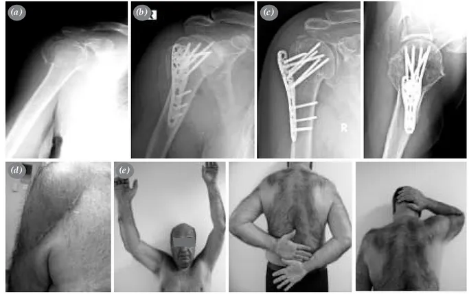
Fractures of the proximal humerus can be treated conservatively in old or young patients if the bone qua-lity is sufficient and the fracture is nondisplaced. Open reduction and internal fixation should be preferred in patients who will not comply with conservative mana-gement, who have comorbidities or osteoporotic and open fractures. In the 1980s, T-plates and 1/3 tubular plates were the preferred fixation material in proximal humerus fractures.[14,15] Plate osteosynthesis was rep-laced with minimal osteosynthesis.[8,14] Numerous tech-niques for minimal osteosynthesis including K wires applied with open or percutaneous technique, cerclage or tension band application with wire or PDS (polydi-oxanone suture), screws, cannulated screws, and intra-medullary nails.[8,16-18,19-21] Also numerous techniques including prosthetic replacement, double tubular pla-tes, Polarus nails (Acumed, Inc., Beaverton,OR), Plan Tan humerus fixator plate (Plan Tan Medizintechnik GmbH, Lambrechtshagen, Germany) are described. [22-24] Minimal invasive methods such as closed reduction and percutaneous pinning require good bone quality, Figure 3. 38 year old male patient. The fracture is 1.1.C.1 according to AO classification. Preoperative (a) and
postoperative (b) X rays (c) Deltoid split incision scar. (d) Postoperative joint range of motion is shown (Constant score: 100 )
(a)
(c) (d)
102 Acta Orthop Traumatol Turc
fractures with minimal fragmentation, and compliant patients.[25] The results of this method are poor in el-derly patients with ostoeporosis.[26] Early rehabilitation and early motion was not possible in this age group. Several complications have been reported with the traditional methods of open reduction and internal fi-xation, including loosening or failure of the plates and
screws, nonunion, malunion, migration of the nails, rotator cuff injury and impingement syndrome.[15,23,24] In osteoporotic individuals, the risk of implant loo-sening and failure is higher due to poor bone quality. [24,27] Excellent results began to be reported after the introduction of the PHILOS plate, a new internal fixa-tion system developed by the AO/ASIF group for the Figure 4.(a) 60 year old female patient. She has a 1.1.B.1 fracture according to AO classification. A-Preoperative
X ray. (b) The patient was operated with the deltopectoral approach. Early postoperative X ray shows that the screw heights are good. During follow up the proximal screws were not loosened, however the proximal head fragment collapsed, causing the screws to dislodge into the joint cavity. (c) These screws were chan-ged 5 mpnths later with deltoid split approach. The defect in the head fragment was grafted with allograft. Postrevision X ray is shown. (d) Range of motion after revision surgery is shown (Constant score: 74 ).
(a)
(d)
(b) (c)
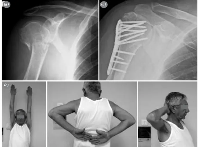
Figure 5.(a) 74 year old female patient.
The patient was operated 3 weeks after the occurrence of fracture, and the frac-ture was reduced intraoperatively. The fracture was 1.1 B.1 according to AO classification. Late postoperative X ray showed the displacement of the major tuberculum. A-Preoperative X ray (b),
(c) Postoperative range of motion. (d)
Deltoid incision scar. (e) Postoperative range of motion. F-Bony union is seen in X rays.(Constant Score: 100) (a) (d) (b) (e) (c)
treatment of proximal humerus fractures which enables angled fixation using multiple interlocking screws.[28, 29] The screws in the humeral head are locked to the pla-te and cannot move backwards, a significant advantage in osteoporotic bones. It also enables the placement of screws in different directions (converging or diverging). The low profile minimizes the risk of impingement.
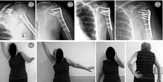
[30-33] Koukakis et al. reported early results in a series of 20 patients, and stated that the design of the plate achieves stable fixation, yields good results, and prevents failure. [32] In a series of 25 cases, Charalambous et al. reported that in 20 of the cases, the fractures united, and none of the patients required a revision due to implant fai-lure or nonunion. Five cases required revision due to Figure 6. (a) Preoperative X ray of a 62 year old male patient with 1.1.C.1 fracture according to AO classification.
(b) Early postoperative imaging after deltopectoral approach revealed that the major tubercle was
displaced, the head was in varus, and the plate was placed obliquely. (c) Late postoperative X rays shows healing of the fracture (d) The deltopectoral incision scar (e) Range of motion of the patient (Constant score 93).
(a)
(d) (e)
(b) (c)
Figure 7. Case example of the fixation of the head in varus position. (a), Preoperative (b) Postoperative
X rays.
104 Acta Orthop Traumatol Turc
nonunion or implant failure. The head screw entered the glenohumeral joint in 4 cases, loosened in another 4 and extruded, and the plate fracutred in 1 case. They stated that PHILOS is an effective system in providing stabilization in these fractures, however cautioned aga-inst the potential complications of the implant.[33] Siffri et ak compared the angled plate and interlocking plate fixation in a biomechanical study performed on cada-vers and synthetic models. The studies on synthetic humerus showed that the torsional stability of the ang-led and interlocking plates were similar. In the cadaver humeral neck model, interlocking plate system provi-ded greater torsional stability in periodic loading, ho-wever the bending stability of both plates were similar. [34] The use of screws loacted on the 4th and 5th level when there is no medial support, and reduction using indirect minimal invasive methods when the bone frag-ments of the medial support is adequte is one of the mpst critical points of this system. In proximal humerus fractures, the protection of the inferomedial support is critical in the preservation of reduction. It is important in stabilizing the nedial column that the lower interloc-king screws directed superiorly must pass through the inferior aspect of the proximal fracture fragment. In comminuted fractures, failure to achieve adequate me-dial column support may result in early reduction loss and failure.35 In our patient group, the primary reason for not encountering plate or screw failure or nonuinon is our meticulous adherence to this technical princip-le from the beginnig of this prospective study. Bone grafts or synthetic materials used for bone defects, e.g. calcum phosphate cement (Norian, SRS) is used in a limited number of patients. Among these 4 patients, 3 are valgus impacted fractures and the other is a proxi-mal humeral enchondroma who is excluded from the study. We do not advocate use of bone grafts except for valgus impacted displaced fractures and postreduction major bone defects. The advantage of deltoid splitting surgical approach is the easy access to supraspinatus, infraspinatus and teres minor. This approach gives bet-ter exposure of the major tuberculum fragment that is displaced posterosuperiorly and the head fragment. In cases with valgus impaction, elevation and grafting can be applied more anatomically. This incision also allows the release of the axillary nerve impinged in the frac-ture line. Our study showed that if the axillary nerve is protected, there will not be a postoperative axillary nerve dysfunction. In the deltopectoral approach, there is greater risk of injury as the deltoid muscle is
retrac-ted laterally. In ther deltoid split approach, there is very little risk of injury to the axillary nerve when it is under the retractor. The axillary nerve can be also be comp-ressed by the retractor if care is not taken. The nerve is directly within sight in deltoid split approach, therefore has a lower risk of injury. Particularly in AO/ASIF Type B and C fractures, the deltoid split approach allows 270 degree control of the proximal humerus, reduction with sutures passing through the tubercular fragments and rotator cuff tendons and fixation with plates. The del-toid split approach that is applied simultaneously with axillary nerve exploration is a useful surgical technique that does not risk deltoid muscle fuction and axillary nerve. We are now conducting more comprehensive studies on this approach and comparison with delto-pectoral approach. PHILOS plate is made of titanium, and therefore lighter than other implants. It has a good biocompatibility. The locking screw and plate system is a reliable internal fixation method for all age groups, if attention is paid to technical details and the tubercular fragments are reduced with sutures fixed to the plate. Easy applicability, biologic property due to the lack of interference with blood supply of the humeral head, no requirement to shape the plate and the achievement of stabilization at constant angles are the benefits of this plate. All screws and the plate move as a single structu-re. Complications related to the plate are very few, the-refore it is possible to avoid most of the complications of traditional plating. In the treatment of osteoporotic fractures, it is superior to other osteosynthesis techniues since it allows early motion and there is no implant in-sufficiency. We therefore believe that the PHILOS plate is a good internal fixation material in the osteosynthe-sis of proximal humerus fractures in patients above age 65. Another aspect of this study is that all patients in this group were operated by 2 orthopedics attendings who were trained on the use of this plate, and when the surgeon is experienced on all technical details and the anatomic approaches it is possible to state that compli-cations rates will be lower than the number reported in the literature. We believe that the reason for high Cons-tant scores in this group is related to adequate surgical technique, good follow up and rehabilitation.
References
1. Nayak NK, Schickendantz MS, Regan WD, Hawkins RJ. Operative treatment of nonunion of surgical neck fractures of the humerus. Clin Orthop Relat Res 1995;(313):200-5. 2. Volgas DA, Stannard JP, Alonso JE. Nonunions of the
3. Neer CS II, Rockwood CA. Fractures and dislocations of the shoulder. In: Rockwood CA Jr,, Green DD editors. Fractures. Vol. 1, Philadelphia: J. B. Lippincott; 1975. p. 686-7.
4. Neer CS II. Displaced proximal humeral fractures. Part I. Classification and evaluation. J Bone Joint Surg [Am] 1970;52:1077-89.
5. Scheck M. Surgical treatment of nonunions of the surgical neck of the humerus. Clin Orthop Relat Res 1982;(167): 255-9.
6. Court-Brown CM, Garg A, McQueen MM. The epidemi-ology of proximal humeral fractures. Acta Orthop Scand 2001;72:365-71.
7. Nordqvist A, Petersson CJ. Incidence and causes of shoul-der girdle injuries in an urban population. J Shoulshoul-der El-bow Surg 1995;4:107-12.
8. Wijgman AJ, Roolker W, Patt TW, Raaymakers EL, Marti RK. Open reduction and internal fixation of three and four-part fractures of the proximal part of the humerus. J Bone Joint Surg [Am] 2002;84:1919-25.
9. Goldman RT, Koval KJ, Cuomo F, Gallagher MA, Zucker-man JD. Functional outcome after humeral head replace-ment for acute three- and four-part proximal humeral frac-tures. J Shoulder Elbow Surg 1995;4:81-6.
10. Tanner MW, Cofield RH. Prosthetic arthroplasty for frac-tures and fracture-dislocations of the proximal humerus. Clin Orthop Relat Res 1983;(179):116-28.
11. Demirhan M, Kilicoglu O, Altinel L, Eralp L, Akalin Y. Prognostic factors in prosthetic replacement for acute prox-imal humerus fractures. J Orthop Trauma 2003;17:181-8. 12. Demirhan M. Factors affecting the results of
hemiarthro-plasty for proximal humerus fractures. [Article in Turkish] Acta Orthop Traumatol Turc 2000;34:463-74.
13. Müller ME, Nazarian S, Koch P, Schatzker J, editors. The comprehensive classification of fractures of long bones. Berlin: Springer; 1990.
14. Constant CR, Murley AH. A clinical method of func-tional assessment of the shoulder. Clin Orthop Relat Res 1987;(214): 160-4.
15. Kuner EH, Siebler G. Dislocation fractures of the proximal humerus-results following surgical treatment. A follow-up study of 167 cases. Unfallchirurgie 1987;13:64-71. [Ab-stract]
16. Wanner GA, Wanner-Schmid E, Romero J, Hersche O, von Smekal A, Trentz O, et al. Internal fixation of displaced proximal humeral fractures with two one-third tubular plates. J Trauma 2003;54:536-44.
17. Rowles DJ, McGrory JE. Percutaneous pinning of the proximal part of the humerus. An anatomic study. J Bone Joint Surg [Am] 2001;83:1695-9.
18. Zyto K, Ahrengart L, Sperber A, Tornkvist H. Treatment of displaced proximal humeral fractures in elderly patients. J Bone Joint Surg [Br] 1997;79:412-7.
19. Speck M, Regazzoni P. 4-fragment fractures of the proxi-mal humerus. Alternative strategies for surgical treatment.
Unfallchirurg 1997;100:349-53. [Abstract]
20. Park MC, Murthi AM, Roth NS, Blaine TA, Levine WN, Bigliani LU. Two-part and three-part fractures of the proximal humerus treated with suture fixation. J Orthop Trauma 2003;17:319-25.
21. Sehr JR, Szabo RM. Semitubular blade plate for fixation in the proximal humerus. J Orthop Trauma 1988;2:327-32. 22. Seidel H. Humeral locking nail: a preliminary report.
Or-thopedics 1989;12:219-26.
23. Robinson CM, Page RS, Hill RM, Sanders DL, Court-Brown CM, Wakefield AE. Primary hemiarthroplasty for treatment of proximal humeral fractures. J Bone Joint Surg [Am] 2003;85:1215-23.
24. Rajasekhar C, Ray PS, Bhamra MS. Fixation of proximal humeral fractures with the Polarus nail. J Shoulder Elbow Surg 2001;10:7-10.
25. Sadowski C, Riand N, Stern R, Hoffmeyer P. Fixation of fractures of the proximal humerus with the PlantTan Hu-merus Fixator Plate: early experience with a new implant. J Shoulder Elbow Surg 2003;12:148-51.
26. Herscovici D Jr, Saunders DT, Johnson MP, Sanders R, DiPasquale T. Percutaneous fixation of proximal humeral fractures. Clin Orthop Relat Res 2000;(375):97-104. 27. Cordasco F A, Bigliani LU. Complications of proximal
hu-merus fractures. Tech Orthop 1997;12:42-50.
28. Lill H, Hepp P, Korner J, Kassi JP, Verheyden AP, Josten C, et al. Proximal humeral fractures: how stiff should an implant be? A comparative mechanical study with new implants in human specimens. Arch Orthop Trauma Surg 2003;123:74-81.
29. Frigg R. Development of the locking compression plate. Injury 2003;34 Suppl 2:B6-10.
30. Ring D, Jupiter JB. Internal fixation of the humerus with locking compression plates. Tech Shoulder Elbow Surg 2003;4:169-74.
31. Bernard J, Charalambides C, Aderinto J, Mok D. Early failure of intramedullary nailing for proximal humeral fractures. Injury 2000;31:789-92.
32. Peter CS, Wolfgang K, Norbert PS. Locking plate fixation of proximal humerus fractures. Tech Shoulder Elbow Surg 2005;6:8-13.
33. Koukakis A, Apostolou CD, Taneja T, Korres DS, Amini A. Fixation of proximal humerus fractures using the PHILOS plate: early experience. Clin Orthop Relat Res 2006;(442):115-20.
34. Charalambous CP, Siddique I, Valluripalli K, Kovacevic M, Panose P, Srinivasan M, et al. Proximal humeral inter-nal locking system (PHILOS) for the treatment of proximal humeral fractures. Arch Orthop Trauma Surg 2007;127:205-10.
35. Siffri PC, Peindl RD, Coley ER, Norton J, Connor PM, Kel-lam JF. Biomechanical analysis of blade plate versus locking plate fixation for a proximal humerus fracture: comparison using cadaveric and synthetic humeri. J Orthop Trauma 2006;20:547-54.