ETS-domain transcription factor Elk-1 mediates neuronal survival: SMN as a
potential target
Ozlem Demir
a, Nese Aysit
c,3, Zeynep Onder
a,1, Nezaket Turkel
a,2, Gurkan Ozturk
c,3,
Andrew D. Sharrocks
b, Isil Aksan Kurnaz
a,⁎
a
Yeditepe University, Department of Genetics and Bioengineering, 26 Agustos Yerlesimi, 34755, Kayisdagi, Istanbul, Turkey
b
University of Manchester, Faculty of Life Sciences, Michael Smith building, Oxford Rd, M13 9PT, Manchester, UK
c
Van 100. Yil University, Faculty of Medicine, Neuroscience Research Unit, Van, Turkey
a b s t r a c t
a r t i c l e i n f o
Article history:
Received 9 September 2010
Received in revised form 11 February 2011 Accepted 23 February 2011
Available online 9 March 2011
Keywords: Neuronal apoptosis Neuroprotection SMA SMN Survival Transcription factor
Elk-1 belongs to the ternary complex factors (TCFs) subfamily of the ETS domain proteins, and plays a critical role in the expression of immediate-early genes (IEGs) upon mitogen stimulation and activation of the mitogen-activated protein kinase (MAPK) cascade. The association of TCFs with serum response elements (SREs) on IEG promoters has been widely studied and a role for Elk-1 in promoting cell cycle entry has been determined. However, the presence of the ETS domain transcription factor Elk-1 in axons and dendrites of post-mitotic adult brain neurons has implications for an alternative function for Elk-1 in neurons other than controlling proliferation. In this study, possible alternative roles for Elk-1 in neurons were investigated, and it was demonstrated that blocking TCF-mediated transactivation in neuronal cells leads to apoptosis through a caspase-dependent mechanism. Indeed RNAi-mediated depletion of endogenous Elk-1 results in increased caspase activity. Conversely, overexpression of either Elk-1 or Elk-VP16 fusion proteins was shown to rescue PC12 cells from chemically-induced apoptosis, and that higher levels of endogenous Elk-1 correlated with longer survival of DRGs in culture. It was shown that Elk-1 regulated the Mcl-1 gene expression required for survival, and that RNAi-mediated degradation of endogenous Elk-1 resulted in elimination of the mcl-1 message. We have further identified the survival-of-motor neuron-1 (SMN1) gene as a novel target of Elk-1, and show that the ets motifs in the SMN1 promoter are involved in this regulation.
Crown Copyright © 2011 Published by Elsevier B.V. All rights reserved.
1. Introduction
The ternary complex factor (TCF) subfamily of ETS domain transcription factors, including Elk-1, SAP-1 and SAP-2/ERP/Net, have been extensively studied with respect to the serum response at the immediate-early gene (IEG) promoters, most notable that of c-fos (reviewed in[1]). Within minutes of serum stimulation, phosphorylation of the ETS domain factor Elk-1 takes place, which can then form a ternary complex with the serum response factor (SRF) on the serum response element (SRE) of the c-fos promoter, thereby activating its transcription
[2,3]. In addition to their role in the immediate growth response, ETS domain transcription factors have been either implicated or shown to
function in many developmental processes, from hematopoiesis to forming functional neural circuits (reviewed in[1,4]).
Interestingly, Elk-1 has been shown to reside in axons and dendrites of adult rat brain cells, and to be phosphorylated upon stimulation in these largely post-mitotic neurons[5]. In these neurons Elk-1 was found to regulate transcription of immediate early genes such as c-fos and zif268, possibly regulating synaptic plasticity[5]. Furthermore, full-length Elk-1 and an alternatively-translated iso-form, short Elk-1, have been shown to have opposing roles in PC12 cells. The short Elk-1 isoform (sElk-1) is primarily involved in neurite extension in NGF-induced differentiation of PC12s[6]. More recently, Elk-1 was shown to interact with neuronal microtubules [7], and phosphorylated Elk-1 was shown to translocate to the nucleus upon stimulation[7,33]. Phosphorylation of Elk-1 on Serine 383/389 was further shown to regulate dendritic elongation and cytoskeletal dynamics, as well as SRF and actin levels[33]. SUMOylation mutant of Elk-1, on the other hand, was shown to both cause faster nuclear translocation of Elk-1, and consequent neuronal differentiation[22]. Taken together, thesefindings suggest that Elk-1 can play additional roles in neurons beyond a cell cycle response[7,8,22,33]. Indeed, Elk-1 and TCFs were indicated to have an anti-apoptotic role in HEK293 cells
[8], and as such a similar role for Elk-1 could be envisaged in neurons.
⁎ Corresponding author. Tel.: +902165780619; fax: +902165780400.
E-mail addresses:[email protected](Z. Onder),[email protected]
(N. Turkel),[email protected](I.A. Kurnaz).
1
Present address: Biology Department, Boston College, Chestnut Hill, MA 02467, USA. Tel.: +1 617 552 3540.
2
Present address: Cell Cycle and Development Laboratory, Peter MacCallum Cancer Centre, East Melbourne, Victoria, Australia. Tel.: +61 613 96563759.
3
Present address: Istanbul Medipol University, Medical School, Physiology Depart-ment, Istanbul, Turkey.
0925-4439/$– see front matter. Crown Copyright © 2011 Published by Elsevier B.V. All rights reserved. doi:10.1016/j.bbadis.2011.02.012
Contents lists available atScienceDirect
Biochimica et Biophysica Acta
j o u r n a l h o m e p a g e : w w w. e l s e v i e r. c o m / l o c a t e / b b a d i sIn this study we have investigated the potential function of Elk-1 in neuronal survival in a range of neuronal model cells including both rat pheochromacytoma (PC12) and human neuroblastoma (SH-SY5Y) model systems, as well as primary dorsal root ganglia (DRG) cultures. We present evidence that Elk-1 can enhance survival of these cells, either by depleting elk-1 mRNA in SH-SY5Y cells through RNA interference experiments, or by overexpressing a dominant negative form, ElkEN (engrailed fusion) in PC12 cells. We further show that exogenous expression of Elk-1 in PC12 cells can protect the cells from chemically-induced apoptosis. This suggests a potential role of Elk-1 in protecting the cells from apoptosis, or maintaining survival. To that end, we have investigated two candidate targets for Elk-1, the anti-apoptotic gene MCL-1, which was previously reported to be regulated by Elk-1 in other cell types[8,9], and a potential novel target human SMN1. Our results show that MCL-1 and SMN1 are Elk-1 target genes in SH-SY5Y neuroblastoma cells and hence may contribute to the anti-apoptotic activities of Elk-1 in neuronal cells.
2. Materials and methods 2.1. Materials
Epidermal growth factor (EGF, Sigma E4127) was typically used at 100 nM concentration. Nerve growth factor (2.5 S, Promega) was used at 50 ng/ml, unless otherwise noted. Cobalt chloride (Sigma) was used at afinal concentration of 750 μM. 1-methyl-4-phenylpyrinidium iodide (MPP+iodide, Sigma) was used at afinal concentration of 1 μM. Caspase inhibitor Z-VAD-fmk was supplied from Promega and used at afinal concentration of 20μM.
pSRE-Luc (pAS821) contains two copies of the c-fos SRE (nucleo-tides −357 to −275, containing both an SRF binding site and an adjacent ets motif) upstream of a minimal tk promoter and the luciferase gene[8]. pMcl-1-Luc (pAS2156) contains the human Mcl-1 promoter (−3893 to +25) upstream from the firefly luciferase gene (kindly provided by Steve Edwards). Renilla luciferase was used as an internal control plasmid. pRSV–Elk-1–VP16 (pAS348) is a Rous sarcoma virus (RSV) promoter-driven vector encoding full-length wildtype human Elk-1 fused to residues 410 to 490 of a VP16 C-terminal sequence
[10]. His-FLAG-tagged Elk-1 (pAS278) was reported previously[8]. For in vivo expression in mammalian cells, pAS1402 (encoding RSV-driven Elk-(L158P)VP16), pElk-EN, and pElk-(L158P)EN have been described elsewhere[8,11]. pCDNA3 rIAP-1 construct was kindly obtained from Dr. M. Holcik. The expression plasmid encoding human wild-type SMN gene was constructed as follows: Total RNA was isolated from SH-SY5Y cells with High Pure RNA Isolation Kit (Roche) and 1μg RNA was converted into cDNA by High Fidelity cDNA Synthesis Kit (Roche) using oligo-dT primers. Then the SMN coding region was amplified by using 5′-AGATC GGA TCC TTT GCT ATG GCG ATG AGC AG-3′ and 5′-AGATC AAG CTT ATT TAA GGA ATG TGA GCA CCT TCC-3′ primers and the resulting 884 bp product was inserted into BamHI/HindIII site of the pCMV-3-tag-6 vector (Stratagene).
The SMN promoter region spanning−553 to +100 with respect to transcriptional initiator was amplified from the human genomic DNA as follows: genomic DNA from SH-SY5Y neuroblastoma cells was isolated using Wizard SV Genomic DNA Purification System (Promega). A 663 bp PCR fragment of SMN promoter was amplified by 5′-ACGAGACGGTACC-CATTCTGACGACAGAGCGAG-3′ and 5′-ACGAGAC AAGCTTTTCTGG-GAGCGGAACAGTAC primers using Pfu DNA polymerase (Fermentas) using the following PCR protocol: 95 °C for 5 min, followed by 30 cycles of 94 °C for 1 min, 60 °C for 1 min and 72 °C for 2 min, and afinal elongation at 72 °C for 5 min. The resulting promoter DNA was cloned into KpnI and HindIII restriction sites of pGL2 plasmid (Promega). Two predicted ets binding domains were deleted using the Gene-Tailor Mutagenesis System (Invitrogen). The SMN promoter, cloned into pGL2 was methylated with DNA methylase and this methylated DNA was used as the template for the subsequent PCR reactions performed with Long PCR Enzyme Mix
(Fermentas). 5′-AGGATCTGCCTTCCCCTGCCCCATGTT-3′ and 5′-AAGGCA-GATCCTTAAACACTAGAAG AT-3′ primers were used for the deletion at +47 to +50 (deletion 1) and 5 ′-CAAACAAAAAAAAAAAGGGGAAATA-TAACACAGTG-3′ and 5′-TTTTTTTTT TTGTTTGTTTGTTTTGAGAC-3′ pri-mers were used for the deletion at +457 to +459 TT (deletion 2). The resulting clones carrying the relevant mutations were confirmed by sequence analysis.
2.2. Tissue culture, cell transfection and reporter gene assays
Rat pheochromacytoma PC12 cells were routinely maintained in high-glucose DMEM supplemented with 10% Horse Serum, 5% Fetal Calf Serum, 1× Penicillin/Streptomycin antibiotics and 1× L-glutamine. SH-SY5Y
human neuroblastoma cells were maintained in low-glucose DMEM supplemented with 10% Fetal Calf Serum, 1× Penicillin/Streptomycin antibiotics and 1×L-glutamine. SH-SY5Y cells were maintained in DMEM
with 10% Fetal Bovine Serum, supplemented with L-glutamine and antibiotics Penicillin/Streptomycin, at 5% CO2.
Transient transfections with PC12 cells were performed using 500 ng total DNA with Effectene reagent (Qiagen) or more routinely using 1μg total DNA with TransFast reagent (Promega) following the manufacturers' instructions. SH-SY5Y cells were plated into 24 well plates at 5 × 10⁴cells 1 day before transfection and the next day they were transfected using the TransFast transfection reagent (Promega) according to the manufacturer's instructions. Usually 400 ng reporter construct, 200 ng pRL-TK Renilla luciferase plasmid as internal control, and varying amounts of expression plasmids were transfected into cells plated as above. After transfection, the cells were maintained in growth medium for a further 48 h. Then SH-SY5Y cells were scraped from the plates, spun down, and washed once with ice-cold PBS. For the luciferase assays, cells were lysed in 100μl passive lysis buffer (Promega) and 50μl of the lysate was placed in 96-well plates, and luciferase reporter assays were carried out using the dual luciferase assay system (Promega) according to the manufacturer's instructions. The assay was monitored and quantified in Thermo Luminoskan Ascent and analyzed in MS Excel. Assays were commonly performed in triplicates, and repeated at least twice.
2.3. Cell survival scoring
The cells were commonly seeded on coverslips in 24-well or 12-well plates at 5×10⁴cells/ml. For immunofluorescence-based cell survival experiments, the coverslips werefixed in methanol and mounted on glass slides using mowiol mounting medium containing Hoechst stain (Sigma). Visualization and scoring of nuclear staining was carried out in pseudo-confocalfluorescence microscope (Leica). Pictures were generated using Picture Publisher software (IPLab). For differentiation, neurite extension beyond almost 2 cell body length was assessed qualitatively and the number of differentiated cells was counted versus total cells, in at least 3 differentfields per experiment; % differentiation was then reported. For apoptosis scoring, apoptotic and total nuclei were counted on duplicate coverslips, for at least three differentfields per coverslip, and % apoptosis was calculated. For mitotic index estimation, cells in various stages of mitosis were counted on duplicate coverslips, at least three differentfields per coverslip. Mitotic index (MI) was calculated as the average % of mitotic cells in the population, and average and standard deviation calculated using MS Excel software. For surviving cell counts, Trypan Blue exclusion assay was used for counting live cells over a period of days and reported in MS Excel software.
2.4. RNA interference
For RNA interference of elk-1 message, psiSTRIKE hMGFP vector was used as per manufacturer's instructions (Promega), and the constructs siElk-1 (targeting human Elk-1) and scrRNA (scrambled RNA) have been described previously [12]. These resulting plasmids were
transfected into the SH-SY5Y human neuroblastoma cells using TransFast transfection reagent as described by the manufacturer (Promega). Transfection was repeated 2 consecutive days in a row so as to ensure complete degradation of the message. Cells were co-transfected with 1 to 4μg Elk-1 plasmid, as indicated in text. Transfection efficiency was confirmed by fluorescence microscopy by observing GFP expression from the RNAi cassette (data not shown). Seventy-two hours after transfection, cells were collected for RNA isolation or protein extraction for subsequent analysis.
2.5. Caspase assay
For the measurement of caspase-3 activity, the CaspACE colorimetric assay system was used as per manufacturer's instructions (Promega). SH-SY5Y neuroblastoma cells were seeded and transfected with scrRNA and siElk-1 plasmids (2μg per well) with or without pCMV-SMN (0, 2 or 4 μg per well). The total DNA amount was equilibrated with the empty vector, pCDNA3. Forty-eight hours later, cells were collected by scraping into cold PBS and spun down at 1000 rpm. The pellet was washed once with PBS and collected by centrifugation. Cells were lysed in 100μl Cell Lysis Buffer and incubated on ice for 15 min. They were frozen and thawed twice and then centrifuged at maximum speed for 15 min at 4 °C. The supernatant was taken and the protein concentration was determined by the Bradford assay (Sigma). One hundred microgramsμg total protein was incubated with DEVD-pNA substrate and buffer at 37 °C for 4 h and the activity of caspase-3 was monitored by measuring absorbances at 405 nm. Mean intensities were compared using Student's t-test.
2.6. cDNA preparation and Reverse Transcription-Polymerase Chain Reaction
RNA was isolated from SH-SY5Y cells using the Mini RNeasy isolation kit (Qiagen), and 1μg was used for cDNA preparation (MMLTV Reverse Transcription kit, Fermentas). cDNAs were then normalized for semi-quantitative RT-PCR analysis using GoTaq polymerase (Promega) and GAPDH-specific primers ADS 733 [5′-GCATTGCTGATGATCTTGAGG-3′] and ADS 734 [5′-TCGGAGTCAACGGATT TG-3′]). Primers used in RT-PCR reactions were as follows: mcl-1 forward: 5′-GCT CCGGAAACTGGACATTA; mcl-1 reverse: 5′-CCCAGTTTGTTACGCCATCT; SMN forward: 5′-GCTGATGCTTTGGGAAGTATGTTA-3′; SMN reverse: 5′-ATTCCAGATCTG-TCTGATCG-3′. PCR reactions were typically performed at annealing tem-peratures of 50 °C (GAPDH), 48 °C (MCL-1) and 58 °C (SMN) for 30 cycles. 2.7. Primary DRG cultures and axotomy
Young adult (6–8 weeks) Balb-C mice were anaesthetized by an I.P. injection of ketamin (100 mg/kg, Ketalar, Pfizer), killed by cervical transection and 15–20 DRGs were quickly and aseptically removed under a stereomicroscope. After trimming all attached nerves in RPMI 1640 medium (Sigma) they were transferred to Neurobasal A medium supplemented with 2% B27 (NBA-B27) (Invitrogen) containing 2 mM Glutamax-I (Invitrogen), 100 units of penicillin, 100 mg streptomycin and 250 ng amphotericin B per ml (Sigma) and 100 U/ml collagenase (Sigma). After 50 min of incubation (37 °C, 5% CO2) DRGs were washed three times in Hank's buffered salt solution (Sigma) and they were subjected to further enzymatic digestion with trypsin (1 mg/ml) in NBA-B27 for 15 min in the incubator. DRGs were then triturated for about 15 min by gently and repeatedly pipetting through the tips of narrowing bores (from 2 mm diameter down) andfinally through a 26 gauge injector needle. DNAse (50 mg/ml) (Sigma) was added to the cell suspension obtained, which was then returned to the incubator and kept there for another 30 min, this time on a custom made agitator hortizontally vibrating at 50 Hz. After this, the suspension was spun at 120g for 3 min, the supernatant was discarded and the pellet was resuspended in NBA-B27 containing 10% fetal calf serum (Sigma) and 700 mg/ml trypsin inhibitor (Sigma) to neutralize the activity of the
residual digestive enzymes. The cell suspension was then carefully pipetted on top of a three-layer percol (Sigma) gradient (60%, 35% and 10% from bottom to top) prepared with NBA-B27 in a plastic tube and spun at 3000g for 20 min in a centrifuge cooled down to 4 °C. Neurons were collected from 35% layer, washed with NBA-B27 and spun once more at 120g for 3 min; the supernatant was discarded and the pellet was resuspended in NBA-B27. Thisfinal cell suspension was seeded on 35 mm glass-bottomed Petri dishes (WPI), which had been previously covered with poly-L-lysine (1.8 mg/cm2, 3 h at RT) and then laminin (40 ng/mm2, overnight at 37 °C). The dishes were left in the incubator for 2 h to let the neurons attach to the bottom, after which they were gently washed to remove unattached cells and remaining debri andfinally filled with NBA-B27 and returned to the incubator.
After 48 h of incubation some axons extended by the cultured neurons were transected with a laser beam using a laser microdissection microscope (PALM Microbeam/Zeiss); this method is routinely employed in the lab so as to monitor what happens to the injured neuron in real time. For this study, 2, 5, 10 or 30 min after axotomy, the samples werefixed in 4% paraformaldehyde and processed for Elk-1 or phospho-Elk-1 immu-noreactivities with indirect immunofluorescence technique (primary antibodies: rabbit polyclonal anti-Elk-1, rabbit polyclonal anti-phospho-Elk-1, both from Cell Signalling; secondary antibody: Alexafluor 488 goat anti-rabbit IgG, Invitrogen). In a parallel set of experiments, preparations were incubated with propidium iodide (PI) for 24 h following axotomy, after which they were washed thoroughly with PBS,fixed and processed for Elk-1 immunoreactivity to compare the Elk-1 contents of dead and live neurons. Fluorescence imaging in both sets of experiments were performed using a laser scanning confocal microscope (Zeiss LSM 510 Meta) and staining intensities of neuronal somata were measured with LSM 3.0 software. Mean intensities were compared using Student's t-test. 2.8. Chromatin immunoprecipitation
Cells were plated in 15 cm dishes at 2 × 10⁶cells/ml. On the next day, they were transfected with 5μg ElkVP16-Flag and 5 μg SMN-Luc promoter constructs (wildtype or deletions) using TransFast trans-fection reagent as described above. After 2 days, DNA and proteins were crosslinked in 1% formaldehyde for 10 min and then quenched with 0.1 M glycine solution. Cells were scraped and suspended in hypotonic nuclei isolation buffer and incubated for 10 min on ice for swelling. 1%NP-40 was added to swollen cells, and vortexed to separate nuclei from cytoplasm. Nuclei were pelleted at 5000 rpm for 3 min in a cold centrifuge. The pelleted nuclei were digested with micrococcal nuclease (New England Biolabs) at 37 °C for 10 min and the reaction was stopped with 10 mM EDTA. After centrifugation at 15,000 rpm, the supernatant containing digested chromatin was collected and diluted to 1 ml with the dilution buffer (see Millipore EZ-ChIP kit for recipe). The chromatin was pre-cleared with 50μl protein-G agarose beads for 1 h. Immunoprecipitations were carried out overnight at 4 °C with mouse monoclonal anti-FLAG (Sigma) and mouse monoclonal anti-IgG (Upstate) antibodies. Antibody–protein– DNA complexes were precipitated with 50μl protein G-agarose beads. Beads were washed with low-salt buffer, high-salt buffer, LiCl buffer and TE buffers (see Millipore EZ-ChIP kit for recipes) consecutively. After elution of proteins from beads, samples were incubated with NaCl at 65 °C for overnight to reverse the cross-link. Then after RNase A and proteinase K treatments as per manufacturer's instructions, immunoprecipitated DNA was purified by a spin column in 20 μl water. PCR was performed with the primers (ets2 forward) 5′-CGA CAG AGC GAG ATT CCG TTT C-3′ and (ets2 reverse) 5′-CCT TGG AAA AGT AAA TGT AAG CTC CTA-3′ at 55 °C annealing temperature for 35 cycles , or with primers (ets1 forward) 5′-AAA AAT AGC TGA GCT TGG TGG CG-3′ and (ets1 reverse) 5′ ACC GCT TGT AGT AAC TTC TCA CGC-3′ at 58 °C annealing temperature for 35 cycles, using Taq polymerase (Fermen-tas). PCR products (176 bp for ets2 region, and 293 bp for ets1 region) were run in 1.5% agarose gels and visualized by ethidium bromide
using a ChemiDoc (Bio-Rad). Quantifications were carried out using QuantityOne software, by measuring band intensities and subtracting the IgG background bands, then calculating Flag IP⁄input values. The average data are reported where input is assumed to be 1 unit. 3. Results
3.1. The dominant repressive Elk-EN fusion interferes with PC12 survival To investigate the function of Elk-1 in neurons, we have used a set of different model systems, including the PC12 pheochromocytoma cell line. PC12 cells are tumors of the chromaffin cells of the adrenal medulla, and having originally derived from the neural crest during development, they can be used as model systems for neuronal differentiation. When treated with epidermal growth factor (EGF), these cells proliferate without differentiating; on the other hand, if treated with nerve growth factor (NGF), they undergo neuronal differentiation as monitored by the expression of neuronal markers as well as electrical activity (reviewed in[13]).
Initially the effect of the dominant repressive Elk-Engrailed (Elk-EN) fusion protein in PC12 cells was tested. This fusion contains full-length Elk-1 and the potent transcriptional repression domain from the engrailed transcription factor [8]. Since Elk-EN can bind to the ets motifs on DNA equally well as Elk-1 and other TCFs, it can displace endogenous Elk-1 and hence repress all endogenous targets of the TCF subfamily ([8],Fig. 1A). In PC12 cells, Elk-EN can repress an Elk-VP16-activated SRE-Luc reporter construct in a dose-dependent manner (Fig. 1B). Having shown the functionality of Elk-EN in PC12 cells, we have next monitored the differentiation of NGF-stimulated cells in the presence of increasing amounts of Elk-EN (Fig. 1C). While EGF gave low level of differentiation response, treatment of NGF led to much higher levels of neuronal differentiation in PC12 cells as determined by morphological changes (seeSection 2.3). Transfection of PC12 cells with increasing amounts of Elk-EN could to a certain extent repress neuronal differentiation in a dose-dependent manner (Fig. 1C). In parallel sets of experiments we could show that while EN expression plasmid in the presence of NGF stimulation did not reduce the ratio of differentiating cells, increasing amounts of exogenous ElkEN expression did result in some decrease in the overall differentiation scores in the presence of NGF (Fig. 1D). Differentiation in these assays was scored as neurite extensions more than twice the length of cell soma (see arrows inFig. 1E), and presence of exogenous ElkEN reduced this differentiation and at most short neurite extensions were observed (those not counted as differentiated cells, see arrowheads inFig. 1E). Expression of Elk(LP) EN, the SRF-interaction mutant that will be explained later in the text, however, did not cause as much reduction in differentiation (Fig. 1E).
This result could be interpreted in two ways: either Elk-EN could be inhibiting differentiation itself (i.e., Elk-1 could be actively promoting differentiation) (as argued in[6]; also seeSection 4), or Elk-EN simply reduces the survival of differentiating cells through promoting cell death (i.e., Elk-1 facilitates survival). In order to address this critical question, we initially carried out a very straightforward survival assay, where the number of surviving cells was monitored over a period of 4 days in response to increased levels of transfected Elk-EN plasmid (seeSection 2.3). Our results showed that the higher the exogenous Elk-EN, the lower the number of surviving cells (Fig. 1D).
3.2. Elk-EN expression causes an increased amount of caspase-dependent apoptosis in PC12 cells
In order to distinguish whether the decreased survival was due to apoptotic cell death or some other means of elimination of the cells, we have stained the PC12 nuclei using Hoechst dye. When PC12 cells were transfected with control plasmid, nuclei were healthy, mostly in interphase, and a few in mitosis (Fig. 2A, long arrow). Transfection of Elk-EN plasmid to PC12 cells led to apoptotic blebbing of many nuclei
(Fig. 2A, short arrows). When Elk-EN was co-transfected with the constitutively active Elk-VP16 construct, apoptotic blebbing was lost. On the contrary, when an SRF-interaction mutant (Elk-L158P, where the Leucine in the SRF-interaction domain, B-box, is mutated[11]) fused to Engrailed repression domain (Elk-(LP)-EN) was used, apoptotic blebbing was almost negligible in PC12 nuclei (Fig. 2A). When this set of data was scored for apoptotic levels and mitotic index (MI), they were seen to be inversely correlated (Fig. 2B).
In order to convincingly show that this apoptotic blebbing was indeed due to caspase-mediated apoptosis, a similar set of experi-ments was performed in rat PC12 cells, using two different means of inhibiting apoptosis: we have either transfected the cells with an empty control plasmid or the Elk-EN expression plasmid as inFig. 2A and B, but in addition we have either co-transfected the cells with both Elk-EN and rat inhibitor of apoptosis protein (rIAP-1) plasmid, or treated the cells with a chemical caspase inhibitor Z-VAD-fmk prior to transfection (Fig. 2C). Elk-EN was unable to cause apoptosis in the presence of either rIAP-1 or Z-VAD-fmk (Fig. 2C), indicating that the death response was indeed caspase-dependent.
3.3. Exogenous Elk-1 can protect neurons from chemically-induced apoptosis, while degradation of endogenous Elk-1 promotes caspase activation
Since the experiments above were conducted using a dominant repressive form of Elk-1, one could argue that the apoptotic response we have observed was due to the artificial overexpression of this construct, and thus not correlated to the real function of endogenous Elk-1. To address these concerns, we have carried out two sets of experiments.
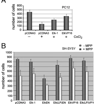
Firstly, we wanted to address whether overexpression of either Elk-1 or a constitutively active form, Elk-VP16, could rescue PC12 cells from apoptotic cell death. To that end, we have used a known chemical inducer of apoptosis, cobalt chloride [14,15]. When cells were transfected with an empty control plasmid, treatment with cobalt chloride resulted in a significant decrease in the surviving cell population (Fig. 3A). However, when cells were priorly transfected with expression plasmids encoding either Elk-1 or Elk-VP16, the decrease in cell number was nearly 3-fold lower (Fig. 3A).
Using a different apoptotic inducer, MPP, in SH-SY5Y neuroblastoma cells, we have shown that this pro-survival function of Elk-1 could be, at least to some extent, SRF-dependent. In the absence of MPP, the ectopic expression of Elk-EN decreased the number of surviving cells, while the SRF interaction mutant Elk(LP)-EN showed less decrease. Expression of Elk-VP16, and to a lesser extent Elk(LP)VP16, showed a slight increase, if any, in the number of surviving cells in culture (Fig. 3B, dark bars), while Elk-1 expression did not significantly alter the number of surviving cells. On the other hand, the treatment of control cells with MPP reduced cell survival by nearly half, which was only partly rescued by Elk-1, but significantly enhanced by Elk-VP16 (Fig. 3B, light bars). Elk(LP)-VP16 was also able to rescue the cells to a certain extent. Elk-EN reduced cell survival even further in the presence of MPP, but survival in MPP-treated cells was not significantly changed in the presence of Elk(LP)-EN (Fig. 3B, light bars).
3.4. Higher levels of endogenous Elk-1 correlates with higher survival rates in primary DRG neurons, with or without axotomy
In order to understand how Elk-1 might affect cellular function in healthy versus injured neurons, we studied Elk-1 in an in vitro neuronal injury model, axotomy of primary neuronal cells. To that end, we have employed primary DRG cultures, and analyzed the survival of cells in culture, with or without axotomy, in relation to endogenous Elk-1 levels and its phosphorylation (seeSection 2.7). It should be noted that 24 h following axotomy, approximately 50% of axotomized neurons die; this rate is only 5% under control conditions (data not presented).
When cells that were in the process of dying or those that were surviving were analyzed for their Elk-1 immunofluorescence inten-sities (24 h after axotomy), it was seen that the“dead” cells exhibited
essentially similar levels of Elk-1, however those that survived, or “live” cells, exhibited higher levels of Elk-1 irrespective of axotomy (Fig. 4A). The axotomized “live” cells (mimicking survival after
Fig. 1. A dominant repressive form of Elk-1, namely ElkEN, can repress transcription from the ets motif and result in decreased differentiation and cell survival in PC12 cells. (A) A schematic diagram showing that ElkEN can bind to ets motifs, interact with its partner protein SRF and repress transcription of TCF-responsive genes. (B) Transfection of a constitutively active fusion, ElkVP16, into PC12 cells in increasing amounts (50 and 100 ng) leads to increased luciferase activity from an SRE-Luc reporter plasmid. Co-transfection of increasing amounts of ElkEN represses ElkVP16-induced transcription up to 10-fold in PC12 cells. (C) PC12 cells were either stimulated by EGF (100μM) or NGF (50 ng/ml) for 3 days and scored for differentiation. Cells were co-transfected with GFP along with either empty control plasmid (pCDNA3) or Elk-EN expression plasmid in increasing amounts (50, 100, 250 ng/well) 1 day prior to growth factor treatment. The differentiation scores are quantified in Excel and shown in graphs; (D) In a parallel set of experiments, cells were co-transfected with GFP along with either empty control plasmid (pCDNA3, 250 ng), EN or Elk-EN expression plasmid in increasing amounts (100 and 250 ng per well) 1 day prior to growth factor treatment and treated with NGF for 4 days. The differentiation scores are quantified in Excel and shown in graphs (*pb0.16; **pb0.2; both compared to NGF control); (E) a parallel set of transfections for NGF, NGF + ElkEN or NGF + Elk(LP)EN were studied under epifluorescence microscope (representative images are shown; arrows indicate representative images quantified as differentiation; arrowheads indicate neurite extensions that are smaller than the accepted threshold and thus not quantified as differentiation). (F) Average number of surviving PC12 cells upon increasing amounts of Elk-EN transfection. Live cells were counted in a minimum of threefields per well, in at least two wells per assay. Data shown are from day 3 after transfection and represent cell survival for days 1–4.
neuronal injury) showed less Elk-1 immunofluorescence than the non-axotomized “live” cells (mimicking survival of healthy, un-damaged neurons), which could be due to the relatively shortened length of the axons, and loss of microtubule-associated Elk-1[7]. These results appear to be specific for Elk-1, since immunoreactivity of another ETS domain transcription factor, PEA3, does not show any significant change in fluorescence intensities (data not shown).
We then investigated the kinetics of Elk-1 phosphorylation following axotomy in these primary DRGs over a period of 30 min after axotomy. In order to provide statistically significant semi-quantification of immuno-fluorescence intensities, we have measured Elk-1 and phospho-Ser383-Elk-1 immunofluorescences in non-axotomized cells within the same culture, and normalized those from axotomized neurons accordingly. When data were analyzed, it was observed that the amount of total Elk-1 peaked at around 5 min following axotomy, after which it reached a plateau, while the amount of phospho-Ser-383-Elk-1 started to increase 10 min after axotomy up until 30 min (Fig. 4B). This relatively fast accumulation of Elk-1 could be due to potential local translation events, since bioinformatics analysis indicates presence of putative dendritic and axonal localization motifs in the untranslated regions (UTRs) of the elk-1 mRNA (seeSection 4). Both axotomized and uninjured neurons had significantly higher Elk-1 immunoreactivity if they had survived, compared to the ones that had died during 24 h of incubation (Fig. 4A). This change in immunofluorescence intensities appears to be significant and specific to Elk-1, since immunofluorescence intensities for another ETS domain factor, PEA3, does not change before and after axotomy (data not shown).The serum-induced nuclear translocation of phospho-Ser383-Elk-1 was previously shown[7]. Our hypothesis is that in this neuronal injury model, axotomy could serve as another signal generating
an increase in phospho-Elk-1, leading to enhanced survival of the injured neuron.
3.5. MCL-1 is a target of Elk-1 in neuronal survival
If the function of phospho-Elk-1 is to ensure survival of the neuron involved, then it is likely that this transcription factor would induce expression of related genes. There are a number of survival-related promoter sequences with putative ets motifs to which Elk-1 can bind, as well as some known Elk-1 targets in other cell systems.
Mcl-1, a member of the Bcl-2 family, was previously shown to be an anti-apoptotic target of Elk-1 in the HEK293 cell line as well as in hematopoietic cells[8,9]. We therefore wanted to address whether Mcl-1 is regulated by Elk-1 in a neuronal context. To that end, we initially performed luciferase reporter assays using MCL-1-Luc reporters with a constitutively active Elk-VP16 fusion protein, and observed a significant increase in luciferase activity (Fig. 5A). No reporter activity was observed when an SRF interaction-deficient Elk (LP)-VP16 construct was used (Fig. 5A), and repression was observed by Elk-EN in PC12 cells (data not shown). However, to be more certain about the validity of this regulatory correlation, we performed RNAi-mediated depletion of endogenous Elk-1 in SH-SY5Y cells. These cells were selected because of their higher transfection efficiency when compared to PC12 cells, so as to make sure that we have a substantial knock-down. When cells were transfected with a scrambled RNA (scrRNA) control plasmid, where Elk-1 protein was still present[12], human MCL-1 mRNA levels were unaffected in RT-PCR experiments. However, when increasing amounts of a plasmid expressing siRNA duplex directed against Elk-1 (siElk-1) were transfected into SH-SY5Y
Fig. 2. Elk-EN-dependent cell death is via caspase-dependent apoptosis. (A) PC12 cells were either transfected with control vector pCDNA3 or with the dominant repressive form, Elk-EN, and nuclei were stained with Hoechst dye. Representative images of mitotic cells (long arrows) and apoptotic nuclei (short arrows) are indicated. (B) Quantification of percent apoptosis and mitotic index (MI) in the experiments presented in (A). (C) PC12 cells were either transfected with a control plasmid, ElkEN expression plasmid on its own, or in the presence of either a rat IAP-1 (rIAP1) expression plasmid or the caspase inhibitor Z-VAD-fmk. Quantification of percent apoptosis and mitotic index (MI) was scored.
cells, there was a significant loss of the MCL-1 mRNA (Fig. 5B), indicating that presence of endogenous Elk-1 is important for maintenance of MCL-1 in these cells.
3.6. Survival motor neuron 1 (SMN1) as a potential novel target of Elk-1 The SMN proteins are components of a large complex that regulate the biogenesis of components of the RNA splicing machinery, and reduced levels of SMN1 correlates with a neurological disorder known as spinal muscular atrophy, SMA, where motor neurons selectively degenerate [16]. The homozygous absence of which leads to spinomuscular atrophy (SMA), a neurodegenerative disorder with progressive loss of motor neurons. SMN1 and SMN2 are two copies of the same gene on different chromosomal locations (telomeric and centromeric regions of chromosome 5q, respectively; [17]). SMN2 codes for a shorter copy due to exon skipping, and cannot compensate for the absence of SMN1 in SMA patients. The promoter sequences for both SMN copies appear to be identical[17], and contain two ets motifs (see also [5], and Section 4). We therefore investigated whether the SMN promoters are genuine targets for regulation by Elk-1. When endogenous Elk-1 levels were knocked down by RNAi in SH-SY5Y cells, we have observed a decrease in the levels of not only the SMN1 message, but also of SMN2 (Fig. 6A). This was further confirmed at the protein level (Fig. 6B). To analyze whether this is a direct effect of transcriptional regulation by Elk-1, we have cloned the human SMN promoter upstream from a luciferase reporter gene. When SH-SY5Y cells are transfected with the constitutively active Elk-VP16 expression plasmid, there was a nearly two-fold increase in the promoter activity, which was reduced when cells were transfected
Fig. 3. Elk-1 can protect neuronal cell lines from apoptosis. (A) PC12 cells were transfected either with control plasmid, Elk-1 or Elk-VP16, and treated with cobalt chloride, a known apoptosis-inducing agent. Live cells were scored and results are representative of surviving cells at the end of day 2. (B) SH-SY5Y neuroblastoma cells were transfected either with plasmid control plasmid, Elk-1, ElkEN, Elk(LP)EN, ElkVP16 or Elk(LP)VP16 expression plasmids, and treated with MPP, a known apoptosis-inducing agent. Live cells were scored and results are representative of surviving cells at the end of 48 h in the presence or absence of 1μM MPP. (*pb0.1 vs relevant control in −MPP and +MPP conditions).
Fig. 4. High levels of Elk-1 expression are correlated with higher rates of survival in primary DRG cultures. (A) Elk-1 immunofluorescence intensities in live and dead cells. PI stains the nuclei of dead neurons. The bold arrow points to an axotomized dead neuron while thin arrows indicate the cells that survived the axotomy at the time of analysis. (seeSection 2; *pb0.05 live vs dead within groups; Elk-1 in green, PI in red, merged image labeled PI+Elk-1). (B) Elk-1 and Phospho-Elk-1 (Elk-1p) levels in cultured DRG neurons following axotomy. The images in the panels show three typical axotomized neurons 5 and 30 min after axotomy. Changes in thefluorescence intensity relative to control preparations were expressed as percentages. (*pb0.05 vs control for Elk-1, and #pb0.05 vs control for Elk-1p).
with the plasmid expressing siRNA directed against Elk-1 (siElk-1) plasmid (Fig. 6C).
To provide further evidence for a role of Elk-1 in regulating the human SMN promoter, we performed the opposite study, i.e., we overexpressed exogenous Elk-1 in SH-SY5Y cells to see whether SMN levels change accordingly. The results showed that the level of SMN transcript was increased in a dose-dependent manner with increasing levels of Elk-1–VP16 (Fig. 7A).
To show whether siElk-1-mediated cell death could be rescued by SMN, and if SMN is a likely target of Elk-1 in relation to its survival function, we co-transfected the SH-SY5Y cells with either scrRNA or siElk-1 along with increasing amounts of SMN expression plasmid. In either scrRNA- or siElk-1-transfected cells, co-transfection of the SMN resulted in a decrease in caspase activity in a dose-dependent manner (Fig. 7B), indicating loss of SMN is at least one of the reasons behind siElk-1-induced apoptosis and that SMN can behave as a pro-survival factor, at least to a certain extent.
We also created single or double mutations of the ets motifs on the SMN promoter to identify the importance of these sequences to the transactivation of this promoter via Elk-1. Elimination of either one or both of those motifs results in a decrease in the basal levels of SMN promoter activation, however when the reporter constructs were co-transfected with the Elk-VP16 expression plasmid it was observed that the second ets motif (ets2) has a more pronounced impact on the activated levels of transcription from this promoter (Fig. 8A). Therefore, we wanted to investigate binding of Elk-1 to this second ets motif in chromatin immunoprecipitation assays (ChIP). We transfected the cells with either wildtype (SMN-Luc) or deleted (SMNΔ1-Luc or SMNΔ2-Luc) reporter constructs, along with the Elk-Flag expression plasmid. Exogenous Elk-1 was immunoprecipitated, and the subsequent PCR for the ets2 region showed that Elk-1 physically binds to this region in the wildtype reporter or the SMN Δ1-Luc construct, but not in the SMNΔ2-Luc construct. This provides further evidence that ets2 motif in the SMN promoter is a genuine direct target for regulation by Elk-1 in neuroblastoma cells (Fig. 8B). When a similar PCR was performed for the ets1 region, however, very little, if any, binding to Elk-1 was observed either in the wildtype reporter or the deletion mutants (around 20% in ets1 PCR as opposed to around 80% in ets2 PCR for wildtype sequence;Fig. 8B). The SMNΔ2 deletion caused nearly total loss of binding by Elk-1 to the ets2 motif as well as the ets1 motif, whereas SMNΔ1 deletion caused very little, if
Fig. 5. 1 can regulate MCL-1, a pro-survival gene, in SH-SY5Y neuroblastoma cells. (A) Elk-VP16, but not Elk(LP)-Elk-VP16, can activate an mcl1-Luc reporter construct in SH-SY5Y cells. Cells were co-transfected with mcl1-Luc reporter construct along with either pCDNA3 empty control plasmid, or expression plasmids encoding either Elk-VP16 or Elk(LP)-VP16. Luminometric measurements were normalized to Renilla luciferase internal activity and reported as relative luminescence intensities. (B) SH-SY5Y cells were transfected with siElk-1 plasmids in the indicated amounts, and RT-PCR assays were carried out for mcl-1 message. cDNAs were normalized for GAPDH expression as shown.
Fig. 6. The Survival-of-motor-neuron (smn) promoter is a novel target for Elk-1 in mediating neuronal survival. (A) SH-SY5Y cells were transfected with either scrambled RNA (scrRNA) plasmid or siElk-1 plasmid (4μg each), and RT-PCR assays were carried out for the SMN1 message. The same primers also amplify an alternatively-spliced isoform, SMN2 (see text for details). cDNAs were normalized for GAPDH expression as shown. (B) SH-SY5Y cells were transfected with either scrambled RNA (scrRNA) plasmid or siElk-1 plasmid (4μg each), and the cell lysates were analyzed for anti-SMN reactivity. Anti-actin antibody was used as a control. (C) The activation of the SMN promoter by Elk-1. The SMN promoter containing two ets motifs was cloned into a luciferase reporter construct. SH-SY5Y cells were co-transfected with the SMN-Luc reporter construct along with either pCDNA3 empty control plasmid, expression plasmid encoding Elk-VP16, or siElk1 plasmid. Luminometric measurements were normalized to Renilla luciferase internal activity and reported as relative luminescence intensities (*pb0.03; **pb0.2).
Fig. 7. ElkVP16 regulates SMN transcription and SMN overexpression can rescue siElk1-induced apoptosis. (A) SH-SY5Y neuroblastoma cells were transfected with either a control plasmid or increasing amounts of an expression plasmid encoding ElkVP16, as indicated, and RT-PCR was performed to analyze the levels of elk-1 and smn1 expression. (B) SH-SY5Y cells were transfected with plasmids encoding either scrambled RNA (scrRNA) or siElk-1, in the presence of either a control plasmid or increasing amounts of an expression plasmid encoding SMN. Caspase activation as an indicator of apoptosis was quantified using CaspACE Assay System (Promega). Statistical analyses are employed as described inSection 2.5. (*pb0.1 vs control).
any, change in either ets1 or ets2 binding, indicating that the deleted region in this construct is probably not a primary binding site for Elk-1 (Fig. 8B). This result also explains why very little decrease is observed in the luciferase assays when Elk-1–VP16 is expressed in the presence of the SMNΔ1-Luc construct, but almost a complete inhibition of activation is found when cells are transfected with Elk-1–VP16 and the SMNΔ2-Luc constructs (Fig. 8A).
4. Discussion
ETS domain transcription factors are characterized by a winged helix–turn–helix DNA binding domain that recognizes the consensus core sequence GGA, known as the ets motif[4,18,19]. One of the subfamilies within the ETS domain transcription factors is the ternary complex factor (TCF) subfamily, which is comprised of Elk-1, Sap-1a, Sap-1b, Fli1 and Net [1]. TCF factors bind to their respective recognition motifs as partners with serum response factor (SRF), which recognizes a nearby CArG box [20], the two motifs collectively known as the serum response element (SRE). This definition of TCF
factors often resulted in their association with mitogenic response of cells. However, TCF proteins have been correlated with various developmental events. We and others have shown that Elk-1, for example, is associated with neurons and the neuronal cytoskeleton
[5,7,21,22].
In order to further understand the function of Elk-1 in healthy and injured neurons, we studied the impact of Elk-1 expression both in cell line models and in a primary neuronal culture system. In PC12 cells, the expression of a dominant negative form of Elk-1 resulted in apoptosis, while overexpression of wild-type Elk-1 rescued these cells from cobalt chloride-induced cell death. Furthermore, RNAi-mediated downregulation of Elk-1 expression resulted in increased caspase activity in SH-SY5Y cells. In the primary DRG culture system, within minutes after axotomy Elk-1 immunoreactivity increased significantly within the cell body while its phosphorylated form accumulated with slower kinetics. When the 5′- and 3′-UTRs within the elk-1 mRNA were bioinformatically analyzed, putative dendritic and axonal localization motifs were found (currently under investigation), which may explain the relatively fast accumulation of Elk-1 protein in DRGs upon axotomy.
It should be noted that the results of our neuronal injury experiments does not conclusively show whether Elk-1 rescues neurons from death, or whether those neurons in the process of dying simply cease to express high levels of Elk-1; however combined with the results from cell lines and the novel target genes identified in this study (see below), our working model is that Elk-1 acts as a neuroprotective agent. In this study we provide evidence that human Elk-1 exerts its neuroprotective role through the regulation of a pro-survival gene MCL-1 and a novel target SMN.
Reduced levels of SMN protein are known to affect lower motor neurons in Spinal muscular atrophies (SMA), an autosomal recessive disorder. This disorder mainly affects the SMN1 gene, but SMN2 cannot effectively compensate for this loss. In mice, disruption of the mouse Smn gene is embryonic lethal[23]. Exogenous SMN was shown to prolong PC12 cell survival under serum-deprived conditions, and to protect NGF-differentiated PC12 cells, but it could not by itself protect against etoposide- or UV-induced apoptosis in these cells[24]. SMN proteins are ubiquitous proteins that function in snRNP assembly, thus reduced levels may affect RNA splicing of genes required for neuronal function, although this hypothesis is still under investigation (reviewed in [25]). Injection of wildtype SMN to smn-deficient zebrafish has been shown to rescue the motor axon defects; whereas some mutations failed to rescue the motor axon defects while retaining snRNP function. This brings to mind the question of whether SMN has an RNA transport role in neurons, in addition to involvement in snRNP biogenesis [26]. A pro-survival role for SMN has been indicated for neurons, in several studies where delivery of SMN to SMA model mice resulted in prolonged survival and restored motor function[27–29], making SMN a particularly important target for Elk-1 in neuronal survival response.
The promoter sequences on both telomeric and centromeric copies (SMN1 and SMN2, respectively) are almost identical [17]. When the promoter for SMN was examined we have observed that they contain two ets motifs to which TCF proteins could potentially bind; a previous report on both human and mouse SMN promoters have also indicated putative Ets-1 binding motifs [30]. Reporter constructs with either wildtype sequence or single or double ets motif deletions have confirmed that Elk-1 could indeed regulate this promoter, and RNAi-mediated degradation of endogenous Elk-1 reduced levels of SMN transcript (either both SMN1 and SMN2, or an alternatively transcribed SMN1; our experiments are not designed to distinguish between these two possibilities since it is not the primary aim of this study), as well as protein (Fig. 6), in spite of the fact that the levels of transcription from these promoters have previously been shown to be higher in primary neuronal cultures than in cell lines[17]. Furthermore, chromatin IP assays indicate that the ets2 motif present within the SMN1 promoter is bound by Elk-1 and this binding can activate
Fig. 8. Elk-1 binds to ets motifs on the SMN promoter. (A) The ets motifs on the SMN promoter were deleted either singly (SMNΔ1- and SMNΔ2-Luc) or doubly (SMNΔ1Δ2-Luc), and constructs were analyzed for transactivation by Elk-VP16. The SH-SY5Y cells were co-transfected with the luciferase reporter constructs, either pCDNA3 empty control plasmid or Elk-VP16 expression plasmid, and Renilla luciferase control plasmid. Luminometric measurements were normalized to Renilla luciferase internal activity and reported as relative luminescence intensities. (B) Chromatin IP assays in SH-SY5Y cells cotransfected with Elk-Flag expression plasmid with either wildtype or deleted smn-Luc reporter plasmids. Quantification is done by measuring intensities of bands (QuantityOne software, BioRad) and normalizing as discussed inSection 2, and reported with respect to input bands. Flag-agarose beads were used to precipitate Elk-1 in the cells, and binding to ets2 motif in all reporter constructs was measured (deletions were 3-bp deletions, while the ets2-containing region amplified in the ChIP assay spanned around 200 bp). The input shows the amplification of DNA isolated before immunoprecipitation. Anti-IgG antibody was used as a control antibody. The gels are representative images of duplicate samples.
transcription from a luciferase reporter (Fig. 8). Importantly, over-expression of SMN can rescue siElk-mediated apoptosis in SH-SY5Y cells. We therefore believe that SMN is a genuine novel target for regulation by Elk-1 in protecting neuronal cells from apoptosis.
Elk-1 is involved in the regulation of many neurodegeneration-related genes, or is regulated by them. It is interesting to note that overexpression ofα-synuclein, the insoluble aggregates of which is known to accumulate in Parkinson's disease, affects Elk-1 phosphorylation through MAPK signaling, which restores decreased cell viability [31]. Moreover α-synuclein forms a complex with Elk-1 in cultured cell lysates, most likely through ERK/MAPK, and Elk-1 reactivity is present in glial cytoplasmic inclusions (GCIs)[32]. Furthermore, it was shown that the ets motif in the promoter sequence of presenilin 1 gene (mutations of which are associated with Alzheimer's disease) was regulated by two ETS domain factors: ER81 activated from the sequence, whereas Elk-1 was shown to repress it at high doses[33]. Ets motifs in the promoter region of the Cu/Zn superoxide dismutase gene SOD1, the only known gene associated with the neurodegenerative disorder amyotrophic lateral sclerosis (ALS), was shown to be activated by Elk-1[34]. It would be interesting to see if the ELK-1 gene in humans carries any loss-of-function mutations in spinomuscular atrophy (SMA) and the above-mentioned neurodegener-ative diseases, and whether Elk-1 is, or could be used as, a general neuroprotective regulator against these degenerative processes.
Elk-1 phosphorylation was shown to increase in hippocampal regions of mice upon fear conditioning[35]. Whether this phosphorylation is related to survival of certain neurons in the circuitry is not yet known. Phospho-Elk-1 was shown to interact with neuronal microtubules, including those in the axon, but most phospho-Elk-1 species were shown to translocate to the nucleus upon serum stimulation[7], while ERK-dependent Elk-1 phosphorylation on Serines 383 and 389 was shown to drive the nuclear translocation of Elk-1, followed by SRE-dependent transcription in neurons [36]. Together with the data presented in this study, we hypothesize that both of these events are likely to enhance the survival of neurons.
However, there have been some contradictory reports, indicating a potential role for Elk-1 in promoting apoptosis in neurons. In one study, Elk-1 was found to be associated with mitochondria in the adult rat brain, and in particular the mitochondrial permeability transition pore complex (PTP). Electroporation of primary neural cultures led to a decrease in cell viability in these experiments, and a PTP inhibitor effectively blocked cell death, indicating that Elk-1 transfected into these cells caused apoptosis
[37]. Another study by the same group further showed that photo-transfection of Elk-1 mRNA to neurites resulted in increased cell death, whereas phototransfection into cell bodies does not[38]. Elk-1 in this study is assumed to be translated in the dendrites and translocated to the nucleus to cause cell death in a transcription-dependent manner. It was shown previously that an alternative translation product called short Elk-1 (sElk-1,[6]) had opposing role to long Elk-1, and that sElk-1 can regulate neuronal differentiation of PC12 cells[22], without any significant apoptosis or cell death. Therefore it is quite possible that these results are not contradictory, but simply that specific intracellular localization of various isoforms of Elk-1 in combination with different upstream signals may dictate whether the outcome for the neuron will be survival, proliferation or cell death. In other words Elk-1 may present a critical decision-making point for the neuron where various signals converge, although the exact mechanism of how and when such a decision is made is yet to be investigated, and is critical for a potential neuroprotective role for Elk-1 in therapeutics.
Acknowledgements
We would like to thank members of the Kurnaz, Ozturk and Sharrocks laboratories for helpful discussions and Prof. M.L. Kurnaz for help with statistics. We also would like to thank our anonymous reviewers for their helpful criticisms for the improvement of this study. This work was supported by TUBITAK-SBAG project no
106S247 as a partner of the ESF COST B-30 Action (neural plasticity and neuroregeneration, NEREPLAS), and TUBA Young Investigator Award to I.A.K. Funding for ADS was from the Wellcome Trust and a Royal Society-Wolfson award.
References
[1] A.D. Sharrocks, The ETS-domain transcription factor family, Nat. Rev. Mol. Cell Biol. 2 (2001) 827–837.
[2] R.A. Hipskind, M. Baccarini, A. Nordheim, Transient activity of Raf-1, MEK and ERK2 coincides kinetically with Ternary Complex Factor phosphorylation and immediate-early gene promoter activity in vivo, Mol. Cell. Biol. 14 (1994) 6219–6231.
[3] H. Gille, M. Kortenjann, O. Thomae, C. Moomaw, C. Slaughter, M.H. Cobb, P.E. Shaw, ERK phosphorylation potentiates Elk-1-mediated ternary complex formation and transactivation, EMBO J. 14 (1995) 951–962.
[4] J.S. Yordy, R.C. Muise-Helmericks, Signal transduction and the Ets family of transcription factors, Oncogene 19 (2000) 6503–6513.
[5] V. Sgambato, P. Vanhoutte, C. Pages, M. Rogard, R. Hipskind, M.-J. Besson, J. Caboche, In vivo expression and regulation of Elk-1, a target of the extracellular-regulated kinase signaling pathway, in the adult rat brain, J. Neurosci. 18 (1998) 214–226.
[6] P. Vanhoutte, J.L. Nissen, B. Brugg, B. Della Gaspera, M.-J. Besson, R.A. Hipskind, J. Caboche, Opposing roles of Elk-1 and its brain-specific isoform, short Elk-1, in NGF-induced PC12 differentiation, J. Biol. Chem. 276 (2001) 5189–5196. [7] O. Demir, S. Korulu, A. Yildiz, A. Karabay, I. Aksan Kurnaz, Elk-1 interacts with
neuronal microtubules and relocalizes to the nucleus upon phosphorylation, Mol. Cell Neurosci. 40 (2009) 111–119.
[8] E.R. Vickers, A. Kasza, I. Aksan Kurnaz, A. Seifert, L. Zeef, A. O'Donnell, A. Hayes, A.D. Sharrocks, Ternary TCF-SRF complex-regulated gene activity is required for cellular proliferation and inhibition of apoptotic cell death, Mol. Cell. Biol. 24 (2004) 10340–10351.
[9] K.J. Townsend, P. Zhou, L. Qian, C.K. Bieszczad, C.H. Lowrey, A. Yen, R.W. Craig, Regulation of MCL1 through a serum response factor/Elk1-mediated mechanism links expression of a viability-promoting member of Bcl2 family to the induction of hematopoietic cell differentiation, J. Biol. Chem. 274 (1999) 1801–1813. [10] M.A. Price, A.E. Rogers, R. Treisman, Comparative analysis of the ternary complex
factors Elk-1, SAP-1a and SAP-2 (ERP/NET), EMBO J. 14 (1995) 2589–2601. [11] Y. Ling, A.G. West, E.C. Roberts, J.H. Lakey, A.D. Sharrocks, Interaction of
transcription factors with serum response factor. Identification of the Elk-1 binding surface, J. Biol. Chem. 273 (1998) 10506–10514.
[12] O. Demir, I. Aksan Kurnaz, Wildtype Elk-1, but not a SUMOylation mutant, represses egr-1 expression in SH-SY5Y neuroblastomas, Neurosci. Lett. 437 (2008) 20–24.
[13] C.J. Marshall, Specificity of receptor tyrosine kinase signaling: transient versus sustained extracellular signal-regulated kinase activation, Cell 80 (1995) 179–185. [14] W. Zou, J. Zeng, M. Zhou, W. Xu, L. Sun, J. Wang, X. Liu, Involvement of caspase-3 and p38 mitogen-activated protein kinase in cobalt chloride-induced apoptosis in PC12 cells, J. Neurosci. Res. 67 (2002) 837–843.
[15] J.Y. Jung, W.J. Kim, Involvement of mitochondrial- and Fas-mediated dual mechanism in CoCl2-induced apoptosis of rat PC12 cells, Neurosci. Lett. 371 (2004) 85–90.
[16] L. Pellizzoni, Chaperoning ribonucleoprotein biogenesis in health and disease, EMBO Rep. 8 (2007) 340–345.
[17] B. Boda, C. Mas, C. Giudicelli, V. Nepote, F. Guimiot, B. Levacher, A. Zvara, M. Santha, I. LeGall, M. Simonneau, Survival motor neuron SMN1 and SMN2 gene promoters: identical sequences and differential expression in neurons and non-neuronal cells, Eur. J. Hum. Genet. 12 (2004) 729–737.
[18] B. Wasylyk, S.L. Hahn, A. Giovane, The Ets family of transcription factors, Eur. J. Biochem. 211 (1993) 7–18.
[19] P. Shore, A.J. Whitmarsh, R. Bhaskaran, R.J. Davis, J.P. Waltho, A.D. Sharrocks, Determinants of DNA-binding specificity of ETS-domain transcription factors, Mol. Cell. Biol. 16 (1996) 3338–3349.
[20] R. Treisman, Ternary complex factors: growth factor regulated transcriptional activators, Curr. Opin. Genet. Dev. 4 (1994) 96–101.
[21] E. Valjent, C. Pages, M. Rogard, M.-J. Besson, R. Maldonado, J. Caboche, Delta-9-tetrahydrocannabinol-induced MAPK/ERK and Elk-1 activation in vivo depends on dopaminergic transmission, Eur. J. Neurosci. 14 (2001) 342–352.
[22] S. Salinas, A. Briancon-Larjollet, G. Bossis, M.A. Lopez, M. Piechaczyk, I. Jariel-Encontre, A. Debant, R.A. Hipskind, SUMOylation regulates nucleo-cytoplasmic shuttling of Elk-1, J. Cell Biol. 165 (2004) 767–773.
[23] B. Schrank, R. Goetz, J.M. Gunnersen, J.M. Ure, K.V. Toyka, A.G. Smith, M. Sendtner, Inactivation of the survival motor neuron gene, a candidate gene for human spinal muscular atrophy, leads to massive cell death in early mouse embryos, Proc. Natl Acad. Sci. USA 94 (1997) 9920–9925.
[24] S. Vyas, C. Bechade, B. Riveau, J. Downward, A. Triller, Involvement of survival motor neuron (SMN) protein in cell death, Hum. Mol. Genet. 11 (2002) 2751–2764. [25] A.H.M. Burghes, C.E. Beattie, Spinal muscular atrophy: why do low levels of
survival motor neuron protein make motor neurons sick? Nat. Rev. Neurosci. 10 (2009) 597–609.
[26] T.L. Carrel, M.L. McWhorter, E. Workman, H. Zhang, E.C. Wolstencroft, C. Lorson, G.J. Bassell, A.H.M. Burghes, C.E. Beattie, Survival motor neuron function in motor axons is independent of functions required for small nuclear ribonucleoprotein biogenesis, J. Neurosci. 26 (2006) 11014–11022.
[27] C.F. Valori, K. Ning, M. Wyles, R.J. Mead, A.J. Grierson, P.J. Shaw, M. Azzouz, Systemic delivery of scAAV9 expressing SMN prolongs survival in a model of spinal muscular atrophy, Sci. Transl. Med. 2 (2010) 35ra42.
[28] M.A. Passini, J. Bu, E.M. Roskelley, A.M. Richards, S.P. Sardi, C.R. O'Riordan, K.W. Klinger, L.S. Shihabuddin, S.H. Cheng, CNS-targeted gene therapy improves survival and motor function in a mouse model of spinal muscular atrophy, J. Clin. Invest. 120 (2010) 1253–1264.
[29] K.D. Foust, X. Wang, V.L. McGovern, L. Braun, A.K. Bevan, T.T. Le, P.R. Morales, M.M. Rich, A.H. Burghes, B.K. Kaspar, Rescue of the spinal muscular atrophy phenıtype in a mouse model by early postnatal delivery of SMN, Nat Biotech 28 (2010) 271–274. [30] R. Rouget, F. Vigneault, C. Codio, C. Rochette, I. Paradis, R. Drouin, L.R. Simard,
Characterization of the SMN promoter provides evidence for complex combinatorial regulation in undifferentiated and differentiated P19 cells, Biochem J 385 (2005) 433–443.
[31] A. Iwata, M. Maruyama, I. Kanazawa, N. Nukina,α-synuclein affects the MAPK patwhay and accelerates cell death, J. Biol. Chem. 272 (2001) 45320–45329. [32] A. Iwata, S. Miura, I. Kanazawa, M. Sawada, N. Nukina,α-synuclein forms a
complex with transcription factor Elk-1, J. Neurochem. 77 (2001) 239–252.
[33] M. Pastorcic, H.K. Das, Ets transcription factors ER81 and Elk-1 regulate the transcription of the human presenilin 1 gene promoter, Mol. Brain Res. 113 (2003) 57–66.
[34] M.S. Chang, H.Y. Yoo, H.M. Rho, Positive and negative regulatory elements in the upstream region of the rat Cu/Zn superoxide dismutase gene, Biochem. J. 339 (1999) 335–341.
[35] F. Sananbenesi, A. Fischer, C. Schrick, J. Spiess, J. Radulovic, Phosphorylation of hippocampal ERK1/2, Elk-1 and p90-Rsk1 during contextual fear conditioning: interactions between ERK1/2 and Elk-1, Mol. Cell. Neurosci. 21 (2002) 463–476. [36] J. Lavaur, F. Bernard, P. Trifilieff, V. Pascoli, V. Kappes, C. Pages, P. Vanhoutte, J. Caboche, A TAT-DEF-Elk-1 peptide regulates the cytonuclear trafficking of Elk-1 and controls cytoskeleton dynamics, J. Neurosci. 27 (2007) 14448–14458. [37] L.E. Barrett, J.-Y. Sul, H. Takano, E.J. Van Bockstaele, P.G. Haydon, J.H. Eberwine,
Region-directed phototransfection reveals the functional significance of a dendritically synthesized transcription factor, Nat. Meth. 3 (2006) 455–460. [38] L.E. Barrett, E.J. Van Bockstaele, J.-Y. Sul, H. Takano, P.G. Haydon, J.H. Eberwine,
Elk-1 associates with the mitochondrial permeability transition pore complex in neurons, Proc. Natl Acad. Sci. USA 103 (2006) 5155–5160.