Original research-Orijinal araştırma
http://dx.doi.org/10.7197/1305-0028.1970First and second trimester hCG levels have no
value in predicting small for gestational age infants
Birinci ve ikinci trimester hCG değerleri gebelik yaşına göre küçük bebekleri
tahmin etmede kullanmaya uygun değildir
Nilgün Güdücü*, Gökçenur Gönenç, Herman İşçi, Alin Başgül Yiğiter, İlkkan Dünder
Department of Obstetrics and Gynecology (Assist. Prof. N. Güdücü, MD, Assist. Prof. G. Gönenç, MD, Assist. Prof. H. İşçi, MD, Assoc. Prof. A. B. Yiğiter, MD, Prof. İ. Dünder, MD), İstanbul Bilim Üniversitesi Avrupa Hospital, TR-34394 İstanbul
Abstract
Aim. The aim of this study was to compare high hCG levels in the first and second trimester in
predicting small-for-gestational age (SGA) infants. This is the first study that compares hCG levels of first and second trimesters. Methods. Patients with both first and second trimester Down syndrome screening tests were investigated retrospectively and human chorionic gonadotropin (hCG) values were recorded. Patients were divided into groups as SGA and non-SGA. High hCG values (≥2 MoM) were compared with low hCG values (<2MoM). Results. There was no significant difference in the hCG levels in women delivering SGA and non-SGA infants. Sensitivity and specificity of both tests were the same (9.09 and 89.95 respectively). Conclusions. Neither first nor second trimester hCG values were predictive in detection of SGA infants
Keywords: Small-for-gestational-age, hCG, Down syndrome screening tests
Özet
Amaç. Bu çalışmada amaç birinci ve ikinci trimester human chorionic gonadotropin (hCG)
değerlerinin gebelik yaşına göre küçük bebekleri tahmin etmede faydalı olup olmadığını araştırmaktı. Yöntem. Hem birinci, hem de ikinci trimester Down sendromu tarama testi yaptırmış olan hastaların sonuçları retrospektif olarak tarandı ve hCG MoM değerleri tespit edildi. Hastalar gebelik yaşına göre küçük ve gebelik yaşına göre küçük (GYK) olan ve olmayan bebek doğuranlar olarak iki gruba ayrıldı. Yüksek hCG değerleri (≥2MoM) ile düşük hCG değerleri (<2MoM) karşılaştırıldı. Bulgular. GYK olan ve olmayan bebek doğuranların hCG değerleri arasında istatiksel olarak anlamlı bir fark görülmedi. Sensitivite ve spesifisite her iki grupta da aynı bulundu (sırasıyla 9.09 ve 89.95). Sonuç. Birinci ve ikinci trimester hCG değerlerinin GYK bebekleri tahmin etmede yeri yoktur.
Anahtar sözcükler: Gebelik yaşına göre küçük, hCG, Down sendromu tarama testleri Geliş tarihi/Received: January 31, 2013; Kabul tarihi/Accepted: May 20, 2013 *Corresponding author:
Dr. Nilgün Güdücü, Kadın Hastalıkları ve Doğum Anabilim Dalı, İstanbul Bilim Üniversitesi Avrupa Hastanesi, TR-34394 İstanbul. E-mail: [email protected]
Introduction
Fetal growth restriction is an important contributor to perinatal mortality and morbidity, as well as being a modulator of abnormal neurodevelopment [1]. Early identification and appropriate management of these fetuses at risk was shown to decrease the associated mortality by four-fold [2]. Human chorionic gonadotropin (hCG) was first used in 1987 to predict pregnancies with Down syndrome [3]. Both first trimester and second trimester screening tests for Down syndrome use hCG. HCG secretion begins very early in
pregnancy and reaches peak values at about 10 weeks of gestation, than the values decrease gradually and stay stable after the twentieth week of pregnancy. In pregnancies with small for gestational age (SGA) fetuses the placental content is also small, so previously an association was suggested between low first trimester hCG levels and delivering a SGA infant [3-7], which was rejected by other studies [8-10]. Goetzinger et al suggested an association between high first trimester hCG levels and the delivery of a SGA infant [11]. The aim of this study was to compare the efficacy of high hCG levels in the first and second trimester in predicting SGA infants. To the best of our knowledge this is the first study that compares hCG levels at first and second trimesters.
Materials and methods
This was a retrospective study performed by searching the data of women attending to our obstetrics outpatient clinic for their routine pregnancy visit between January 2006 and April 2010. The patients that underwent Down syndrome screening tests were searched and the results of first trimester screening tests (11-13 weeks) and second trimester screening tests (16-18 weeks) were investigated. All patients had their last menstrual period confirmed by a first trimester ultrasound. Serum levels of the analytes were then converted into multiples of median (MoM). Only the patients delivering in our institute after 34 weeks of pregnancy were included. Patients of our clinic are from a rather good socioeconomic status. The research project has been approved by the Ethics Committee of our University and it conforms to the ethical guidelines of the Declaration of Helsinki (revised Tokyo 2004). Exclusion criteria included pregnancies with incomplete information, known abnormal fetal karyotype, congenital malformations, multiple pregnancies, maternal illnesses as hypertension and preeclampsia, gastrointestinal malabsorption and also smoking. We did not exclude any case on the basis of abnormal fetal biometry or birth weight. We did not take the gender of the fetus and the method of conception into consideration.
Pregnancies were divided into two groups, SGA was defined as below and equal to the 10th percentile for that gestational age, other infants were defined as non-SGA. Gestational age was calculated according to the last menstrual period and was confirmed by the first trimester crown-rump length measurement.
For statistical analysis we used NCSS (Number Cruncher Statistical System) 2007 and PASS (Power Analysis and Sample Size) 2008 statistical Software (Utah, USA). Data showing the anthropometric parameters were presented with standard descriptive statistical calculations (mean±standard deviation). Sensitivity, specificity, positive and negative predictive values were calculated. Odds ratios were given. Within 95% confidence interval p-values <0.05 were considered as statistically significant. The results were considered statistically significant when the p-value was calculated less than 0.05 at a confidence interval of 95%.
Results
We identified 451 patients during our study period. Mean maternal age was 30±4 years (18-43 years), mean maternal height was 164±5.8cm (150-180cm), mean maternal weight before pregnancy was 62.5±10 kg (40-104kg), mean maternal weight at delivery was 78±10kg (56-117kg), mean maternal weight gain during pregnancy was 15±5kg (-2 to +45kg), mean gestational age at delivery was 38.9±1.2weeks (34-42weeks), mean birth weight was 3377±413 grams (1800-4560 grams). Forty-five infants were classified as SGA and 406 as non-SGA.
Table 1. First and second trimester hCG levels (MoM).
Min-Max Mean ±SD Median
hCG first trimester 0.18-4.73 1.12±0.69 0.95 hCG second trimester 0.04-5.83 1.12±0.65 1.02 Paired Samples Test p 0.937
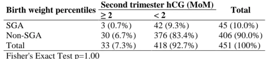
Table 1 shows the mean hCG levels in the first and second trimester, 1.12±0.69 (0.18-4.73) and 1.12±0.64 (0.04-5.83) respectively. Table 2 shows the results of second trimester screening tests and Table 3 shows the results of first trimester screning tests. There was no statistically significant relationship between first and second trimester hCG levels and birth weight percentiles. The results of both tests were the same: sensitivities were 9.09%, specificities were 89.05%, positive predictive values were 6.67%, negative predictive values were 92.61%, odds ratios were 0,895 (0,26-3,06).
Table 2. Second trimester hCG levels (MoM) compared to birth weight percentiles. Birth weight percentiles Second trimester hCG (MoM) Total
≥ 2 < 2
SGA 3 (0.7%) 42 (9.3%) 45 (10.0%) Non-SGA 30 (6.7%) 376 (83.4%) 406 (90.0%) Total 33 (7.3%) 418 (92.7%) 451 (100%) Fisher's Exact Test p=1.00
Table 3. First trimester hCG levels (MoM) compared to birth weight percentiles. Birth weight percentiles First trimester HCG (MoM) Total
≤ 2 > 2
SGA 41 (9.1%) 4 (0.9%) 45 (10%) Non-SGA 365 (80.9%) 41 (9.1%) 406 (90%) Total 406 (90%) 45 (10%) 645 (100%) Fisher’s Exact test p=1.000
Discussion
HCG is a well characterized hormone primarily produced by the placenta and this peptide hormone is important during implantation [12]. It is an angiogenic factor for uterine endothelial cells [13, 14] and it also induces vascular endothelial growth factor (VEGF) production in the placenta [15]. Increased hCG levels were suggested to be the result of placental immaturity [16] and were also proposed as a compensatory hypersecretory response of trophoblasts to hypoxia that results from abnormal placentation [17]. Some previous studies have shown a trend toward growth restriction with decreasing levels of hCG in the first trimester, which did not reach statistically significant levels [4-7]. Spencer et al. [10] detected no association with low hCG levels and SGA infants in the first trimester. Canini et al found decreased hCG levels to be associated only with hypertensive diseases of pregnancy but not with birth weight or growth restriction [18]. In a recent study where the first trimester MoMs for hCG were converted to 5th, 10th, 90th and 95th percentiles, only hCG levels higher than 90th percentile, which was nearly equal to 2 MoM, were associated with delivering a SGA infant [11]. In our study we did not detect an association between first trimester hCG levels higher than 2 MoM and fetal growth restriction. A recent meta-analysis showed that hCG predicted SGA fetuses better in the second trimester when compared to first trimester [19]. Studies performed in the second trimester also reported contraversial results about the association of hCG with fetal growth restriction [20-25], one possible explanation may be the association between increased hCG and hypertensive diseases of the pregnancy, where growth restriction is common. A previous study showed no association between hCG levels and growth retarded fetuses, but women with preeclampsia had higher hCG values [26]. To avoid this bias we excluded patients with hypertensive diseases of the pregnancy in our study. Placental bed biopsies from pregnancies with fetal growth restriction but without preeclampsia demostrated partial absence of the physiologic changes in the spiral arteries [27]. Probably more than one mechanism played role in the defective placentation, but all resulted in ischemia [28]. The findings of this study confirm that in pregnancies that subsequently deliver SGA infants there is no evidence of increased hCG levels in the first or second trimester.
Extremely high hCG levels in the second trimester may still draw attention [29, 30], we used a lower cut-off level than suggested in the mentioned studies and the maximum hCG level detected in our study was 5,8MoM. As far as we know there is no study that controlled the effect of extremely high hCG levels in delivering a SGA infant in the first trimester. HCG is used both in the first and second trimester Down syndrome screening tests but one potential disadvantage of screening in the second trimester is the completion of invasive activity of the extravillous trophoblastic cells [31], after this time intervention with aspirin treatment is probably too late. In our study where first and second trimester tests were performed using the same patient population, the results of first and second trimester hCG were nearly equal. Measurement of hCG levels in the first or second trimester did not seem to contribute to the early prediction of fetal growth restriction. Our limitations were the retrospective nature of the study and the possible gender differences that we did not take into consideration.The predictive value of one marker of the Down syndrome screening tests could be biased by another high or low marker, we did not exclude those patients. It is also possible that the use of population based growth chart percentiles instead of customized growth chart percentiles adjusted for maternal weight, height, sex and gender might have obscured the results. This may have prevented the selection of constituonally small fetuses that do not have any risk of peripartum asphyxia. Patients with high hCG levels might have undergone more amniocentesis and this might have been the reason for some of the adverse pregnancy outcomes, but we do not expect such a bias to complicate the etiology of a SGA infant.
In conclusion we failed to detect an association between delivering a SGA infant and high levels of hCG in the first or second trimester, but to take extremely high levels of hCG into consideration may still be beneficial.
References
1. Kok JH, den Ouden AL, Verloove-Vanhorick SP, Brand R. Outcome of very preterm small for gestational age infants: the first nine years of life. BJOG 1998; 105: 162-8.
2. Lindqvist PG, Molin J. Does antenatal identification of small-for-gestational age fetuses significantly improve their outcome? Ultrasound Obstet Gynecol 2005; 25: 258-64.
3. Bogart MH, Pandian MR, Jones OW. Abnormal maternal serum chorionic gonadotropin levels in pregnancies with fetal chromosome abnormalities. Prenat Diagn 1987; 7: 623-30.
4. Smith GC, Stenhouse EJ, Crossley JA, Aitken DA, Cameron AD, Connor JM. Early pregnancy levels of pregnancy-associated plasma protein a and the risk of intrauterine growth restriction, premature birth, preeclampsia, and stillbirth. J Clin Endocrinol Metab 2002; 87: 1762-7.
5. Krantz D, Goetzl L, Simpson JL, Thom E, Zachary J, Hallahan TW, Silver R, Pergament E, Platt LD, Filkins K, Johnson A, Mahoney M, Hogge WA, Wilson RD, Mohide P, Hershey D, Wapner R; First Trimester Maternal Serum Biochemistry and Fetal Nuchal Translucency Screening (BUN) Study Group. Association of extreme first-trimester free human chorionic gonadotropin-beta, pregnancy-associated plasma protein A, and nuchal translucency with intrauterine growth restriction and other adverse pregnancy outcomes. Am J Obstet Gynecol 2004; 191: 1452-8.
6. Ong CY, Liao AW, Spencer K, Munim S, Nicolaides KH. First trimester maternal serum free beta human chorionic gonadotrophin and pregnancy associated plasma protein A as predictors of pregnancy complications. BJOG 2000; 107: 1265-70.
7. Tul N, Pusenjak S, Osredkar J, Spencer K, Novak-Antolic Z. Predicting complications of pregnancy with first-trimester maternal serum free-betahCG, PAPP-A and inhibin-A. Prenat Diagn 2003; 23: 990-6.
8. Morssink LP, Kornman LH, Hallahan TW, Kloosterman MD, Beekhuis JR, de Wolf BT, Mantingh A. Maternal serum levels of free beta-hCG and PAPP-A in the first trimester of pregnancy are not associated with subsequent fetal growth retardation or preterm delivery. Prenat Diagn 1998; 18: 147-52.
9. Kavak ZN, Basgul A, Elter K, Uygur M, Gokaslan H. The efficacy of first-trimester PAPP-A and free beta hCG levels for predicting adverse pregnancy outcome. J Perinat Med 2006; 34: 145-8.
10. Spencer K, Cowans NJ, Avgidou K, Molina F, Nicolaides KH. First-trimester biochemical markers of aneuploidy and the prediction of small-for-gestational age fetuses. Ultrasound Obstet Gynecol 2008; 31: 15-9.
11. Goetzinger KR, Singla A, Gerkowicz S, Dicke JM, Gray DL, Odibo AO. The efficiency of first-trimester serum analytes and maternal characteristics in predicting fetal growth disorders. Am J Obstet Gynecol 2009; 201: 412.e1-6. 12. Tao YX, Lei ZM, Hofmann GE, Rao CV. Human intermediate trophoblasts
express chorionic gonadotropin/luteinizing hormone receptor gene. Biol Reprod 1995; 53: 899-904.
13. Lei ZM, Reshef E, Rao V. The expression of human chorionic gonadotropin/luteinizing hormone receptors in human endometrial and myometrial blood vessels. J Clin Endocrinol Metab 1992; 75: 651-9.
14. Zygmunt M, Herr F, Keller-Schoenwetter S, Kunzi-Rapp K, Münstedt K, Rao CV, Lang U, Preissner KT. Characterization of human chorionic gonadotropin as a novel angiogenic factor. J Clin Endocrinol Metab 2002; 87: 5290-6.
15. Rodway MR, Rao CV. A novel perspective on the role of human chorionic gonadotropin during pregnancy and in gestational trophoblastic disease. Early Pregnancy 1995; 1: 176-87.
16. Fitzgerald B, Levytska K, Kingdom J, Walker M, Baczyk D, Keating S. Villous trophoblast abnormalities in extremely preterm deliveries with elevated second trimester maternal serum hCG or inhibin-A. Placenta 2011; 32: 339-45.
17. Hsu CD, Chan DW, Iriye B, Johnson TR, Hong SF, Repke JT. Elevated serum human chorionic gonadotropin as evidence of secretory response in severe preeclampsia. Am J Obstet Gynecol 1994; 170: 1135-8.
18. Canini S, Prefumo F, Pastorino D, Crocetti L, Afflitto CG, Venturini PL, De Biasio P. Association between birth weight and first-trimester free beta-human chorionic gonadotropin and pregnancy-associated plasma protein A. Fertil Steril 2008; 89: 174-8.
19. Morris RK, Cnossen JS, Langejans M, Robson SC, Kleijnen J, Ter Riet G, Mol BW, van der Post JA, Khan KS. Serum screening with Down's syndrome markers to predict pre-eclampsia and small for gestational age: systematic review and meta-analysis. BMC Pregnancy and Childbirth 2008; 8: 33.
20. Spencer K. Second-trimester prenatal screening for Down syndrome and the relationship of maternal serum biochemical markers to pregnancy complications with adverse outcome. Prenat Diagn 2000; 20: 652-6.
21. Sayin NC, Canda MT, Ahmet N, Arda S, Süt N, Varol FG. The association of triple-marker test results with adverse pregnancy outcomes in low-risk pregnancies with healthy newborns. Arch Gynecol Obstet 2008; 277: 47-53. 22. Toal M, Chaddha V, Windrim R, Kingdom J. Ultrasound detection of placental
insufficiency in women with elevated second trimester serum alpha-fetoprotein or human chorionic gonadotropin. J Obstet Gynaecol Can 2008; 30: 198-206. 23. Odibo AO, Sehdev HM, Stamilio DM, Macones GA. Evaluating the thresholds of
abnormal second trimester multiple marker screening tests associated with intra-uterine growth restriction. Am J Perinatol 2006; 23: 363-7.
24. Karsidag AY, Buyukbayrak EE, Kars B, Suyugul U, Unal O, Turan MC. The relationship between unexplained elevated serum markers in triple test, uterine artery Doppler measurements and adverse pregnancy outcome. J Pak Med Assoc 2010; 60: 181-6.
25. Gagnon A, Wilson RD, Audibert F, Allen VM, Blight C, Brock JA, Désilets VA, Johnson JA, Langlois S, Summers A, Wyatt P; Society of Obstetricians and Gynaecologists of Canada Genetics Committee. Obstetrical complications associated with abnormal maternal serum markers analytes. J Obstet Gynaecol Can 2008; 30: 918-49.
26. Luckas MJ, Sandland R, Hawe J, Neilson JP, McFadyen IR, Meekins JW. Fetal growth retardation and second trimester maternal serum human chorionic gonadotrophin levels. Placenta 1998; 19: 143-7.
27. Brosens I, Dixon HG, Robertson WB. Fetal growth retardation and the arteries of the placental bed. Br J Obstet Gynaecol 1977; 84: 656-63.
28. Romero R, Kusanovic JP, Kim CJ. Placental disorders in the genesis of the great obstetrical disorders. In: Pijnenborg R, Brosens I, Romero R, eds. Placental bed disorders. Cambridge University Press; 2010: 271-89.
29. Ganapathy R, Lamont RF, Bassett P. Unexplained elevated maternal serum beta-HCG concentration and adverse pregnancy outcome. Prenat Diagn 2007; 27: 995-9.
30. Tavor O, Shohat M, Lipitz S. The relationship between perinatal outcome of singleton pregnancies and isolated highly elevated levels of maternal serum human chorionic gonadotropin at mid-gestation. Isr Med Assoc J 2007; 9: 509-12.
31. Caniggia I, Winter J, Lye SJ, Post M. Oxygen and placental development during the first trimester: implications for the pathophysiology of pre-eclampsia. Placenta 2000; 21 Suppl A: S25-30.