Tremor onset with acute frontal infarct and disappearance
with the second stroke
Deniz Yerdelen, MD, Selin Yetkinel, MD, Ali Doğan, MD.
ABSTRACT
ىلع اهيلإ رظني ةكرلحا تابارطضاب ةطبترلما ةيغامدلا ةتكسلا نإ
.اهروهظ رخأت وأ ةيغامدلا ةتكسلا تاملاع نم ةملاع لوأ اهنأ
تابارطضا ينب ةردان ةجيتن يه ةيغامدلا ةتكسلا دعب شاعرلا
شاعرلا ءافتخا نع ةروشنلما تانايبلاو تامولعلما رفوتتو .ةكرلحا
نع ةروشنلما ريراقتلا ىلإ ةفاضلإاب كلذو يغامدلا ءاشتحلاا دعب
ةلاح مدقن ،ريرقتلا اذه يف .يغامدلا ءاشتحلاا نع تجانلا شاعرلا
ةيرشقلا ءاشتحا اهيلت ديلل لباقلما ءزلجا يف دالحا شاعرلا نم
قطانم مضيو يناثلا ءاشتحلاا دعب شاعرلا نم ءافتخاو يماملأا
.ةركلا فصن سفن يف ةعساو
Ischemic stroke associated movement disorders can be seen as the first sign of a stroke or as a delayed onset development. Tremor after a stroke is a rare finding among movement disorders. In addition to reports of tremor caused by cerebral infarction of varied locations, data on the disappearance of existing tremor following infarction is also available. In this report, we present a case with acute tremor in the contralateral hand following frontal cortical infarction, and disappearance of the tremor after the second infarction comprising large areas in the same hemisphere.Neurosciences 2015; Vol. 20 (2): 164-166 doi: 10.17712/nsj.2015.2.20140588 From the Department of Neurology (Yerdelen, Yetkinel), Faculty of Medicine, Baskent University, Adana, and the Department of Neurology (Dogan), Bandırma Public Hospital, Balıkesir, Turkey. Received 16th September 2014. Accepted 16th March 2015.
Address correspondence and reprint request to: Prof. Deniz Yerdelen, Department of Neurology, Faculty of Medicine, Baskent University, Adana, Turkey. Fax. +90 (322) 3271274. E-mail: [email protected]
Disclosure. The authors have no affiliation or financial involvement with organizations or entities with a direct financial interest in the subject matter or materials discussed in the manuscript. No funding was received for this work from any organization.
Case Reports
I
nvoluntary abnormal movements caused by strokes are relatively common. Chorea, tremor, dystonia, Parkinsonism, and myoclonus associated with both infarctions and cerebral hemorrhage may occur delayed or progressive as part of the symptomatology of acute stroke.1 Patients with hand tremor associated with cerebralinfarct are not encountered frequently. In addition to reports of tremor caused by cerebral infarction of varied locations, data on the disappearance of existing tremor following infarction is also available.2-5 Kim2 reported
hand tremor that developed 3-21 months after infarct of the caudate nucleus, internal capsule, and thalamus in 3 patients. Kim et al4 also reported a case with
essential tremor, which improved 5 weeks after frontal infarct. There is a scarcity of reports in the literature on the development of, and improvement of hand tremor following a subsequent stroke. In this report, we present a case with acute tremor in the contralateral hand following frontal cortical infarction and disappearance of the tremor after the second infarction comprising large areas in the same hemisphere. Our objective in presenting this particular case is to report the interesting development and disappearance of tremor as a result of ischemic stroke.
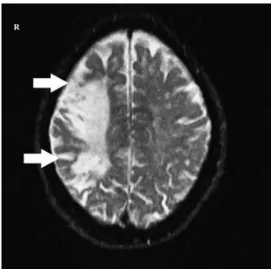
Case Report. A 78-year-old male was admitted to the emergency department with a complaint of sudden onset of tremor in his left hand fingers. His medical and family histories were unremarkable. Vital signs were within normal limits. Neurological examination revealed resting and postural, continuous, rhythmic, involuntary movements like tremor, prominent in left first and second fingers of the hand. The frequency of the tremor was the same at rest and during action. He did not have rigidity at the time of the tremor. He did not have a tremor in his leg. As the complaint’s started acutely, cerebral and diffusion MRI were carried out, and the results showed an acute infarction in the right frontal region (Figure 1). Examinations for the etiology of the stroke (carotid-vertebral artery color Doppler, electrocardiography, and echocardiography) were not remarkable. The EEG revealed normal findings. Electromyographic assessment of the left
OPEN ACCESS
165 Neurosciences 2015; Vol. 20 (2) Tremor associated with frontal infarct … Yerdelen et al
www.neurosciencesjournal.org
first metacarpal region with surface electrodes showed resting and postural involuntary rhythmic activity at a frequency of 6-7 Hz, 20-50 ms duration and 0.3 to one millivolt amplitude (Figure 2). He was started on 100 mg acetylsalicylic acid. Due to the acute nature of the stroke, no treatment for the tremor was given and we planned to examine the patient after 2 weeks. However, he did not attend for examination.
His second presentation to our clinic was one year after the first event. He had developed left hemiparesis syndrome 7 months after the first event. According to the information given by the patient and his relatives, the tremor in the left hand had disappeared following the second stroke. On neurological examination, left spastic hemiparesis (upper extremity 0/5, lower extremity 4/5) was determined. The tremor was not detected. Cerebral MRI showed chronic ischemia of right fronto-parietal and parietal-occipital regions (Figure 3).
Discussion. Many brain regions play a role in the development of tremor. These regions that control
voluntary and involuntary commands are the main parts of the critical circuits. Each of the loops has specific anatomical connections, and they interact with numerous sensory feedback signals. These circuits are the loop between the motor cortex and basal ganglia, the circuit between the cerebellum and brain stem especially Guillain-Mollaret triangle, the circuit among the cerebellum, the thalamic nucleus and the motor cortex (cerebello-thalamocortical pathways and cortico-ponto-cerebellar pathways), the peripheral circuits from muscle spindles to the alpha-motor neurons (spinal loop), and from the peripheral recipients to the motor cortex (transcortical loop).6 Clinically, tremor may
be divided into resting, postural, kinetic, and task-specific forms. Action tremor results from voluntary muscle contractions and includes postural, kinetic, and isometric tremor forms. Major tremor causes include Parkinson’s disease and Parkinson-Plus syndromes, stroke, drug-induced tremor, psychogenic tremor, essential tremor, multiple sclerosis, cerebellar diseases, dystonic tremor, and writing tremor.7 In a study by
Alarcón et al,1 the frequency of movement disorders after
stroke was reported as 3.7%, with 14% tremor.Ischemic stroke associated movement disorders can be seen as the first sign of a stroke or as a delayed onset development. Tremor after a stroke is a rare finding among movement disorders. Holme’s tremor, which accompanies different types of tremor occurring after a stroke, has been reported as the most common tremor type.8 Tremor associated
with involvement of many regions of the brain has been reported. Tremor disappearance has also been reported as a result of lesions of these regions. In the literature,2 3
cases have been reported with developed delayed onset (ranging from 3-21 months) hand tremors caused by
Figure 1 - Cerebral and diffusion MRI showing acute infarction in the
right frontal region (arrows).
Figure 2 - Electromyographic assessment of the left first metacarpal
region with surface electrodes showing resting and postural involuntary rhythmic activity at a frequency of 6-7 Hz, 20-50 ms duration and 0.3 to 1 millivolt amplitude (arrows).
Figure 3 - Second cerebral MRI showing chronic ischemia of right
166
Tremor associated with frontal infarct … Yerdelen et al
Neurosciences 2015; Vol. 20 (2) www.neurosciencesjournal.org
References
1. Alarcón F, Zijlmans JC, Due-as G, Cevallos N. Post-stroke movement disorders: report of 56 patients. J Neurol Neurosurg
Psychiatry 2004; 75: 1568-1574.
2. Kim JS. Delayed onset hand tremor caused by cerebral infarction. Stroke 1992; 23: 292-294.
3. Ferbert A, Gerwig M. Tremor due to stroke. Stroke 1993; 24: 2094-2096.
4. Kim JS, Park JW, Kim WJ, Kim HT, Kim YI, Lee KS. Disappearance of essential tremor after frontal cortical infarct.
Mov Disord 2006; 21: 1284-1285.
5. Constantino AE, Louis ED. Unilateral disappearance of essential tremor after cerebral hemispheric infarct. J Neurol 2003; 250: 354-355.
6. Manto M. Tremorgenesis: a new conceptual scheme using reciprocally innervated circuit of neurons. J Transl Med 2008; 6: 71.
7. Bhidayasiri R. Differential diagnosis of common tremor syndromes. Postgrad Med J 2005; 81: 756-762.
8. López Domínguez JM, Rojas-Marcos I, Sanz Fernández G, Robledo Strauss A. Frontal cortical infarction and contralateral postural and international tremor. Neurologia 2008; 23: 62-64. 9. Dethy S, Luxen A, Bidaut LM, Goldman S. Hemibody tremor
related to stroke. Stroke 1993; 24: 2094-2096.
stroke.Also, in 2 patients, tremor reportedly occurred immediately at the onset of the stroke and lasted only a few days.3 In another patient, right arm, and leg tremor
developed 3 months after the left centrum semiovale and caudate lacunar infarction.9 In addition, patients
whose tremor resolved following a frontal infarct have been reported.4,5
In our case, the tremor was localized to the hand and seen at rest and posturally, and developed immediately at the onset of the frontal infarct. Seven months later, as a consequence of the development of new infarct areas comprising the frontal, parietal, and occipital regions, the upper extremity became plegic, and the tremor disappeared. However, the disappearance of the tremor may be associated with the complete power loss.
In this report, we wanted to draw attention to tremor developing after a stroke, and disappearing after a second stroke in the same patient. The initial presentation with only a mild hand tremor is another interesting aspect of this case. As a result, we can suggest that stroke should be suspected in a patient with only a mild hand tremor, if the onset is acute and the tremor may disappear after a new subsequent stroke.
ILLUSTRATIONS, FIGURES, PHOTOGRAPHS
Four copies of all figures or photographs should be included with the submitted manuscript. Figures submitted electronically should be in JPEG or TIFF format with a 300 dpi minimum resolution and in grayscale or CMYK (not RGB). Printed submissions should be on high-contrast glossy paper, and must be unmounted and untrimmed, with a preferred size between 4 x 5 inches and 5 x 7 inches (10 x 13 cm and 13 x 18 cm). The figure number, name of first author and an arrow indicating “top” should be typed on a gummed label and affixed to the back of each illustration. If arrows are used these should appear in a different color to the background color. Titles and detailed explanations belong in the legends, which should be submitted on a separate sheet, and not on the illustrations themselves. Written informed consent for publication must accompany any photograph in which the subject can be identified. Written copyright permission, from the publishers, must accompany any illustration that has been previously published. Photographs will be accepted at the discretion of the Editorial Board.