94
Pressure Treatment on Electrocardiographic, Echocardiographic and
Overnight Polysomnographic Parameters in Newly Diagnosed
Moderate/Severe Obstructive Sleep Apnea Patients
Davran Cicek,
1MD, Akif Serhat Balcio
ğ
lu,
1MD,
Hüseyin Lakadamyali,
2MD, and Haldun Müderriso
ğ
lu,
3MD
Summary
The objective of the study was to determine the effects of nasal continuous positive airway pressure (nCPAP) thera-py on left ventricular (LV) function and electrocardiographic parameters in newly diagnosed moderate/severe obstructive sleep apnea (OSA) patients without cardiovascular comorbidities and medical treatments. We examined 44 patients who underwent overnight polysomnography together with 24-hour Holter electrocardiography, cardiopulmonary exercise testing including heart rate recovery at 1 minute (HRR-1), echocardiography, surface electrocardiography, and those who were diagnosed with moderate/severe OSA apnea—hypopnea index ≥ 15. After 3 months of nCPAP treatment, the above-mentioned examinations were repeated. Forty-four patients completed the treatment period. Twelve weeks on ef-fective nCPAP induced a significant increase in the mitral E/A ratio (P = 0.001), as well as reductions in isovolumic re-laxation time (P = 0.001) and mitral deceleration time (DT) (P = 0.002). There were no significant differences in LV ejection fraction, LV mass index, and pulsed wave Doppler parameters. Mean heart rate was 79.2 ± 12.5 pulses/minute, maximum P-wave duration 117.5 ± 8.6 msec, P-wave dispersion (PWd) 54.6 ± 10.2 msec, corrected QT interval (QTc) 436.5 ± 40.5 msec, and QT dispersion (QTd) 46.3 ± 7.1 msec, which significantly decreased to 70.4 ± 9.6 pulses/minute (P < 0.001), 111.5 ± 8.7 msec (P < 0.001), 51.6 ± 8.9 msec (P < 0.001), 418.4 ± 31.2 msec (P < 0.001), and 33.8 ± 3.4 msec (P < 0.001), respectively. Exercise capacity at baseline determined as 10.5 ± 2.2 metabolic equivalents (METS) and HRR-1 (20.6 ± 11.7 bpm) significantly increased (12.1 ±1.5 METS and 27.4 ± 8.6 bpm). There was no significant difference in aortic root parameters. Three-month nCPAP therapy significantly increased LV shortening fraction, with no effect on systolic function or aortic root diameters and a positive effect on heart rate, PWd, HRR-1, QTc and QTd time following nCPAP therapy. (Int Heart J 2015; 56: 94-99)
Key words: Apnea-hypopnea index, Heart rate recovery time, Left ventricular function, P-wave dispersion, QT correct-ed interval
T
he prevalence of OSA is increasing, presently affecting at least 5% of the adult population 1,2) and is associated with several cardiovascular disorders and increased morbidity/mortality.3) The physiologic links between obstruc-tive airway events and cardiac disorders are multifactorial and are summarized in a consensus document from the American College of Cardiology and American Heart Association 4) as well as in several recent review articles.5,6) Currently, nCPAP is a standard treatment in OSA.7-9) The effects of nCPAP treat-ment on LV systolic 10,11) and diastolic function 12) as well as ar-rhythmic electrocardiographic parameters 13-16) were investigat-ed in a number of studies. However, the effect of nCPAP treatment on moderate/severe OSA patients has not been fully investigated because of coexisting factors affecting these pa-rameters such as obesity, hypertension, coronary artery disease,and rhythm disorders.
In the present study, we attempted to evaluate the effects of nCPAP treatment on electrocardiographic, echocardiograph-ic, and overnight polysomnographic parameters in newly diag-nosed well selected moderate/severe OSA patients without cardiovascular comorbidities and medical treatments.
Methods
A total of 1112 patients who had undergone overnight polysomnography at our institute from November 2009 to March 2012 were eligible for inclusion in the present study. Among these patients, those who met the following conditions that might have had an influence on the results of
echocardio-From the Departments of 1 Cardiology and 2 Chest Disease, Medical and Research Center of Alanya, Başkent University, Antalya, and 3 Department of Cardiology,
Faculty of Medicine, Başkent University, Ankara, Turkey.
Address for correspondence: Davran Cicek, MD, Başkent Üniversitesi Alanya Uygulama ve Araştırma Merkezi, Kardiyoloji AD. Saray Mh. Yunus Emre Cad. No: 1, 07400 Alanya, Antalya, Turkey. E-mail: [email protected]
Received for publication May 17, 2014. Revised and accepted June 21, 2014. Released in advance online on J-STAGE December 11, 2014.
graphic and electrocardiographic parameters were excluded; chronic atrial fibrillation, second or third atrioventricular block, all bundle branch blocks, a permanent pacemaker, symptoms or signs of congestive heart failure, pericarditis, valvular heart disease, pulmonary emboli, abnormal thyroid function, cardio-myopathies, pulmonary hypertension, abnormal serum electro-lyte values, neurological disease, use of digitalis, use of an-tiarrhythmic agents, use of beta-blockers, or use of calcium antagonists affecting heart rate (HR), including verapamil and diltiazem. Finally, from 654 patients who were clinically sus-pected of an OSA (apnea-hypopnea index, AHI ≥ 5) diagnosis; 46 patients without our exclusion parameters were prospec-tively analyzed using overnight polysomnography and cardi-opulmonary exercise testing, including HRR-1, echocardiogra-phy, 24-hour Holter electrocardiograechocardiogra-phy, surface electrocardio- gram, and measurement of several metabolic parameters. Two patients were non-compliant with continuous positive airway pressure (CPAP) and discharged from the study group. Follow-ing 3 months of CPAP treatment, the above-mentioned exami-nations were repeated. During follow-up, we did not make any changes in the patients’ medical treatment. The study protocol was approved by the ethics committee at the listed institutions, and informed consent was obtained from each patient prior to the study.
Polysomnography: All study participants underwent nography at a sleep laboratory using a computerized polysom-nography device (Compumedics, E series, 44 channel, Aus-tralia). Sixteen channels were used to document the following parameters: four channel electroencephalogram, electrooculo-gram, submental and leg electromyoelectrooculo-gram, electrocardioelectrooculo-gram, nasal airflow using nasal pressure cannula, airflow at the nose and mouth (thermistors), chest and abdominal respiratory movement, oxygen saturation (pulse oximetry), snoring micro-phone, and body position. All studies were interpreted by a sleep specialist who was blinded to participant characteristics. Apnea was defined as the absence of airflow for 10 sec, and hypopnea was defined as a discernible reduction of airflow as-sociated with a reduction in oxygen saturation by 4% from baseline. The AHI was defined as the average number of ap-neic and hypopap-neic events per sleep hour. Sleep staging was performed according to American Academy of Sleep Medicine criteria.17) Moderate/severe OSA was defined as an AHI ≥ 15/ AHI ≥ 30.
Echocardiography: Transthoracic echocardiography was per-formed with a Vivid 4 system (GE Vingmed, Norway) using a 3.5 MHz transducer, according to the method of the American Society of Echocardiography.18) The echocardiograms were re-viewed and were excluded from the analysis if they had inade-quate two-dimensional images. Imaging was performed by one experienced echocardiographer, who was unaware of which group the subjects belonged to.
LV end-diastolic and end-systolic diameters were ob-tained by M-mode measurements during two-dimensional echocardiography, according to the method of the American Society of Echocardiography.19) The LV mass index was calcu-lated according to Devereux, et al.20) Systolic function was as-sessed by left ventricular shortening fraction (LVSF) and left ventricular ejection fraction (LVEF).21)
LV diastolic function was assessed with both two-dimen-sional and Doppler echocardiography in accordance with the American Society of Echocardiography method. The mitral
in-flow pattern of the sample volume between the leaflet tips was recorded using the pulsed-wave Doppler technique. The fol-lowing variables were measured: peak flow velocity in early diastole (E wave), peak velocity at atrial contraction (A wave), isovolumic relaxation time (IVRT), and mitral deceleration time (DT). The modified Bernoulli equation for tricuspid re-gurgitation was used to calculate estimated systolic pulmonary artery pressure.22) Measurements were made in 3 cardiac cy-cles, and the average was calculated for 3 consecutive analy-ses.
Aortic root (Ao) diameter was measured according to the method of Stefanadis, et al.23) Aortic diameters were measured with a caliper in systole and diastole as the distance between the trailing edge of the anterior aortic wall and the leading edge of the posterior aortic wall. Aortic systolic diameter (AoS) was measured at full opening of the aortic valve, and aortic diastolic diameter (AoD) was measured at the QRS peak. Three consecutive beats were measured and averaged. The aortic strain, expressed as the percentage change of the Ao, was calculated as follows: %Ao = 100 × (AoS – AoD)/ AoD. Aortic stiffness index = (SBP/DBP)/(AoS – AoD)/AoD, where DBP is diastolic blood pressure and SBP is systolic blood pressure.
All Doppler and echocardiographic recordings were stored on optical disks and were analyzed off-line by a single experienced investigator blinded to patient status and treat-ment.
Electrocardiographic analyses: All subjects underwent a 12-lead electrocardiography recording following a 20-min resting period in a supine position at a paper speed of 50 mm/sec and 2 mV/cm. The P-wave duration was measured manually in all simultaneously recorded 12 leads of the surface electrocardi-ography by two of the investigators unaware of the study hy-pothesis. The mean values for the three complexes recorded in each lead were calculated. The onset of the P-wave was de-fined as the point of the first visible upward departure from baseline for positive waveforms, and as the point of the first downward departure from baseline for negative waveforms. Return to baseline was considered to be the end of the P-wave. Maximum P-wave duration (Pmax) measured in any of the 12 leads of the surface electrocardiography was used as the long-est atrial conduction time. The difference between Pmax and minimum P-wave duration (Pmin) was calculated and defined as P-wave dispersion (PWd = Pmax – Pmin).
The QT intervals were manually measured from the onset of the QRS to the end of the T wave, which was defined as the return to T-P isoelectric baseline, using a tangential method. Only monophasic well-defined T waves were accepted for measurement. When U waves were present, the QT was meas-ured to the nadir of the curve between the T and U waves, with the aid of a tangent. If the end of the T wave was not reliably determined and the T waves were isoelectric or of low ampli-tude, then the lead was not included in the analyses. The meas-urements were obtained from precordial leads. The measure-ments were performed manually by an experienced observer blinded to the clinical data of the patients. Bazett’s formula (corrected QT interval, QTc = QT/ √RR) was used to obtain HR-corrected values of QT intervals.24)
Holter electrocardiography: Together with overnight polys-omnography, 24-hour Holter electrocardiography was per-formed using a digital recorder with 7 channels (DMS 300-7
Holter Recorder; MTM Multitechmed GmbH; Germany). Polysomnography and Holter electrocardiography were time-synchronized. Mean HRs during a 24-hour cycle of wakeful-ness and sleep were calculated. Based on the electroencephalo-gram, a sleep period was defined as a period from the start of sleep to the end of sleep.
Cardiopulmonary exercise test: Participants underwent cardiopulmonary exercise testing on a treadmill ergometer (Q-4500 Quinton, San Diego, CA, USA). Prior to testing, the tightness of the face mask was adjusted manually to avoid leakage. To allow familiarization with the test mode, the proto-col started with a warm-up period of 3 minutes at a constant workload (speed 1.6 km/hour, inclination 6%), followed by an individualized ramp protocol. The ramp protocol was selected according to the patient’s subjective estimate of physical fit-ness from a set of 12 ramp protocols for exercise capacities ranging from 4 to 17 METS (as estimated from speed and grade of the treadmill) to achieve a test duration of approxi-mately 10 minutes (ramp portion of the test).25) During the tests, subjects were verbally encouraged to exercise until ex-haustion. A 12-lead electrocardiography was recorded continu-ously, and blood pressure (by indirect arm-cuff sphygmoman-ometry) was assessed every 2 minutes. For assessment of the chronotropic response, peak HR was expressed as the percent of age-predicted HR, where age-predicted HR was defined as ‘220 – age’ in years. In addition, the percentage of HR reserve used was calculated, and it was defined as (peak HR - HR at rest) / (age-predicted HR - HR at rest) × 100.26) Following the achievement of peak workload, participants spent at least 2 minutes in a cool-down period on the treadmill at baseline workload (speed 1.6 km/hour, inclination 6%), and HRR-1 data were obtained. HRR-1 was defined as the difference be-tween HR at peak exercise and after 1 minute of recovery.27) Statistical analyses: Data are presented as the mean ± standard deviation (SD). Categorical variables are presented as numbers and percentages. The Shapiro-Wilk test was used to evaluate whether the distribution of variables was normal. Paired t-test analyses were used to compare the variable at baseline and fol-lowing nCPAP therapy. A two-tailed P < 0.05 was considered significant. SPSS 18.0 software was used for statistical analy-ses (Version 18, SPSS Inc., Chicago, IL, USA).
Results
Forty-four of 46 patients were compliant with nCPAP therapy, which was defined as ≥ 3.5 hours of nightly use. At the end of the study protocol, the mean daily CPAP usage of 44 patients was 6.1 ± 1.3 hours per night. There were 12 male patients and 32 female patients. None of the compliant patients used alcohol, and 56.8% of them were smokers. Thirteen of 44 patients were diabetic and 14 of the 44 patients were dyslipi-demic. The basic characteristics of the compliant OSA patients are shown in Table I. The mean age of the patients was 58.7 ± 12.8 and the mean body mass index was 28.5 ± 5.3 kg/m2. Overnight polysomnographic parameters and blood pressure: Baseline overnight polysomnographic findings and blood pres-sure values of the study group are shown in Table II. The mean AHI and Epworth sleepiness scale were 44.2 ± 13.9 per hour and 8.5 ± 5.1 per hour, respectively. Following 3 months of nCPAP treatment, AHI (37.8 ± 16.7 per hour, P < 0.001) and
the Epworth sleepiness scale (5.8 ± 2.9 per hour, P < 0.001) decreased significantly.
Mean O2night, O2day, and minimum O2 saturation were 89.2 ± 3.2, 95.5 ± 2.5, and 78.9 ± 7.2 respectively. Following treat-ment, O2 saturation (night, daytime and minimum) (91.9 ± 2.4, 96.9 ± 1.4, 79.6 ± 5.7) increased significantly.
We also observed a significant increase in systolic (133.5 ± 6.1 versus 122.4 ± 9.3 mmHg, P = 0.002) and diastolic (86.4 ± 3.3 versus 78.8 ± 10.4 mmHg, P = 0.001) blood pressure following 12 weeks of nCPAP treatment.
Electrocardiographic parameters: Electrocardiographic pa-rameters of the study group are shown in Table III. The mean HR at baseline (70.2 ± 12.5 pulses/minute) significantly de-creased following nCPAP therapy (70.4 ± 9.6 pulses/minute) (P < 0.001). Pmax at baseline (117.5 ± 8.6 msec) significantly decreased following nCPAP therapy (111.5 ± 8.7 msec) (P < 0.001). There was no significant change in mean Pmin
follow-Table I. Baseline Characteristics of the Study Population
Variable Patients*, n 44 Age, years 58.7 ± 12.8 Male/Female, n 12/32 Diabetes mellitus, n (%) 13 (29.5) Smoking, n (%) 25 (56.8) Dyslipidemia, n (%) 14 (31.8)
Body mass index, kg/m2 28.5 ± 5.3
Data are presented as mean ± SD or number (n) (%). *Patients with
apnea-hypopnea index ≥ 15.
Table II. Overnight Polysomnographic Findings and Blood Pressures of
the Study Group Before and After CPAP Treatment
Before CPAP After CPAP P
Apnea-hypopnea index 44.2 ± 13.9 37.8 ± 16.7 < 0.001 Epworth sleepness scale 8.5 ± 5.1 5.8 ± 2.9 < 0.001 Mean O2night saturation, % 89.2 ± 3.2 91.9 ± 2.4 < 0.001
Mean O2day saturation, % 95.5 ± 2.5 96.9 ± 1.4 0.003
Minimum O2 saturation, % 78.9 ± 7.2 79.6 ± 5.7 < 0.001
Total sleep time, hours 6.4 ± 0.4 6.1 ± 0.8 < 0.001 Systolic blood pressure, mmHg 133.5 ± 6.1 122.4 ± 9.3 0.002 Diastolic blood pressure, mmHg 86.4 ± 3.3 78.8 ± 10.4 0.001 Data are presented as the mean ± SD or (%).
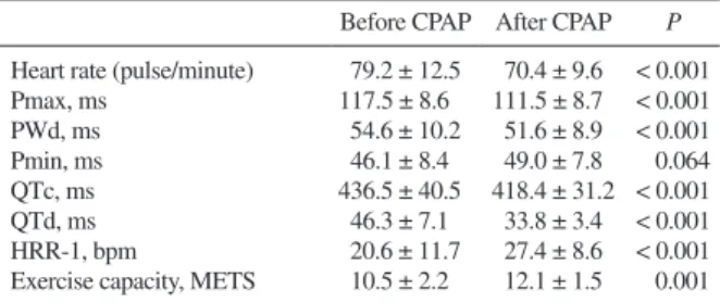
Table III. Electrocardiographic Parameters of the Patients With
Obstruc-tive Sleep Apnea Before and After CPAP Treatment
Before CPAP After CPAP P
Heart rate (pulse/minute) 79.2 ± 12.5 70.4 ± 9.6 < 0.001 Pmax, ms 117.5 ± 8.6 111.5 ± 8.7 < 0.001 PWd, ms 54.6 ± 10.2 51.6 ± 8.9 < 0.001 Pmin, ms 46.1 ± 8.4 49.0 ± 7.8 0.064 QTc, ms 436.5 ± 40.5 418.4 ± 31.2 < 0.001 QTd, ms 46.3 ± 7.1 33.8 ± 3.4 < 0.001 HRR-1, bpm 20.6 ± 11.7 27.4 ± 8.6 < 0.001 Exercise capacity, METS 10.5 ± 2.2 12.1 ± 1.5 0.001 HRR-1 indicates heart rate recovery at 1 minute; METS, metabolic equiv-alents; Pmax, maximum p-wave duration; Pmin, minimum p-wave dura-tion; PWd, P-wave dispersion; QTc, corrected QT interval; and QTd, QT dispersion. Data are presented as mean ± SD.
ing nCPAP therapy. PWd at baseline (54.6 ± 10.2 msec) sig-nificantly decreased following 3 months of nCPAP therapy (51.6 ± 8.9 msec, P < 0.001). The QTc (436.5 ± 40.5 msec) and QTd (46.3 ± 7.1 msec) at baseline significantly decreased following nCPAP treatment (418.4 ± 31.2 msec, P < 0.001 and 33.8 ± 3.4 msec, P < 0.001). The exercise capacity at baseline determined as 10.5 ± 2.2 METS and HRR1-1 (20.6 ± 11.7 bpm) significantly increased (12.1 ± 1.5 METS and 27.4 ± 8.6 bpm) following 3 months of nCPAP therapy.
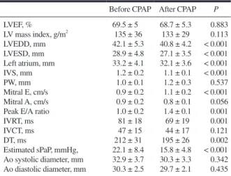
Echocardiographic parameters: At baseline, mean LVEF of the study group was 69.5 ± 5.0 (%). Following treatment, no significant difference existed for LVEF (P = 0.883), LV mass index (g/m2) (P = 0.113), or posterior wall diastolic thickness (P = 0.537). Following 12 weeks of nCPAP treatment, a signif-icant increase in the E/A ratio (1.0 ± 0.2 versus 1.4 ± 0.1, P = 0.001) and a significant reduction in IVRT (81 ± 18 versus 69 ± 19, P = 0.001) and DT (212 ± 31 versus 195 ± 26, P = 0.002) were observed.
AoS and AoD at baseline were 32.9 ± 3.7 mm and 30.3 ± 2.5 mm. There were no significant differences following 3 months of nCPAP treatment in the aortic root parameters (30.3 ± 3.3 mm, P = 0.342 and 29.7 ± 2.1 mm, P = 0.435). Estimat-ed systolic pulmonary artery pressure decreasEstimat-ed significantly following nCPAP treatment (22.1 ± 8.4 mmHg versus 15.8 ± 4.8 mmHg, P < 0.001).
Discussion
This study investigated the effects of nCPAP therapy on LV function and electrocardiographic parameters in moderate/ severe OSA patients. In our last two trials with this well select-ed group, we demonstratselect-ed that HR, PWd, HRR-1, and QTc are correlated with the severity of obstructive sleep apnea syn-drome (OSAS)28) and also we published the association
be-tween OSA and aortic root parameters and LV functions.29) The main findings of the present study are that abnormalities in diastolic function, arrhythmic electrocardiographic parame-ters such as PWd, QTc, QTd, and HRR-1 and deteriorated overnight polysomnographic parameters could be reversible, at least in part, with nCPAP therapy.
Previous studies have demonstrated a positive association between elevated HR at rest and adverse cardiovascular events in both the general population and the population with cardio-vascular disease.30-36) Few studies have investigated the influ-ence of severity of OSA on HR in patients with OSA. Sumi, et al16) reported that mean HR during 24 hours correlated posi-tively with AHI in 62 patients with OSA and that mean HR in the daytime (06:00-22:00 hour) and night-time (22:00-06:00 hour) were significantly reduced following 3 or 4 days of nC-PAP treatment in those patients. However, their study included patients using beta-blockers, which can affect mean HR. Therefore, it is unclear whether mean HR is independently as-sociated with the severity of OSA in patients with OSA. In ad-dition, the long-term effects of nCPAP therapy on mean HR remain unclear. In our study, we found a significant decrease in mean HR in moderate/severe OSA patients following 3 months of nCPAP therapy. Our study group is well selected without cardiovascular comorbidities and medical treatment, which can affect heart rate and nCPAP therapy is required for longer peri-ods than the above-mentioned trial.
Increased activation of the sympathetic nervous system due to nocturnal hypopnea and apnea episodes and arousal re-actions, respectively, is a pathophysiological hallmark of OSAS,37-39) which has been shown to be improved following therapy with CPAP ventilation.40) HRR-1 following exercise termination is a simple and readily available measure of vagal tone that has been found to be of high prognostic value in pa-tients referred for exercise testing for evaluation of coronary artery disease,27) in patients with established severe coronary artery disease,41) and in patients with congestive heart failure.42) Maeder, et al 15) concluded that the severity of OSAS expressed as higher AHI is independently associated with lower HRR-1. The mean AHI was similar in both severe OSAS groups (57.8 ± 22.5 versus 50 ± 11). In our moderate/severe OSAS patients, we found a significant improvement in HRR-1 following 12 weeks of nCPAP treatment as in previous studies.
The prolongation of intra-atrial and inter-atrial conduc-tion time and the inhomogeneous propagaconduc-tion of sinus impuls-es are well-known electrophysiological characteristics of the atrium prone to fibrillate and have been evaluated using two simple electrocardiography markers, Pmax and PWd.43-45) In-creases in the P-wave duration and PWd have been used as predictors of atrial fibrillation development in various clinical settings.44-49) Can, et al found that PWd was greater in patients with OSA than patients without OSA and is associated with severity of the disease.13) To the best of our knowledge, the ef-fects of nCPAP treatment on P-wave duration and PWd have not been evaluated in patients with OSA. In our study, we ob-served a significant decrease in Pmax and PWd following 3 months of nCPAP therapy and there were no significant chang-es in mean Pmin following nCPAP therapy.
QTd reflects inhomogeneity of repolarization. Delayed cardiac repolarization leading to prolongation of the QT inter-val is a well-characterized precursor of arrhythmias. The QTd is increased in patients with a prior myocardial infarction, who
Table IV. Left Ventricular Systolic, Diastolic and Aortic Root Parameters
of the Study Group Before and After CPAP Treatment
Before CPAP After CPAP P
LVEF, % 69.5 ± 5 68.7 ± 5.3 0.883 LV mass index, g/m2 135 ± 36 133 ± 29 0.113 LVEDD, mm 42.1 ± 5.3 40.8 ± 4.2 < 0.001 LVESD, mm 28.9 ± 4.8 27.1 ± 3.5 < 0.001 Left atrium, mm 33.2 ± 4.1 32.1 ± 3.6 < 0.001 IVS, mm 1.2 ± 0.2 1.1 ± 0.1 < 0.001 PW, mm 1.0 ± 0.1 1.2 ± 0.3 0.537 Mitral E, cm/s 0.9 ± 0.2 1.1 ± 0.2 < 0.001 Mitral A, cm/s 0.9 ± 0.2 0.8 ± 0.1 0.056 Peak E/A ratio 1.0 ± 0.2 1.4 ± 0.1 0.001
IVRT, ms 81 ± 18 69 ± 19 0.001 IVCT, ms 47 ± 15 44 ± 17 0.121 DT, ms 212 ± 31 195 ± 26 0.002 Estimated sPaP, mmHg, 22.1 ± 8.4 15.8 ± 4.8 < 0.001 Ao systolic diameter, mm 32.9 ± 3.7 30.3 ± 3.3 0.342 Ao diastolic diameter, mm 30.3 ± 2.5 29.7 ± 2.1 0.435 Data are presented as the mean ± SD. A indicates late diastolic peak flow velocity; Ao, aortic root; DT, mitral deceleration time; E, early diastolic peak flow velocity; IVCT, isovolumic contraction time; IVRT, isovolumic relaxation time; IVS, interventricular septum; LVEDD, left ventricular end diastolic diameter; LVEF, left ventricular ejection fraction; LVESD, left ventricular end systolic diameter; PW, posterior wall; and sPAP, systolic pulmonary artery pressure.
have a susceptibility to ventricular tachyarrhythmias, most ob-viously by re-entry mechanisms.50) Roche, et al 51) reported a significant alteration in the relationship between QT length and HR during CPAP treatment, but they did not find any changes in static repolarization parameters (QT, RT, QTc, and RTc). On the other hand, Yamashita, et al 52) observed more severe auto-nomic nervous dysfunction and increased QTd in hypertensive patients with OSA during episodes of apneas and hypopneas compared to those without OSA, and suggested that OSA might be associated with future development of cardiac events. Also, in our study we observed a significant decrease in QTc and QTd after nCPAP treatment in moderate/severe OSA pa-tients.
Changes in echocardiographic diastolic parameters fol-lowing nCPAP therapy have been reported in several studies, some with different patient selection criteria.53-55) The effects of cardioactive drugs, hypertension, obesity, or possible disease that affect diastolic function could have affected the results in some of the studies.56-59) Arias, et al investigated the effects of nCPAP therapy on LV diastolic function and concluded that chronic application of nCPAP could avoid the progression of diastolic abnormalities, and indeed, it might reserve these al-terations, at least in the initial stages prior to severe structural changes developing.12) In our study, we observed a significant improvement in diastolic function following 12 weeks of nC-PAP therapy in moderate/severe untreated OSA patients with/ without diastolic dysfunction.
The positive effect of nCPAP treatment on LV systolic function in heart failure patients has been well reported in pre-vious studies.60,61) At baseline, the mean LVEF of our study group was normal. Following treatment, we did not observe any significant difference for LVEF (P = 0.883). We can con-clude that nCPAP treatment has a positive effect on deteriorat-ed LV function, but no effect on patients with normal LV func-tion.
Conclusion: We demonstrated that in patients with moderate/ severe OSA, 12 weeks of nCPAP therapy significantly in-creased LV diastolic function and had no effect on systolic function and aortic root diameters. A positive effect on HR, PWd, HRR-1, QTc, and QTd was found following effective nCPAP therapy.
Disclosure
The authors declare that they have no conflict of interests.
References
1. Young T, Palta M, Dempsey J, Skatrud J, Weber S, Badr S. The occurrence of sleep-disordered breathing among middle-aged adults. N Engl J Med 1993; 328: 1230-5.
2. Young T, Peppard PE, Gottlieb DJ. Epidemiology of obstructive sleep apnea: a population health perspective. Am J Respir Crit Care Med 2002; 165: 1217-39. (Review)
3. Butt M, Dwivedi G, Khair O, Lip GY. Obstructive sleep apnea and cardiovascular disease. Int J Cardiol 2010; 139: 7-16. (Review) 4. Somers VK, White DP, Amin R, et al. Sleep apnea and
cardiovas-cular disease: an American Heart Association/American College of Cardiology Foundation Scientific Statement from the American Heart Association Council for High Blood Pressure Research
Pro-fessional Education Committee, Council on Clinical Cardiology, Stroke Council, and Council on Cardiovascular Nursing. J Am Coll Cardiol 2008; 52: 686-717.
5. Bradley TD, Floras JS. Obstructive sleep apnoea and its cardiovas-cular consequences. Lancet 2009; 373: 82-93. (Review) 6. Kasai T, Bradley TD. Obstructive sleep apnea and heart failure:
pathophysiologic and therapeutic implications. J Am Coll Cardiol 2011; 57: 119-27. (Review)
7. Dursunoglu N, Dursunoglu D, Ozkurt S, et al. Effects of CPAP on left ventricular structure and myocardial performance index in male patients with obstructive sleep apnoea. Sleep Med 2007; 8: 51-9.
8. Ryan CM, Usui K, Floras JS, Bradley TD. Effect of continuous positive airway pressure on ventricular ectopy in heart failure pa-tients with obstructive sleep apnoea. Thorax 2005; 60: 781-5. 9. Simantirakis EN, Schiza SI, Marketou ME, et al. Severe
bradyar-rhythmias in patients with sleep apnoea: the effect of continuous positive airway pressure treatment: a long-term evaluation using an insertable loop recorder. Eur Heart J 2004; 25: 1070-6. 10. Kaneko Y, Floras JS, Usui K, et al. Cardiovascular effects of
con-tinuous positive airway pressure in patients with heart failure and obstructive sleep apnea. N Engl J Med 2003; 348: 1233-41. 11. Mansfield DR, Gollogly NC, Kaye DM, Richardson M, Bergin P,
Naughton MT. Controlled trial of continuous positive airway pres-sure in obstructive sleep apnea and heart failure. Am J Respir Crit Care Med 2004; 169: 361-6.
12. Arias MA, Garcia-Rio F, Alonso-Fernández A, Madiano O, Mar-tínez I, Villamor J. Obstructive sleep apnea syndrome affects left ventricular diastolic function: effects of nasal continuous positive airway pressure in men. Circulation 2005; 112: 375-83.
13. Can I, Aytemir K, Demir AU, et al. P-wave duration and disper-sion in patients with obstructive sleep apnea. Int J Cardiol 2009; 133: e85-9.
14. Dursunoglu D, Dursunoglu N. Effect of CPAP on QT interval dis-persion in obstructive sleep apnea patients without hypertension. Sleep Med 2007; 8: 478-83.
15. Maeder MT, Münzer T, Rickli H, et al. Association between heart rate recovery and severity of obstructive sleep apnea syndrome. Sleep Med 2008; 9: 753-61.
16. Sumi K, Chin K, Takahashi K, et al. Effect of nCPAP therapy on heart rate in patients with obstructive sleep apnoea-hypopnoea. QJM 2006; 99: 545-53.
17. Iber C, Ancoli-Israel S, Chesson AL, Quan SF for the American Academy of Sleep Medicine. The AASM Manual for the Scoring
of Sleep and Associated Events: Rules, Terminology and Technical Specifications. 1st ed. Westchester, IL : American Academy of Sleep Medicine; 2007.
18. Sahn DJ, De Maria A, Kisslo J, Weyman A. Recommendations re-garding quantification in M-mode echocardiography: results of a survey of echocardiographics measurements. Circulation 1978; 58: 1072-83.
19. Schiller NB, Shah PM, Crawford M, et al. Recommendations for quantification of the left ventricle by two-dimensional echocardi-ography. American Society of Echocardiography Committee on Standards, Subcommittee on Quantification of Two-Dimensional Echoacrdiograms. J Am Soc Echocardiogr 1989; 2: 358-67. (Re-view)
20. Devereux RB, Alonso DR, Lutas EM, et al. Echocardiographic as-sessment of left ventricular hypertrophy: comparison to necropsy findings. Am J Cardiol 1986; 57: 450-8.
21. Quinones MA, Waggoner AD, Reduto LA, et al. A new, simplified and accurate method for determining ejection fraction with two-dimensional echocardiography Circulation 1981; 64: 744-53. 22. Berger M, Haimowitz A, Van Tosh A, Berdoff RL, Goldberg E.
Quantitative assessment of pulmonary hypertension in patients with tricuspid regurgitation using continuous wave doppler ultra-sound. J Am Coll Cardiol 1985; 6: 359-65.
23. Stefanadis C, Stratos C, Boudoulas H, Kourouklis C, Toutouzas P. Distensibility of the ascending aorta: Comparison of invasive and non-invasive techniques in healthy men and in men with coronary
artery disease. Eur Heart J 1990; 11: 990-6.
24. Bazett HC. An analysis of the time-relations of electrocardio-grams. Ann Noninvasive Electrocardiol 1997; 2: 177-94. 25. Fletcher GF, Balady GJ, Amsterdam EA, et al. Exercise standards
for testing and training: a statement for healthcare professionals from the American Heart Association. Circulation 2001; 104: 1694-740.
26. Mehra R, Benjamin EJ, Shahar E, et al. Association of nocturnal arrhythmias with sleep-disordered breathing: The Sleep Heart Health Study. Am J Respir Crit Care Med 2006; 173: 910-6. 27. Cole CR, Blackstone EH, Pashkow FJ, Snader CE, Lauer MS.
Heart-rate recovery immediately after exercise as a predictor of mortality. N Engl J Med 1999; 341: 1351-7.
28. Çiçek D, Lakadamyali H, Gökay S, Sapmaz I, Muderrisoglu H. Effect of obstructive sleep apnea on heart rate, heart rate recovery and QTc and P-wave dispersion in newly diagnosed untreated pa-tients. Am J Med Sci 2012; 344: 180-5.
29. Cicek D, Lakadamyali H, Yağbasan BD, Sapmaz I, Müderrisoğlu H. Obstructive sleep apnoea and its association with left ventricu-lar function and aortic root parameters in newly diagnosed, un-treated patients: a prospective study. J Int Med Res 2011; 39: 2228-38.
30. Benetos A, Rudnichi A, Thomas F, Safar M, Guize L. Influence of heart rate on mortality in a French population: role of age, gender, and blood pressure. Hypertension 1999; 33: 44-52.
31. Diaz A, Bourassa MG, Guertin MC, Tardif JC. Long-term prog-nostic value of resting heart rate in patients with suspected or proven coronary artery disease. Eur Heart J 2005; 26: 967-74. 32. Gillman MW, Kannel WB, Belanger A, D’Agostino RB. Influence
of heart rate on mortality among persons with hypertension: the Framingham Study. Am Heart J 1993; 125: 1148-54.
33. Greenland P, Daviglus ML, Dyer AR, et al. Resting heart rate is a risk factor for cardiovascular and noncardiovascular mortality: the Chicago Heart Association Detection Project in Industry. Am J Epidemiol 1999; 149: 853-62.
34. Kannel WB, Kannel C, Paffenbarger RS Jr, Cupples LA. Heart rate and cardiovascular mortality: the Framingham Study. Am Heart J 1987; 113: 1489-94.
35. Palatini P, Julius S. Association of tachycardia with morbidity and mortality: pathophysiological considerations. J Hum Hypertens 1997; 11: S19-27. (Review)
36. Tverdal A, Hjellvik V, Selmer R. Heart rate and mortality from cardiovascular causes: a 12 year follow-up study of 379,843 men and women aged 40-45 years. Eur Heart J 2008; 29: 2772-81. 37. Grassi G, Facchini A, Trevano FQ, et al. Obstructive sleep
apnea-dependent and -inapnea-dependent adrenergic activation in obesity. Hy-pertension 2005; 46: 321-5.
38. Narkiewicz K, van de Borne PJ, Cooley RL, Dyken ME, Somers VK. Sympathetic activity in obese subjects with and without ob-structive sleep apnea. Circulation 1998; 98: 772-6.
39. Narkiewicz K, van de Borne PJ, Montano N, Dyken ME, Phillips BG, Somers VK. Contribution of tonic chemoreflex activation to sympathetic activity and blood pressure in patients with obstruc-tive sleep apnea. Circulation 1998; 97: 943-5.
40. Narkiewicz K, Kato M, Phillips BG, Pesek CA, Davison DE, Somers VK. Nocturnal continuous positive airway pressure de-creases daytime sympathetic traffic in obstructive sleep apnea. Circulation 1999; 100: 2332-5.
41. Vivekananthan DP, Blackstone EH, Pothier CE, Lauer MS. Heart rate recovery after exercise is a predictor of mortality, independent of the angiographic severity of coronary disease. J Am Coll Cardi-ol 2003; 42: 831-8.
42. Arena R, Guazzi M, Myers J, Peberdy MA. Prognostic value of heart rate recovery in patients with heart failure. Am Heart J 2006; 151: 851.e7-13.
43. Boriani G, Diemberger I, Biffi M, et al. P wave dispersion and short-term vs. late atrial fibrillation recurrences after
cardiover-sion. Int J Cardiol 2005; 101: 355-61.
44. Dilaveris PE, Gialafos EJ, Sideris SK, et al. Simple electrocardio-graphic markers for the prediction of paroxysmal idiopathic atrial fibrillation. Am Heart J 1998; 135: 733-8.
45. Köse S, Aytemir K, Sade E, et al. Detection of patients with hyper-trophic cardiomyopathy at risk for paroxysmal atrial fibrillation during sinus rhythm by P-wave dispersion. Clin Cardiol 2003; 26: 431-4.
46. Dilaveris PE, Gialafos JE. P-wave dispersion: a novel predictor of paroxysmal atrial fibrillation. Ann Noninvasive Electrocardiol 2001; 6: 159-65. (Review)
47. Dogan A, Acar G, Gedikli O, et al. A comparison of P-wave dura-tion and dispersion in patients with short-term and long-term atrial fibrillation. J Electrocardiol 2003; 36: 251-5.
48. Tezcan UK, Amasyali B, Can I, et al. Increased P wave dispersion and maximum P wave duration after hemodialysis. Ann Noninva-sive Electrocardiol 2004; 9: 34-8.
49. Tsikouris JP, Kluger J, Song J, White CM. Changes in P-wave dis-persion and P-wave duration after open heart surgery are associat-ed with the peak incidence of atrial fibrillation. Heart Lung 2001; 30: 466-71.
50. Perkiömäki JS, Koistinen MJ, Yli-Mäyry S, Huikuri HV. Disper-sion of QT interval in patients with and without susceptibility to ventricular tachyarrhythmias after previous myocardial infarction. J Am Coll Cardiol 1995; 26: 174-9.
51. Roche F, Barthélémy JC, Garet M, Duverney D, Pichot V, Sforza E. Continuous positive airway pressure treatment improves the QT rate dependence adaptation of obstructive sleep apnea patients. Pacing Clin Electrophysiol 2005; 28: 819-25.
52. Yamashita J, Nomura M, Uehara K, et al. Influence of sleep apnea on autonomic nervous activity and QT dispersion in patients with essential hypertension and old myocardial infarction. J Electrocar-diol 2004; 37: 31-40.
53. Alchanatis M, Paradellis G, Pini H, Tourkohoriti G, Jordanoglou J. Left ventricular function in patients with obstructive sleep apnoea syndrome before and after treatment with nasal continuous posi-tive airway pressure. Respiration 2000; 67: 367-71.
54. Chazan R, Najniger MJ, Przybylowski T, Pfejfer DL, Droszcz W. Effect continuous positive airway pressure (CPAP) on left ven-tricular diastolic function in patients with obstructive sleep apnea syndrome--OSAS. Pol Arch Med Wewn 1995; 94: 8-13. (Polish) 55. Konermann M, Sanner B, Klewer J, Kreuser I, Laschewski F,
Bur-mann-Urbanek M. Modification of cardiopulmonary performance parameters in patients with obstructive sleep apnea treated with nCPAP therapy. Wien Med Wochenschr 1996; 146: 340-3. (Ger-man)
56. Alchanatis M, Tourkohoriti G, Kosmas EN, et al. Evidence for left ventricular dysfunction in patients with obstructive sleep apnoea syndrome. Eur Respir J 2002; 20: 1239-45.
57. Fung JW, Li TS, Choy DK, et al. Severe obstructive sleep apnea is associated with left ventricular diastolic dysfunction. Chest 2002; 121: 422-9.
58. Kraiczi H, Caidahl K, Samuelsson A, Peker Y, Hedner J. Impair-ment of vascular endothelial function and left ventricular filling: association with the severity of apnea-induced hypoxemia during sleep. Chest 2001; 119: 1085-91.
59. Niroumand M, Kuperstein R, Sasson Z, Hanly PJ. Impact of ob-structive sleep apnea on left ventricular mass and diastolic func-tion. Am J Respir Crit Care Med 2001; 163: 1632-6.
60. Akar Bayram N, Ciftci B, Durmaz T, et al. Effects of continuous positive airway pressure therapy on left ventricular function as-sessed by tissue Doppler imaging in patients with obstructive sleep apnoea syndrome. Eur J Echocardiogr 2009; 10: 376-82. 61. Ferrier KA, Neill AM, O’Meeghan T, Richards M, Campbell AJ.
Continuous positive airway pressure in heart failure patients with obstructive sleep apnoea. Intern Med J 2008; 38: 829-36.