Original Paper
Med Princ Pract 2019;28:533–538
Evaluation of the Relationship between Type II
Diabetes Mellitus and the Prevalence of Apical
Periodontitis in Root-Filled Teeth Using Cone
Beam Computed Tomography: An Observational
Cross-Sectional Study
Selen Nihal Sisli
Department of Endodontics, Faculty of Dentistry, Baskent University, Ankara, Turkey
Received: November 7, 2018 Accepted: April 18, 2019 Published online: April 19, 2019
Selen Nihal Sisli © 2019 The Author(s)
Significance of the Study
• A possible association between type II diabetes mellitus and apical periodontitis was evaluated in this study using cone beam computed tomographic images of adequately root-filled and restored teeth. A greater prevalence of apical periodontitis and severe bone destruction was observed in patients with diabetes mellitus compared with nondiabetic patients.
DOI: 10.1159/000500472
Keywords
Periapical periodontitis · Diabetes mellitus · Cone beam computed tomography
Abstract
Objective: This study aimed to investigate the prevalence of apical periodontitis (AP) in patients with type II diabetes mel-litus (DM) with either optimal glycemic control (OGC) or poor glycemic control (PGC) compared with nondiabetics using cone beam computed tomography (CBCT). Subjects and Methods: The periapical status of 75 teeth with adequate root canal treatment performed at least 1 year ago that could be visualized in the field of view of CBCT images of 43 DM patients (29 females, 14 males) was compared with a control group consisting of 162 teeth of 86 nondiabetics (56 females, 30 males). In addition, the DM group was divided into
sub-groups according to the patients’ mean glycated hemoglo-bin level as either having OGC or PGC. The periapical status of the teeth was evaluated using the CBCT periapical index (CBCTPAI), and AP was diagnosed as a CBCTPAI ≥1. χ2,
Fish-er’s exact test, and independent-samples t tests were used for the statistical analysis, and p < 0.05 was considered sig-nificant. Results: Significant differences between the DM group and the control group were observed (p < 0.05) in terms of AP (the frequencies of both CBCTPAI ≥1 and CBCTPAI ≥3) and the frequency of cardiovascular disease, while there were no significant differences between the DM subgroups (p > 0.05). Conclusion: The prevalence of AP and severe bone destruction in periapical tissues was significant-ly higher in the DM patients compared with the nondiabetic patients. © 2019 The Author(s)
Introduction
Polymicrobic agents originating from the necrotic dental pulp can cause apical periodontitis (AP), which is characterized by an inflammatory response and bone de-struction in periapical tissues [1, 2]. Bone dede-struction from AP can be identified radiographically as localized radiolucent destruction in the corresponding tooth root apex of the adjacent bone tissue [3]. Ideally, endodontic treatment should clean, disinfect, and fill all the root ca-nals hermetically, aiming to cure the AP. However, inad-equately aseptic environments, procedural errors, and systemic disease can disrupt the recovery mechanism of an endodontic treatment [4] and cause persistent AP.
A systemic disease possibly associated with AP that has been investigated is diabetes mellitus (DM), which results from the absence (type I DM) or deficiency (type II DM) of insulin [5]. DM is a chronic disease that leads to spe-cific complications associated with hyperglycemia and is associated with delayed healing, a decrease in cytokine production, and a diminished tissue repair capacity, in-cluding neutrophil chemotaxis [6]. The level of glycated hemoglobin (HbA1c) has been accepted as the most
im-portant reference for controlling mean glycemia and predicting the risk of the development of DM complica-tions [7]. According to the American Association of Clin-ical Endocrinologists, an HbA1c level of ≤6.5% is
consid-ered the goal for optimal glycemic control in diabetic pa-tients [8].
Investigations of the hypothesis that DM is associated with a higher incidence of AP, using both animal [9–12] and human [5, 6, 13–15] models, have yielded inconclu-sive results. All previous human studies [5, 6, 13–15] have used panoramic or periapical radiographs in their meth-odology, although the limitations of conventional radio-graphic methods (i.e., the 2D nature of the image, ana-tomical noise, geometric distortion, and possible dismiss-al of periapicdismiss-al lesions within cancellous bone) are well established [16, 17]. Recently, cone beam computed to-mography (CBCT) imaging has gained popularity in epi-demiological studies because its 3D nature allows for the detection of AP before it can be recognized radiographi-cally [18, 19]. In addition to the reports of CBCT imaging results in higher detection rates of AP, CBCT scanning produces findings closer to histologic evaluations [20– 22]. Therefore, by having superior validity and reliability compared with radiographs, CBCT minimizes false diag-noses. Despite these outstanding features, the radiation dose is significantly higher compared with conventional radiographic techniques [23]; therefore, the American
Association of Endodontists and the American Academy of Oral and Maxillofacial Radiology recommend that the application of CBCT with limited field of view (FOV) can be the method of choice when the third dimension is needed [23]. Existing CBCT images can be analyzed to obtain additional information from incidental findings that may be discovered within the FOV [24, 25].
This cross-sectional study aimed to compare the prev-alence of AP in root-filled teeth in type II diabetic patients with optimal glycemic control (OGC) and poor glycemic control (PGC) with that of nondiabetic patients from a Turkish subpopulation using CBCT evaluations.
Subjects and Methods
Study Design and Data Source
The records of DM patients who had been followed up in the Endocrine Polyclinic and also attended the Dental Polyclinic of the Adana Baskent University between December 2014 and December 2017 were scanned for this study. Type II DM patients with a his-tory of immunosuppressive medication or chemotherapy and a lack of follow-up information were excluded, and those who coin-cidentally had CBCT scans performed for any diagnostic reasons were listed.
Inclusion Criteria
Patients with at least one tooth with an adequate root canal treatment and coronal restoration within the FOV of an existing CBCT were selected. However, after careful examination of the anamneses, if the root canal treatment of a selected tooth was found to have been performed less than 1 year ago, the subject was excluded. Additionally, root-filled teeth without permanent resto-ration were also excluded. Ultimately, 75 teeth of 43 DM patients with adequate root canal treatment were included in the study.
Information about the most recent HbA1c levels, the presence
of cardiovascular disease (CVD), and the age and gender of the pa-tients (29 females, 14 males; mean age 56.40 ± 10.30 years) were obtained from the hospital’s medical database. Adequate glycemic control was defined according to the American Association of Clin-ical Endocrinologists as an HbA1c level ≤6.5% [8]. Based on HbA1c
levels, the DM patients were divided into two subgroups: those with an HbA1c level ≤6.5% were placed in the OGC group and those with
an HbA1c level >6.5% were placed in the PGC group. Thirty DM
patients (53 teeth in total) were placed in the OGC group, while 13 patients (22 teeth in total) were placed in the PGC group. As a con-trol group, 162 teeth with adequate root canal treatment of 86 pa-tients whose age and gender distribution was compatible with the DM group (56 females, 30 males; mean age 56.28 ± 10.07 years) were selected from the CBCT patient records. The inclusion criteria for the control group were the same as those for the DM group.
CBCT Examination and Evaluation
All of the CBCT scans were performed by two trained techni-cians using a Pax-i3D apparatus (Vatech, Hwaseong, South Korea) with the manufacturer’s recommended exposure conditions (24 s, 90 kVp, and 5.7 mA) with a voxel size of 0.200 mm at 4 different
FOVs: 50 × 50, 80 × 50, 80 × 80, and 120 × 90 mm. EasyDent V4 Viewer (Version 4.1.4.5.) software was used for the reconstruction of the images. The quality of the root canal fillings was examined according to a modified form of the criteria described by De Moor et al. [26]. An adequate root canal filling was defined as a complete filling ending at least 1–2 mm from the apex with no voids. An in-adequate filling was defined as a poorly condensed filling with an over- or under-root filling length. For teeth with multiple canals, the quality of the root canal filling was ranked according to the root canal with the worst quality.
The periapical status of the teeth was independently evaluated by two experienced endodontists that had been previously cali-brated in using the cone beam computed tomography periapical index (CBCTPAI) [27]. This index uses a 6-point scoring system from 0 to 5, with 2 variables describing the expansion (E) and de-struction (D) of cortical bone (Table 1). The CBCTPAI score was determined by the largest extension of the lesion. A CBCTPAI score ≥1 was considered to be a sign of AP. In multirooted teeth, the worst CBCTPAI score of all the roots was used. The kappa co-efficient was used at the end of the evaluation to analyze the agree-ment between the evaluators of the presence and/or absence of AP (k = 0.89). Cases in which the evaluators disagreed were resolved by joint discussion until consensus was achieved.
Statistical Analysis
The minimum sample size for each group of 70 root-filled and restored teeth was estimated according to α = 0.05 and a power of 80% using http://powerandsamplesize.com/Calculators/Com-pare-2-Proportions/2-Sample-Equality. The SPSS (Version 23.0) package program was used for the statistical analysis of the data. Categorical measurements were summarized using numbers and percentages, whereas means and standard deviations (median and minimum-maximum values where necessary) were used for con-tinuous measurements. χ2 or Fisher’s exact tests were used for the
comparisons of categorical variables. Independent-samples t tests were used for the comparisons of continuous measurements be-tween groups. Values of p < 0.05 were considered significant.
Table 1. Cone beam computed tomography periapical index
(CBCTPAI) scores defined by Estrela et al. [27]
Score Quantitative bone alterations in mineral struc-tures
0 Intact periapical bone structures
1 Diameter of periapical radiolucency >0.5–1 mm 2 Diameter of periapical radiolucency >1–2 mm 3 Diameter of periapical radiolucency >2–4 mm 4 Diameter of periapical radiolucency >4–8 mm 5 Diameter of periapical radiolucency >8 mm Score, n + E* Expansion of periapical cortical bone Score, n + D* Destruction of periapical cortical bone
* The variables E (expansion of cortical bone) and D (destruc-tion of cortical bone) were added to each score if either of these conditions was detected in the CBCT analysis.
Table 2. Distribution of CBCTPAI scores of teeth in DM and
con-trol groups
Groups Total control DM
CBCTPAI 0 Count of teeth 116 47 163 % within group 71.6 62.7 68.8 1 Count of teeth 15 4 19 % within group 9.3 5.3 8.0 2 Count of teeth 20 8 28 % within group 12.3 10.7 11.8 3 Count of teeth 4 4 8 % within group 2.5 5.3 3.4 4 Count of teeth 3 3 6 % within group 1.9 4.0 2.5 5 Count of teeth 0 2 2 % within group 0.0 2.7 0.8 5+D Count of teeth 4 7 11 % within group 2.5 9.3 4.6 Total Count of teeth 162 75 237
% within group 100.0 100.0 100.0 CBCTPAI, cone beam computed tomography periapical index; D, destruction of cortical bone.
Table 3. Distribution of CBCTPAI scores of teeth in OGC and
PGC groups
DM Total OGC PGC
CBCTPAI
score 0 Count of teeth% within group 3362.3 1463.6 4762.7 1 Count of teeth 3 1 4 % within group 5.7 4.5 5.3 2 Count of teeth 5 3 8 % within group 9.4 13.6 10.7 3 Count of teeth 4 0 4 % within group 7.5 0.0 5.3 4 Count of teeth 1 2 3 % within group 1.9 9.1 4.0 5 Count of teeth 2 0 2 % within group 3.8 0.0 2.7 5+D Count of teeth 5 2 7 % within group 9.4 9.1 9.3 Total Count of teeth 53 22 75
% within group 100.0 100.0 100.0 CBCTPAI, cone beam computed tomography periapical index; D, destruction of cortical bone; OGC, optimal glycemic control; PGC, poor glycemic control.
Results
CBCTPAI scores for the DM group and the control group are presented in Table 2. The frequency of AP was 37.3% for the DM group and 28.4% for the control group, and this difference was statistically significant (p < 0.05). The frequency of CBCTPAI score ≥3 in the DM group was 21.3%, while it was 6.79% in the control group, and this difference was also statistically signifi-cant (p < 0.05).
There was no significant difference between the DM group and the control group in terms of age, gender, and type of teeth evaluated (teeth with single root canals vs. teeth with multiple root canals). The frequencies of teeth with single root canals were 73.4 and 71.7% for the DM group and the control group, respectively. The frequen-cies of teeth with multiple root canals were 26.6 and 28.3% for the DM group and the control group, respectively.
In the DM group, 23 patients (53.5% of the DM group) had CVD, while 16 patients in the control group (18.6%) had CVD. This difference was found to be statistically significant (p < 0.05).
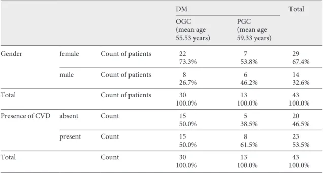
The CBCTPAI scores for the DM subgroups are pre-sented in Table 3. The gender distribution and frequen-cies of CVD are presented in Table 4. No significant dif-ference was found in either the frequency of AP and CVD or in the distribution of gender and age (p > 0.05).
Discussion
According to the results of the present study, the prev-alence of AP was significantly higher in the DM patients compared with the nondiabetics. This result can be inter-preted as DM adversely affecting or delaying the healing process of root canal treatments because they were per-formed at least 1 year previously. Since the preoperative radiologic and clinical evaluation of the treated teeth were absent, it was impossible to discriminate still-heal-ing cases, which is the main limitation of this study. This limitation arose because the effective CBCT radiation dose is higher than that for conventional radiography, and the nature of cross-sectional study design. Accord-ing to the current guidelines, CBCT cannot be used pre-operatively when performing an ordinary root canal treatment [23]. Therefore, study subjects were coinci-dently visualized within the FOV of recorded CBCT im-ages that had already been taken for any diagnostic rea-son. Moreover, in such a cross-sectional study design, data is collected at one point in time to state disease prev-alence and associate the disease to pathogenic factors. However, these data do not capture all the information about how the root canal treatments were performed, and it is not possible to control all the factors affecting the healing process or the outcome [25]. By including only the teeth with adequate root canal fillings and res-torations that were performed at least 1 year earlier and
Table 4. Gender distribution and frequencies of cardiovascular disease (CVD) in DM subgroups
DM Total OGC (mean age 55.53 years) PGC (mean age 59.33 years)
Gender female Count of patients 22 7 29 73.3% 53.8% 67.4% male Count of patients 8 6 14
26.7% 46.2% 32.6% Total Count of patients 30 13 43
100.0% 100.0% 100.0% Presence of CVD absent Count 15 5 20
50.0% 38.5% 46.5%
present Count 15 8 23
50.0% 61.5% 53.5%
Total Count 30 13 43
choosing control subjects who were age- and sex-matched to the DM group, such confounding variables were in-tended to be reduced.
As shown in Table 1, CBCTPAI scores <3 revealed minimal bone destruction of about 2 mm, so the frequen-cy of a CBCTPAI score ≥3 was also statistically evaluated. Consequently, as well as the frequency of a CBCTPAI score ≥1, a CBCTPAI score ≥3 was also significantly higher in the DM group, which indicates that severe bone destruction was significantly more frequent in the DM subjects with AP.
Similar to the findings of the present study, a recent meta-analysis [5] reported a significant association be-tween DM and AP in endodontically treated teeth. Ad-ditionally, the findings of Arya et al. [13] from a prospec-tive study design with a 1-year follow-up period were in accordance with the present study. They observed signif-icantly less periapical healing in the DM group (43%) compared with the control group (80%). However, in a systematic review, Aminoshariae et al. [14] stated that it is impossible to infer a significant relationship between DM and endodontic outcome based on the previous in-conclusive findings.
The current literature indicates that the relationship between DM and endodontic diseases is a paradox. The negative effect of DM on tissue healing and the mecha-nism of bone turnover is well known and supported by animal studies which show that DM induces periapical lesions [9] and has an adverse effect on vital pulp thera-py [10]. On the other hand, AP is not an entirely local-ized periradicular problem; the cell membranes of the anaerobic gram-negative bacteria that cause AP stimu-late the release of proinflammatory cytokines into the systemic circulation, thus increasing the systemic in-flammatory status [28]. In an animal model, Cintra et al. [11] showed that both endodontic and periodontal in-fections may increase the levels of HbA1c in both
dia-betic and nondiadia-betic rats. In addition, Astolphi et al. [12] found that the presence of a periapical lesion can have detrimental effects on insulin signaling and cause insulin resistance. In a cross-sectional clinical study that investigated the possible association between HbA1c
lev-els and the prevalence of AP [15], worse periapical status was found to be significantly correlated with HbA1c
lev-els ≥6.5% in type II DM patients. However, the preva-lence of AP in root-filled teeth was found to be similar in both HbA1c groups, as was reported in the present
study (Table 3).
As 3D images were evaluated in the present study, the CBCTPAI defined by Estrela et al. [27] was preferred over
Ørstavik’s index [29], which has previously been success-fully used in the evaluation of 2D images. CBCTPAI of-fers an accurate diagnosis through the analysis of lesions in 3D high-resolution images, with CT slices obtained in mesiodistal, buccopalatal, and diagonal directions.
In the present study, the prevalence of CVD was sig-nificantly higher in the DM group. Since type II DM is related to clustered risk factors for CVD, and amongst DM patients a prevalence of 75–85% for hypertension, 70–80% for elevated LDL, and 60–70% for obesity has been reported [30], this result agrees with a well-estab-lished association between CVD and DM.
Conclusion
Within the limitations of this cross-sectional study, a significant association was observed between DM and a higher prevalence of AP in root-filled teeth and CVD. Further clinical and prospective epidemiologic investiga-tions are needed to clarify this issue.
Acknowledgment
This study was approved and supported by Baskent University.
Statement of Ethics
The protocol for this cross-sectional study was approved by the Medical Ethics Committee of Baskent University (Project No: D-KA16/15).
Disclosure Statement
There are no conflicts of interest to declare.
References 1 Graves DT, Oates T, Garlet GP: Review of os-teoimmunology and the host response in
end-odontic and periodontal lesions. J Oral
Mi-crobiol. 2011 Jan 17;3.
2 Siqueira JF Jr, Rôças IN. Present status and future directions in endodontic microbiolo-gy. Endod Topics. 2014;30(1):3–22.
3 Bender IB, Seltzer S. Roentgenographic and direct observation of experimental lesions in
bone: I. 1961. J Endod. 2003 Nov;29(11):
702–6.
4 Nair PN. On the causes of persistent apical
periodontitis: a review. Int Endod J. 2006 Apr;
5 Segura-Egea JJ, Martín-González J, Cabanil-las-Balsera D, Fouad AF, Velasco-Ortega E, López-López J. Association between diabetes and the prevalence of radiolucent periapical lesions in root-filled teeth: systematic review
and meta-analysis. Clin Oral Investig. 2016
Jul;20(6):1133–41.
6 Delamaire M, Maugendre D, Moreno M, Le Goff MC, Allannic H, Genetet B. Impaired
leucocyte functions in diabetic patients.
Dia-bet Med. 1997 Jan;14(1):29–34.
7 Expert Committee on the Diagnosis and Clas-sification of Diabetes Mellitus. Report of the expert committee on the diagnosis and
clas-sification of diabetes mellitus. Diabetes Care.
2000 Jan;23 Suppl 1:S4–19.
8 Gionfriddo MR, McCoy RG, Lipska KJ. The 2013 American Association of Clinical Endo-crinologists’ diabetes mellitus management recommendations: improvements needed.
JAMA Intern Med. 2014 Feb;174(2):179–80. 9 Iwama A, Nishigaki N, Nakamura K,
Imai-zumi I, Shibata N, Yamasaki M, et al. The ef-fect of high sugar intake on the development of periradicular lesions in rats with type 2
dia-betes. J Dent Res. 2003 Apr;82(4):322–5.
10 Garber SE, Shabahang S, Escher AP, Torabi-nejad M. The effect of hyperglycemia on
pulp-al hepulp-aling in rats. J Endod. 2009 Jan;35(1):
60–2.
11 Cintra LT, Samuel RO, Facundo AC, Prieto AK, Sumida DH, Bomfim SR, et al. Relation-ships between oral infections and blood glu-cose concentrations or HbA1c levels in
nor-mal and diabetic rats. Int Endod J. 2014 Mar;
47(3):228–37.
12 Astolphi RD, Curbete MM, Colombo NH, Shirakashi DJ, Chiba FY, Prieto AK, et al. Periapical lesions decrease insulin signal and
cause insulin resistance. J Endod. 2013 May;
39(5):648–52.
13 Arya S, Duhan J, Tewari S, Sangwan P, Gha-laut V, Aggarwal S. Healing of Apical Peri-odontitis after Nonsurgical Treatment in
Pa-tients with Type 2 Diabetes. J Endod. 2017
Oct;43(10):1623–7.
14 Aminoshariae A, Kulild JC, Mickel A, Fouad AF. Association between Systemic Diseases and Endodontic Outcome: A Systematic
Re-view. J Endod. 2017 Apr;43(4):514–9.
15 Sánchez-Domínguez B, López-López J, Jané-Salas E, Castellanos-Cosano L, Velasco-Orte-ga E, Segura-Egea JJ. Glycated hemoglobin levels and prevalence of apical periodontitis
in type 2 diabetic patients. J Endod. 2015 May;
41(5):601–6.
16 Patel S, Durack C, Abella F, Shemesh H, Roig M, Lemberg K. Cone beam computed
tomog-raphy in Endodontics - a review. Int Endod J.
2015 Jan;48(1):3–15.
17 Estrela C, Bueno MR, Leles CR, Azevedo B, Azevedo JR. Accuracy of cone beam comput-ed tomography and panoramic and periapical radiography for detection of apical
periodon-titis. J Endod. 2008 Mar;34(3):273–9.
18 Karabucak B, Bunes A, Chehoud C, Kohli MR, Setzer F. Prevalence of apical periodon-titis in endodontically treated premolars and molars with untreated canal: A cone-beam
computed tomography study. J Endod. 2016
Apr;42(4):538–41.
19 Nur BG, Ok E, Altunsoy M, Ağlarci OS, Çolak M, Güngör E. Evaluation of technical quality and periapical health of root-filled teeth by
using cone-beam CT. J Appl Oral Sci. 2014
Nov-Dec;22(6):502–8.
20 Uraba S, Ebihara A, Komatsu K, Ohbayashi N, Okiji T. Ability of Cone-beam Computed Tomography to Detect Periapical Lesions That Were Not Detected by Periapical Radi-ography: A Retrospective Assessment
Ac-cording to Tooth Group. J Endod. 2016 Aug;
42(8):1186–90.
21 de Paula-Silva FW, Wu MK, Leonardo MR, da Silva LA, Wesselink PR. Accuracy of peri-apical radiography and cone-beam computed tomography scans in diagnosing apical peri-odontitis using histopathological findings as a
gold standard. J Endod. 2009 Jul;35(7):1009–
12.
22 Kanagasingam S, Lim CX, Yong CP, Mann-occi F, Patel S. Diagnostic accuracy of periapi-cal radiography and cone beam computed to-mography in detecting apical periodontitis using histopathological findings as a
refer-ence standard. Int Endod J. 2017 May;50(5):
417–26.
23 American Association of Endodontists. American Academy of Oral and Maxillofacial Radiology: AAE and AAOMR joint position statement use of cone beam computed
to-mography in endodontics 2015 update. J
En-dod. 2015;41:1393–6.
24 Oser DG, Henson BR, Shiang EY, Finkelman MD, Amato RB. Incidental Findings in Small Field of View Cone-beam Computed
Tomog-raphy Scans. J Endod. 2017 Jun;43(6):901–4.
25 Gambarini G, Piasecki L, Miccoli G, Gaimari G, Nardo DD, Testarelli L. Cone-beam com-puted tomography in the assessment of peri-apical lesions in endodontically treated teeth
[Erratum in: Eur J Dent 2018;12] [2]. Eur J
Dent. 2018 Jan-Mar;12(1):136–43.
26 De Moor RJ, Hommez GM, De Boever JG, Delmé KI, Martens GE. Periapical health re-lated to the quality of root canal treatment in
a Belgian population. Int Endod J. 2000 Mar;
33(2):113–20.
27 Estrela C, Bueno MR, Azevedo BC, Azevedo JR, Pécora JD. A new periapical index based
on cone beam computed tomography. J
En-dod. 2008 Nov;34(11):1325–31.
28 Henderson B, Wilson M. Cytokine induction
by bacteria: beyond lipopolysaccharide.
Cyto-kine. 1996 Apr;8(4):269–82.
29 Ørstavik D, Kerekes K, Eriksen HM. The peri-apical index: a scoring system for
radiograph-ic assessment of apradiograph-ical periodontitis. Endod
Dent Traumatol. 1986 Feb;2(1):20–34. 30 Preis SR, Pencina MJ, Hwang SJ, D’Agostino
RB Sr, Savage PJ, Levy D, et al. Trends in car-diovascular disease risk factors in individuals with and without diabetes mellitus in the
Framingham Heart Study. Circulation. 2009